4343
Evaluation of Location-Dependent Attenuation of Experimental Autoimmune Encephalomyelitis after Glial-Restricted Progenitor Transplantation using CEST MRI and MRS1Johns Hopkins University, Baltimore, MD, United States, 2Kennedy Krieger Institute, Baltimore, MD, United States
Synopsis
The utility of CEST MRI and MRS as a noninvasive strategy for visualizing and tracking glial-restricted progenitor cells (GRPs) after transplantation was evaluated. GRPs increased signal in on-resonance variable delay multiple pulse (onVDMP) chemical exchange saturation transfer (CEST) MRI maps and 1H MRS at the lactate peak in cell phantoms, in naïve mice, and in an experimental autoimmune encephalomyelitis mouse model of multiple sclerosis at and/or proximal to the initial location of cells.
Introduction
Few methods are approved clinically to visualize and track transplanted cells. Tracking cells labeled with superparamagnetic iron oxide nanoparticles (SPIOs) using T2*-weighted MRI has been explored for nearly 2 decades. This approach is highly sensitive with the potential for single cell detection, but is not specific due to other endogenous sources of hypointense contrast and the prevalence of blooming artifacts in the images that preclude the demarcation of cells1,2. Tracking cells labeled with fluorine nanoparticles using 19F MRI has emerged as a more specific approach that also permits cell quantification due to the lack of background signal, but this approach is limited by its low sensitivity, requiring 105-106 cells for detection2,3. Long-term, the signal from both labeling agents can be retained in bystander cells well after transplant cell death, which can complicate image interpretation3.
Complementing label-based cell tracking strategies with MRI methods that are able to monitor distinguishing features of transplanted cells can circumvent this problem by resolving the cellular source of the label’s signal. The lactate content of glial-restricted progenitors (GRPs) is >1.5x higher than their progeny4-7 and >10x higher than neurons8, microglia9 and macrophages10. We hypothesized that CEST MRI has potential as a complementary cell tracking strategy for transplanted glial progenitors.
Methods
Experimental autoimmune encephalomyelitis (EAE) induction: EAE was induced in C57Bl/6 mice by injection of 250 ng of pertussis toxin (i.p.) and 300 μg of MOG35-55 in IFA supplemented with 4 mg/ml tuberculin (s.c.) on days 0 and 2. Naïve mice were the control.
MRI: Mice were imaged 3, 5, or 17 days after transplantation using a horizontal bore Biospec 11.7T scanner with a 72-mm volume resonator.
On-resonance variable delay multiple pulse (onVDMP) CEST MRI (2 mm image slice), a 2x2 phased array coil was used with a TE=5 ms, TR=5 s, Rare factor=10, NA=1, repetitions=9, a B1=46.8 μT, 32 pulse-exchange modules with a 2 ms pulse width, and eight delays (mixing times from 1.14 to 100 ms), and FOV=128x128.
1H MRS (STEAM): We used TE=3 ms, TR=2.5 s, NA=128, and voxel=4x2x2 mm, with a total scan time of 320 s. Metabolite concentrations were assessed using TARQUIN with the standard 1H brain library.
Cell transplantation: Murine GRPs were isolated from luciferase-transgene expressing neonatal pups (E14.5) and cultured for two weeks. Up to two million cells were injected using a Hamilton 31G microinjection needle and a stereotactic device into immunocompetent (C57Bl/6), immunodeficient (Rag2-/-) or EAE-induced mice.
Statistics: Significance was evaluated using an ANOVA with Tukey post-hoc or Student’s t-test, as appropriate.
Results
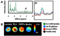
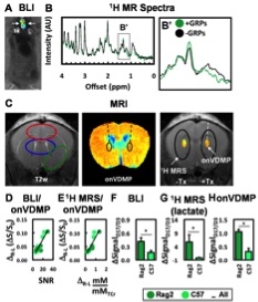
Detecting GRPs using onVDMP CEST MRI and 1H MRS (lactate peak) was first evaluated in phantoms (Figure 1). GRPs enhanced both signals compared to the negative control (media alone). Detecting GRPs in vivo was confirmed initially in naïve mice (Figure 2). onVDMP CEST MRI signal correlated well with the bioluminescence (BLI) used as imaging biomarker for cell survival and the 1H MRS signal on day 3. Changes in MR signal from day 3 to day 17 post-transplantation in C57Bl/6 (immunocompentant) and Rag2-/- (immunodeficient) mice differed (p<0.05) in all imaging modalities.
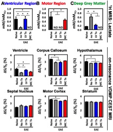
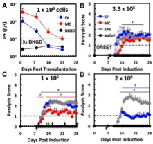
GRP detection was then evaluated in an EAE model of multiple sclerosis (Figure 3), a disease characterized by oligodendrocytes loss, which transplanted GRPs can potentially replenish. onVDMP CEST MRI and 1H MRS signals differed in multiple regions of the cerebral brain of EAE mice compared to naïve mice. Transplantation of 1x10⁶ allogeneic GRPs into the motor cortex or lateral ventricles partially restored MR signatures 5 days later at or proximal to the transplantation site. Transplantation of GRPs at either site attenuated paralysis in EAE mice (Figure 4). Cells were short-lived; however, fewer cells were needed to significantly attenuate paralysis when transplanted into the motor cortex.
Discussion and Conclusion
GRPs enhanced the signal of 1H MRS and onVDMP CEST MRI in phantoms & in vivo. We tentatively assign the onVDMP signal changes to be due at least in part to lactate changes, but other changes, e.g. due to differences in mobile protein content could also play a role. Signal dynamics in immunocompetent and immunodeficient mice indicate transplanted GRPs were the primary source of the in vivo signal, and not a host immune response to the transplantation procedure. Transplantation of GRPs partially restored onVDMP CEST MRI and 1H MRS signatures in EAE mice at and/or proximal to the transplantation site. Lastly, the motor cortex was demonstrated to be a promising transplantation site for attenuating paralysis in EAE and MS using GRPs.Acknowledgements
This work was funded by the National Multiple Sclerosis Society (NMSS RG 4994-A-3) and the TEDCO Maryland Stem Cell Fund for Postdoctoral Fellows (MSCRF-3900).References
1. Masthoff M et al. Scientific Reports. 2018;8:9563.
2. Gaudet JM et al. Magnetic Resonance in Medicine. 2017;78:713.
3. Fink C et al. Scientific Reports. 2018;8:590.
4. Griffin JL et al. NMR Biomed. 2002;15:375.
5. Bhakoo KK and Pierce D. J Neurochem 2000;74:254.
6. Ferraiuolo L et al. PNAS. 2016;113:E6496.
7. Liddell JR et al. J Neurosci Res. 2009;87:2696.
8. Hertz L and Dienel GA. JNR. 2005;79:11.
9. Voloboueva LA et al. FEBS Lett. 2013;587:756.
10. Tan Z et al. J Biol Chem. 2015;290:46.
Figures