4333
Flexible Numerical Simulation Framework for Dynamic PET-MRI1Physikalisch-Technische Bndesanstalt (PTB), Braunschweig and Berlin, Germany, 2Institute of Nuclear Medicine, University College London, London, United Kingdom, 3Scientific Computing Department, Science and Technology Facilities Council, RAL, Didcot, United Kingdom, 4Centre for Medical Imaging, University College London, London, United Kingdom, 5School of Biomedical Engineering and Imaging Sciences, King's College London, London, United Kingdom
Synopsis
A numerical simulation framework for dynamic simultaneous PET- MR is presented, which allows for simulated data acquisition of different anatomy with cardiac and respiratory motion and dynamic contrast changes (due to MR contrast agent or PET tracer changes over time). The output of the simulation framework is provided in ISMRMRD and PET interfile raw data format and can be directly used in a range of available reconstruction packages. The reconstructed PET and MR images of the simulated data were compared to an in-vivo patient scan demonstrating that the simulation framework yields realistic data.
Introduction
Realistic numerical simulations of dynamic processes (e.g. respiratory and cardiac motion and dynamic contrast uptake) play an important role in the development of advanced image reconstruction and post-processing methods1,2,3. To assess the accuracy of motion estimation and correction techniques, ground truth (GT) motion information is required. For simultaneous PET-MR, MR and PET simulations require hardware-dependent parameters (e.g. MR receiver coil sensitivities or PET detector geometry) to ensure realistic output. Several PET and/or MR simulation frameworks have been proposed, which take physiological motion (e.g. breathing, heartbeat) into account4,5. However, they commonly are developed for a very specific task and rely on custom reconstruction software. The input parameters are chosen for a certain application, making them often challenging to adapt for other purposes4. In this study, a novel framework to generate simulated dynamic PET-MR rawdata is presented. It provides simultaneous PET and MR rawdata in standardized MR (ISMRMRD) and PET (Interfile) rawdata format6,7. Cardiac and respiratory motion and dynamic uptake of contrast agents and PET tracers can be simulated and GT motion information is provided. Chemical shifts between fat and water are also correctly simulated.Materials and Methods
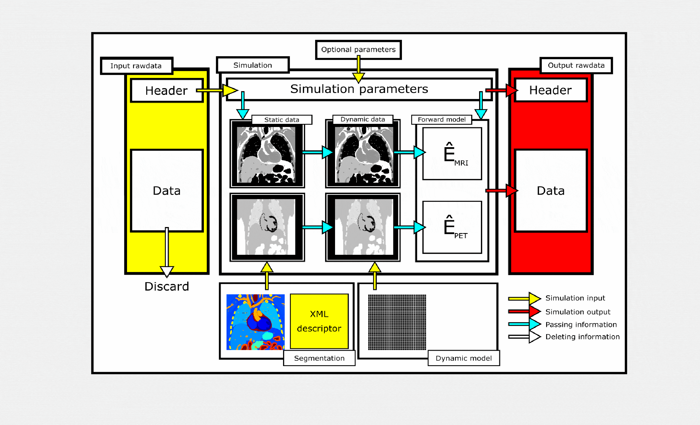
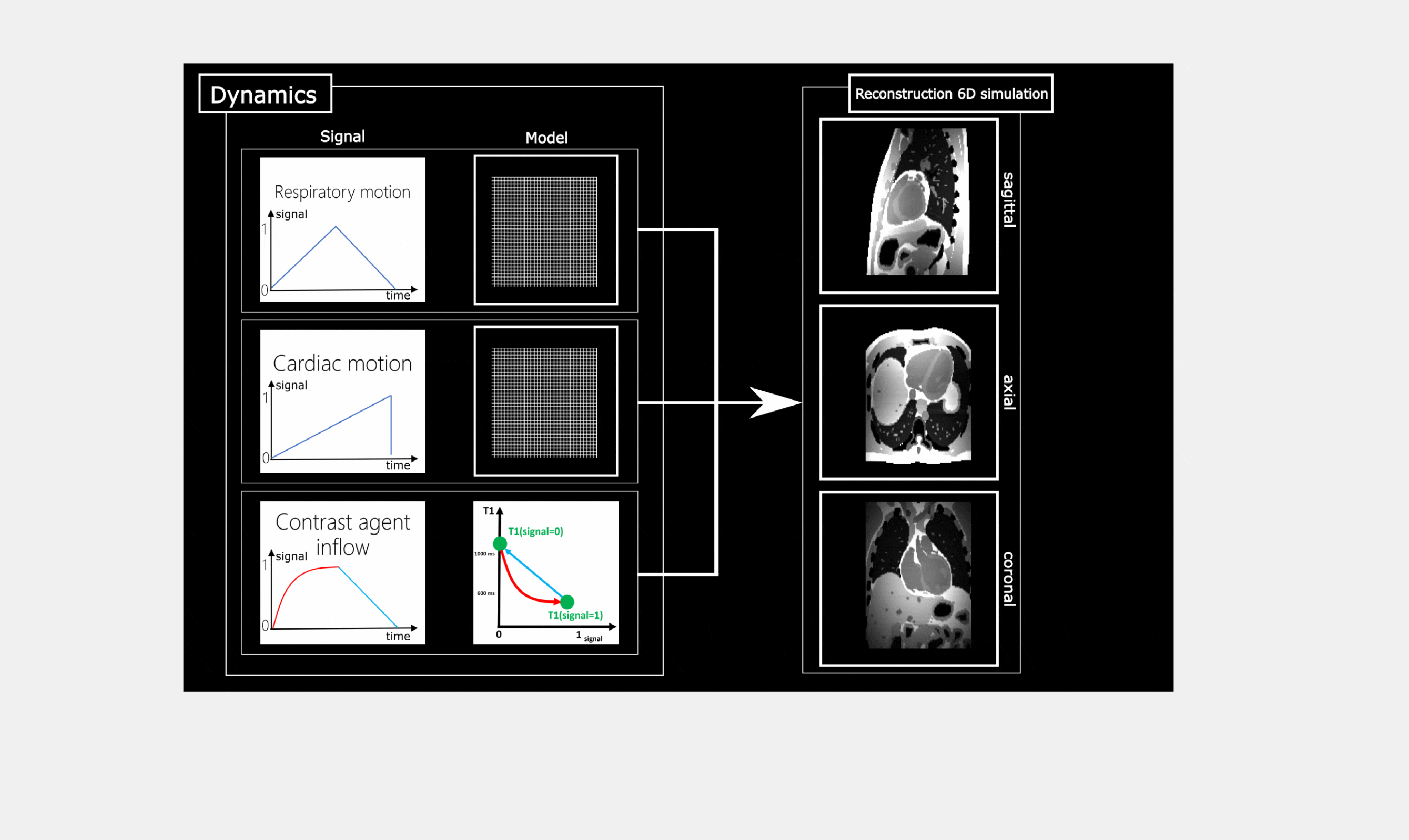
An overview of the framework design is given in Fig. 1. One input for the simulation is a standardized rawdata file (ISMRMRD format for MRI, Interfile for PET). All hardware-related parameters (TE, TR, flip angle, sequence type, number of receiver coils, k-space trajectory for MR or detector geometry for PET) are taken from the header information and the data part is replaced by the generated simulation data to ensure a valid rawdata file is generated. In this manner, the simulation can emulate the acquisition of already available in-vivo data while simultaneously providing GT information. In addition to the rawdata file a tissue segmentation combined with an XML descriptor detailing the tissue parameters in each voxel of the segmentation (T1, T2, spin density, chemical shift for MR, and activity and attenuation values for PET) must be supplied. Based on these parameters combined with those from the input rawdata, the MR simulation generates k-space data using multiple receiver coils, and the PET simulation forward projects the accumulated activity. Motion or contrast changes can be added to the simulation to dynamically modify the segmentation. Each of these contains a model of the dynamic process and its temporal progression which are incorporated into the signal model during the acquisition simulation. An example is given in Fig. 2. These elements are integrated into the open-source software project Synergistic Image Reconstruction Framework8 (SIRF). It is implemented entirely in C++ employing the functionality of the open-source MR and PET reconstruction engines Gadgetron9 and STIR10 and provides a Matlab and Python interface for easy usability. Experiments An XCAT-based tissue segmentation and motion model of the thorax and abdomen were used to simulate an FDG-PET-MR exam on a 3T Siemens Biograph mMR. The simulation was performed using rawdata files from a patient data examination with the patient’s self-navigator and ECG signal as dynamic signal input3. Continuous MR data acquisition during free-breathing was simulated for a triple-echo prototype Dixon-based GRE Golden angle Radial Phase Encoding11 sequence (TE=1.2/2.7/4.2ms, FA=10°). The spatial resolution of MR was 1.9x3.2x3.2mm3 and 2x2.1x2.1mm3 for PET. Fat-water separation was carried out on the MR data using an iterative chemical-shift approach12. The following comparisons were carried out:
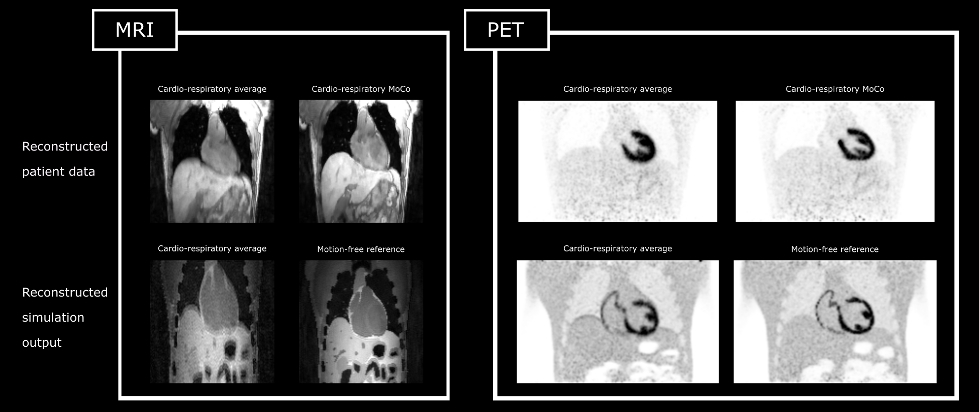
- Free-breathing PET and MR acquisition with and without respiratory and cardiac motion correction3.
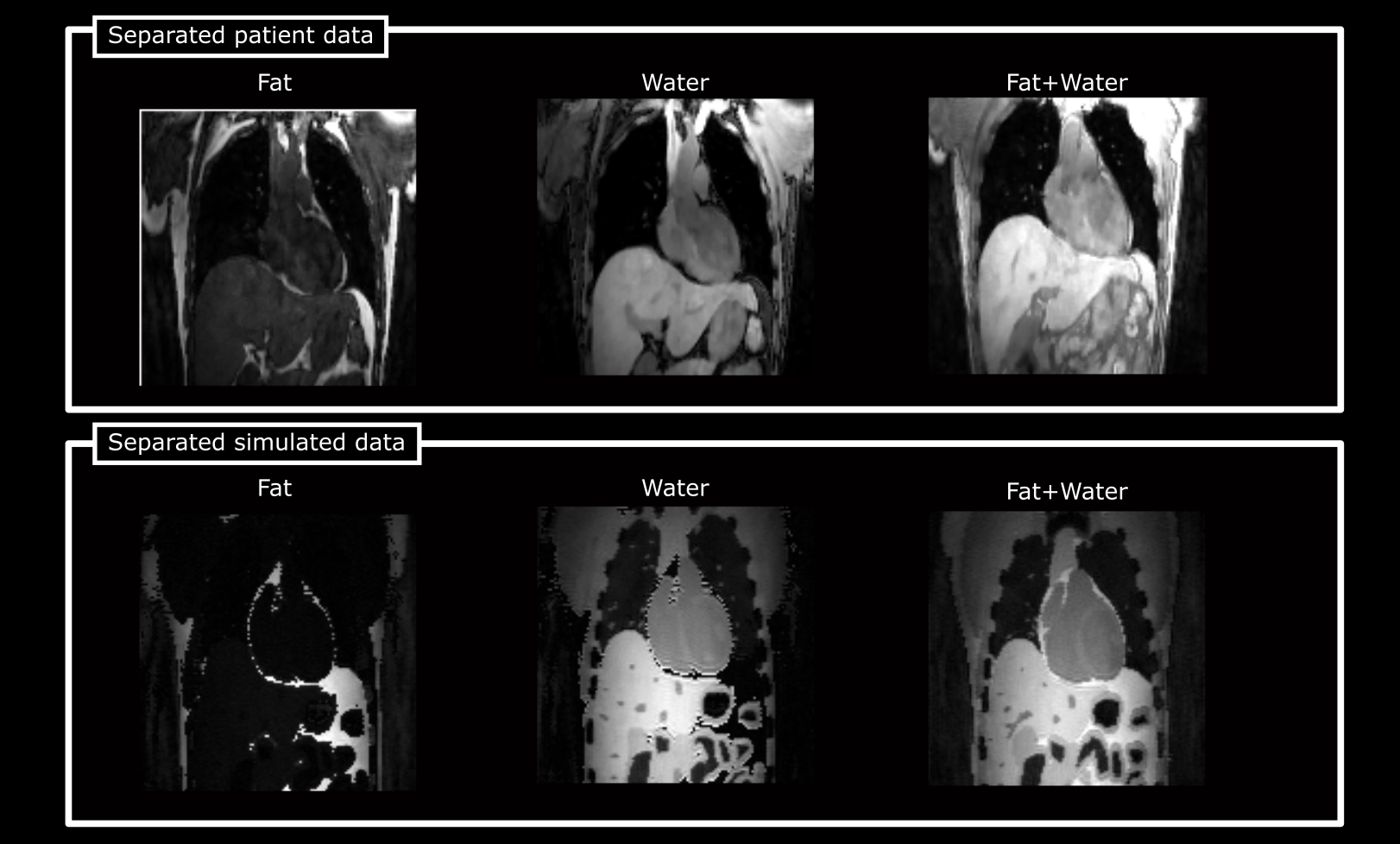
- 3D Fat-water imaging for MR-based AC map calculation.
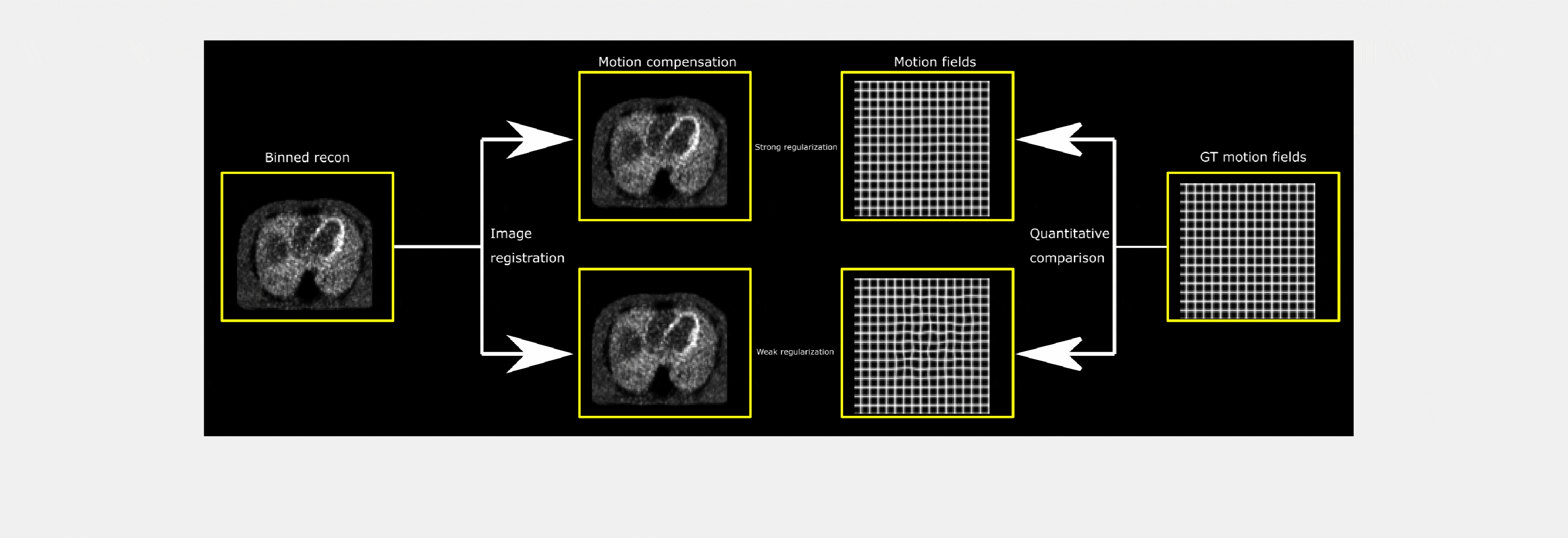
- Evaluation of PET-based motion estimation for different image registration13.
Results
All simulated images displayed in Fig. 3 to 5 are reconstructions from the rawdata output of the simulation using the same reconstruction software as for the in-vivo data. Fig. 3 demonstrates realistic image quality and motion artefacts. A fat-water separated reconstruction of the simulated MR data, commonly used for attenuation correction estimation in PET, is displayed in Fig. 4. Cardiac motion fields obtained from PET images are compared to GT motion fields in Fig. 5.Discussion and Conclusion
A framework to simulate realistic PET-MR data with motion and contrast changes was presented. We demonstrated that this framework can be used to evaluate the effect of different types of motion on MR and PET image quality and assess motion-corrected image reconstruction techniques and fat-water imaging methods. Additionally, GT motion information allowed for assessment of motion estimation approaches. This study used the XCAT model but the framework can be used with any available segmentations and motion information. The standardized output enables reconstruction with open-source reconstruction packages, including BART14, Gadgetron, STIR and other frameworks able to read ISMRMRD or Interfile format.Acknowledgements
Financing from the German Research Foundation (DFG) project number GRT 2260, BIOQIC, and CCPPETMR (UK EPSRC EP/M022587/1 & EP/P022200/1) is acknowledged.References
1. Rank, Christopher M., et al. "4D respiratory motion-compensated image reconstruction of free-breathing radial MR data with very high undersampling." Magnetic resonance in medicine 77.3 (2017): 1170-1183.
2. Küstner, Thomas, et al. "MR-based respiratory and cardiac motion correction for PET imaging." Medical image analysis 42 (2017): 129-144.
3. Kolbitsch, C. et al., 2018. Joint PET-MR image registration for cardiac and respiratory motion correction of simultaneous cardiac PET-MR. In Proceedings of Joint Annual Meeting ISMRM-ESMRMB, Paris, France. p. 478.
4. Polycarpou, Irene, Georgios Soultanidis, and Charalampos Tsoumpas. "Synthesis of Realistic Simultaneous Positron Emission Tomography and Magnetic Resonance Imaging Data." IEEE transactions on medical imaging 37.3 (2018): 703-711.
5. Wissmann, Lukas, et al. "MRXCAT: Realistic numerical phantoms for cardiovascular magnetic resonance." Journal of Cardiovascular Magnetic Resonance 16.1 (2014): 63.
6. Inati, Souheil J., et al. "ISMRM Raw data format: a proposed standard for MRI raw datasets." Magnetic resonance in medicine77.1 (2017): 411-421.
7. Todd-Pokropek, A., T. D. Cradduck, and F. Deconinck. "A file format for the exchange of nuclear medicine image data: a specification of Interfile version 3.3." Nuclear medicine communications 13.9 (1992): 673-699.
8. Ovtchinnikov, Evgueni, et al. "SIRF: Synergistic Image Reconstruction Framework."
9. Hansen MS, Sørensen TS. Gadgetron: An Open Source Framework for Medical Image Reconstruction. Magn Reson Med. 2013 Jun;69(6):1768-76.
10. Thielemans, Kris, et al. "STIR: software for tomographic image reconstruction release 2." Physics in Medicine & Biology 57.4 (2012): 867,
11. Prieto, Claudia, et al. "3D undersampled golden angle radial phase encoding for DCE-MRA using inherently regularized iterative SENSE." Magnetic resonance in medicine 64.2 (2010): 514-526.
12. Berglund J, Kullberg J. Three-dimensional water/fat separation and T2* estimation based on whole-image optimization-Application in breathhold liver imaging at 1.5 T. Magn Reson Med. 2012;67:1684-1693.
13. Rueckert, Daniel, et al. "Nonrigid registration using free-form deformations: application to breast MR images." IEEE transactions on medical imaging 18.8 (1999): 712-721.14. Martin Uecker, Frank Ong, Jonathan I Tamir, Dara Bahri, Patrick Virtue, Joseph Y Cheng, Tao Zhang, and Michael Lustig, Berkeley Advanced Reconstruction Toolbox, Annual Meeting ISMRM, Toronto 2015, In Proc. Intl. Soc. Mag. Reson. Med. 23:2486
14. Martin Uecker, Frank Ong, Jonathan I Tamir, Dara Bahri, Patrick Virtue, Joseph Y Cheng, Tao Zhang, and Michael Lustig, Berkeley Advanced Reconstruction Toolbox, Annual Meeting ISMRM, Toronto 2015, In Proc. Intl. Soc. Mag. Reson. Med. 23:2486
Figures