4324
Mitochondrial redox imaging of NASH animal model using in vivo Dynamic Nuclear Polarization MRI1Gifu University, Gifu, Japan, 2Kyushu University, Fukuoka, Japan, 3Japan Redox Inc., Fukuoka, Japan
Synopsis
Currently available non-invasive imaging technologies,
including CT, MRI and ultrasonography, are only able to assess fat accumulation
in the liver. Therefore, these methods are not suitable for a precise diagnosis
of NASH. The standard technique for diagnosing NASH, liver biopsy, has several
drawbacks, such as the higher risk of complications that accompanies invasive
procedures. Here, we demonstrated that in
vivo mitochondrial redox metabolism was dramatically altered at an early
stage, and NASH could be accurately diagnosed by in vivo dynamic nuclear polarization-magnetic resonance imaging, with
carbamoyl-PROXYL as a molecular imaging probe. In addition, this technique was
feasible for distinguishing between NAFLD and NASH. Our data reveal a novel
method for monitoring the dynamics of redox metabolic changes and assessing the
efficacy of therapeutic agents in NAFLD/NASH.
Introduction
Given the rising incidence of non-alcoholic fatty liver disease (NAFLD)
in both adults and children, the development of a non-invasive diagnostic
method for assessing disease progression to non-alcoholic steatohepatitis
(NASH) has become an important research goal. Currently available non-invasive
imaging technologies, including CT, MRI and ultrasonography, are only able to
assess fat accumulation in the liver. Therefore, these methods are not suitable
for a precise diagnosis of NASH. The standard technique for diagnosing NASH,
liver biopsy, has several drawbacks, such as the higher risk of complications
that accompanies invasive procedures. Here, we demonstrated that in vivo
mitochondrial redox metabolism was dramatically altered at an early stage, and
NASH could be accurately diagnosed by in vivo dynamic nuclear
polarization-magnetic resonance imaging, with carbamoyl-PROXYL as a molecular
imaging probe.
Methods
The feasibility of in vivo DNP-MRI for monitoring the development of NASH was assessed in two types of diet-induced mouse model: a methionine-choline-deficient (MCD) diet and a high-fat (HF) diet. Liver tissues stained with haematoxylin and eosin (H&E). Hepatic fibrosis was evaluated using Masson’s trichrome stain. Liver sections were routinely deparaffinized and immunostained for 4-HNE and8-OHdG, as reliable markers of lipid peroxidation and oxidative DNA damage, respectively. In vivo redox molecular imaging was performed with a low magnetic field, in vivo DNP-MRI system (Keller-Japan redox Inc.). The external magnetic field (B0) for EPR irradiation and MRI was fixed at 15 mT, and the radiofrequencies for EPR irradiation and MRI were 455 MHz and 683 kHz, respectively. In vivo DNP-MRI scanning of the upper abdomen was started immediately after intravenous injection of carbamoyl-PROXYL (150 mM CmP in half saline, 10 μL/g body weight). Pharmacokinetic DNP-MRI images were obtained at 1, 2.5, 4, 5.5, 7, 8.5, 10, 11.5 and 13 min after administration. The reduction rate of Cmp radical probes by isolated mitochondria was measured by X-band EPR at 30 min after adding 5 μM Cmp, 1 μM Rotenone, 0.8 mg/ml mitochondria, 1 mM NADH, 1.2 mM ADP and 10 mM succinate.Results and Discussion
All MCD mice had obvious hepatic steatosis, which gradually worsened, depending on the feeding period. Hepatic vein fibrosis was observed after 4 weeks of feeding, and fibrotic regions started to develop steatosis. Plasma alanine transaminase (ALT) and aspartate transaminase (AST) levels were significantly higher in MCD mice compared with control mice. To monitor liver redox status non-invasively, in vivo DNP-MRI was performed after 1, 2, 4, 6 and 8 weeks of MCD dietary treatment in living mice. Enhanced DNP-MRI was conducted every 90 s from 1 min to 13 min after intravenous injection of CmP. The distribution of the enhanced signal area was observed in the whole liver at 1 min after injection. The intensity of the first image after CmP injection was increased, according to the duration of feeding. For quantitative assessment, the reduction rate of DNP signal enhancement was calculated using pharmacokinetic DNP MR images. Interestingly, even 1 week after starting MCD treatment, the reduction rate was significantly decreased, with further decreases after longer feeding intervals. In the control group, the reduction rate of CmP in liver was stable in all groups for each feeding period. The oxidized form of CmP was significantly decreased in liver homogenates from the 2-week MCD group compared with controls. Total CmP, which was measured after re-oxidative treatment with potassium ferricyanide, was not significantly different between control and MCD liver homogenates. These results suggest that the reduction rate monitored by in vivo DNP-MRI showed, not the difference of liver uptake and excretion, but radical loss of CmP by redox reaction. The EPR signal of CmP was reduced by freshly prepared liver homogenates. The EPR signal change at 5 min was significantly lower in the MCD groups than the control groups. Interestingly, these differences were completely inhibited by the addition of potassium cyanide (KCN), an inhibitor of complex IV in the mitochondrial electron transfer chain (ETC). Furthermore, CmP was not reduced by cytosol in either group. These results suggest that mitochondrial metabolism was responsible for triggering disease progression, and these metabolic changes were identified at an early phase of the change in redox status using in vivo DNP-MRI.Conclusion
This technique might have potential value for acceleration of drug discovery by early detection of NASH, diagnostic imaging techniques and clinical follow-up for NASH patients.Acknowledgements
This work was supported by the Medical Research and Development Programs Focused on Technology Transfer, Development of Advanced Measurement and Analysis Systems (SENTAN) from the Japan Agency for Medical Research and Development, AMED Grant Number 162128; Health Labour Sciences Research Grant (Research on Publicly Essential Drugs and Medical Devices) from the Ministry of Health, Labour and Welfare of Japan; and Special Coordination Funds for Promoting Science and Technology (SCF funding program “Innovation Center for Medical Redox Navigation”). This work was also supported by JSPS KAKENHI (Grant Number 16H05079 and 16H05113).References
Nakata R, Hyodo F, Murata M, Eto H, Nakaji T, Kawano T, Narahara S, Yasukawa K, Akahoshi T, Tomikawa M, Hashizume M. In vivo redox metabolic imaging of mitochondria assesses disease progression in non-alcoholic steatohepatitis. Sci Rep. 2017 Dec 7;7(1):17170. doi: 10.1038/s41598-017-17447-2.Figures
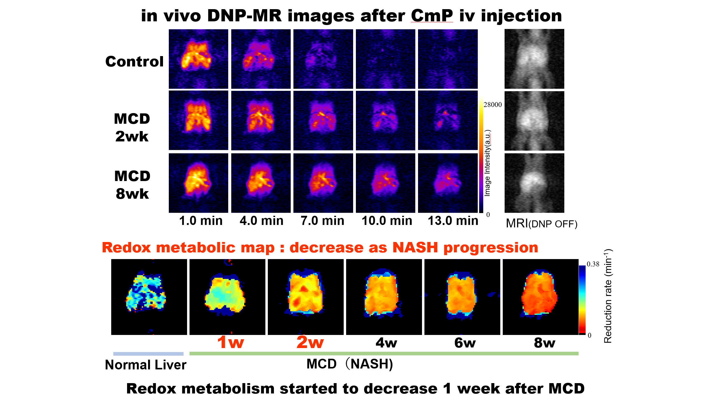
in vivo pharmacokinetic DNP MR images of NASH mice after iv injection of CmP redox probe. Redox map was calculated using pharmacokinetic DNP MR images.