4318
Hyperpolarized Dihydroxyacetone Metabolism in the Mouse Liver Under Physiological Perfusion Conditions1Biochemistry and Mol. Bio., University of Florida, Gainesville, FL, United States, 2Imaging Physics, MD Anderson Cancer Center, Houston, TX, United States
Synopsis
Hyperpolarized [2-13C]dihydroxyacetone is a promising new molecular imaging agent that is sensitive to hepatic gluconeogenesis as well as glycolytic flux. Here, a physiological perfusion condition produces a metabolic profile in keeping with current understanding of hepatic metabolism, as compared to previous results from a model intended to strongly upregulate gluconeogenesis.
Target Audience
Researcher in hepatic intermediary metabolism and molecular imagingIntroduction
Hyperpolarized (HP) [2-13C]dihydroxyacetone (DHAc) has been previously used to assay liver metabolism in vivo, showing sensitivity to a fructose challenge paradigm commonly used to assess hepatic energy charge.1 Work in the perfused mouse liver at 9.4 T showed additional insights regarding hepatic glucose output in gluconeogenic and glycogenolytic states.2 The previous perfusion conditions were intended to induce gluconeogenesis (GNG) from phosphoenolpyruvate (PEP). Here DHAc is used to measure hepatic flux through the Embden-Meyerhof pathway in perfusion conditions that more accurately reflect normal hepatic physiology. Significant changes in lactate production were observed in the fasted condition compared to previous results.Methods
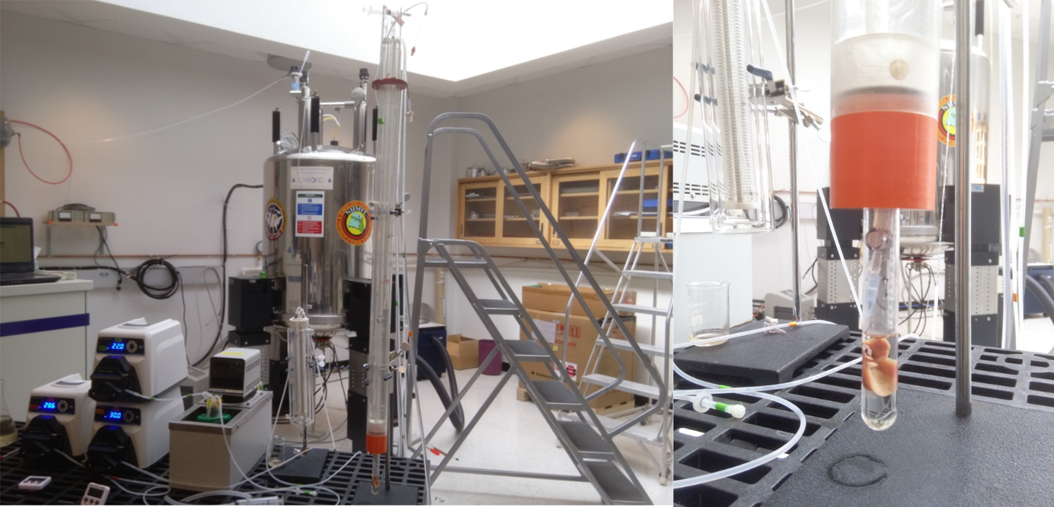
All experiments were approved by the University of Florida IACUC. Mice were fasted overnight prior to surgery, and anesthetized using isoflurane. Livers were excised from C57/BLKS mice and perfused at constant pressure with a Krebs-Henseleit buffer. Metabolic substrates included in the buffer included 0.63 mM mixture of fatty acids complexed to 1.5 % bovine serum albumin, 0.1 mM pyruvate, and 1 mM lactate. The livers were perfused for ~30 minutes to achieve a metabolic steady state prior to examination with HP DHAc. Oxygen consumption was measured at multiple time points throughout to assure proper function of the liver. The perfusion rig (Figure 1) was water jacketed and maintained at 37 oC. The rig was placed into a vertical bore 14.1 T Bruker Avance III HD spectrometer (Bruker Biospin, Billerica, MA) using an 18 mm Doty (Doty Scientific, Columbia, SC) NMR probe optimized for 13C. [2-13C]DHAc was purchased from Millipore-Sigma (Darmstadt, Germany) and used without further purification. Trityl-OXO63 was purchased from Oxford Instruments and used without further purification. Details of the HP procedure were the same as previous.2 Samples were polarized for ~2 hours prior to use, to a final polarization greater than 30%. Upon dissolution, the HP DHAc (3 ml) was rapidly mixed with 20 ml of perfusate and then injected into the liver over the next 90 seconds. NMR data was acquired over the same time period using a pseudo-2D pulse sequence. The excitation angle was 30 degrees, with a 3 s Tr. WALTZ-64 was used for 1H decoupling.
Results and Discussion
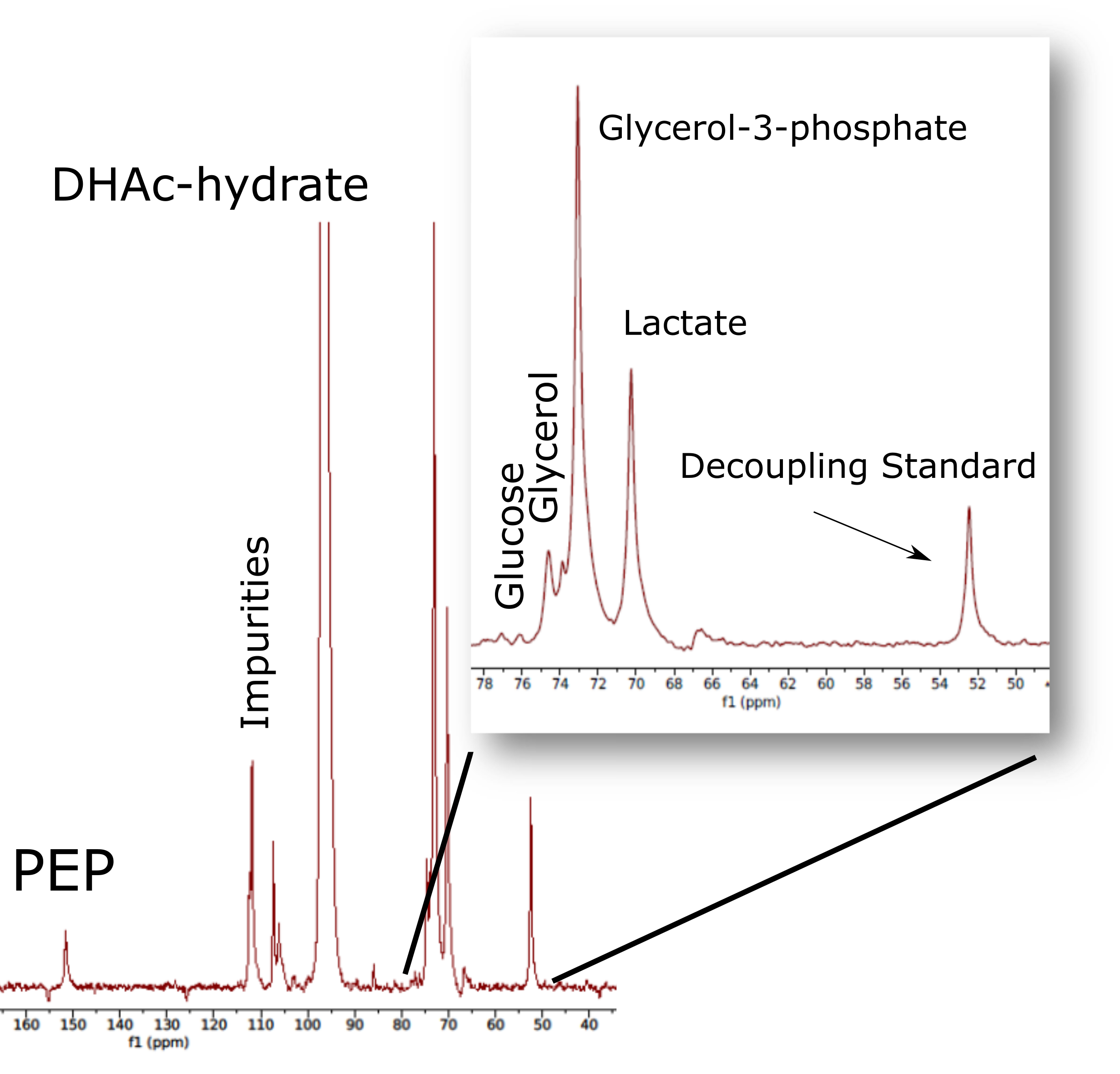
Previous results were acquired using livers from fasted mice and a perfusate containing 0.2 mM octanoate as well as 2 mM pyruvate as a gluconeogenic substrate. This condition was meant to dramatically stimulate GNG from the level of the tricarboxylic (TCA) cycle. This was realized, as the fraction of glucose produced by the liver that was derived from the level of the TCA cycle nearly 80 % in this condition. However, this state was non-physiological as 2 mM pyruvate is excessively high and octanoate is known to flood the mitochondria with reducing equivalents, as its uptake and β-oxidation is not regulated by CPT-I.3 In this case, a physiologically relevant ratio of lactate to pyruvate (10:1) and a mixture of long chain fatty acids was provided. The switch to a more conventional hepatic redox milieu results in a profound shift of the hepatic DHAc handling (Figure 2). While previous results showed a more or less equal ratio of phosphoenolpyruvate (PEP) to lactate, data here shows the ratio is closer to 8 to 1. As compared to the glycerol-3-phosphate (G3P) signal, lactate was previously 1/10th of the G3P signal, where now it is approximately 1/2. G3P is a convenient reference signal, as it is the first observable product of DHAc metabolism, as dihydroxyacetone phosphate is not readily observed due to proximity to the parent DHAc signal at 212 ppm. Glucose production, as assessed by the 2-13C and 5-13C glucose signals in the region 72-76 ppm remained more or less unchanged as compared to the G3P signal. These results suggest that redox balance is more appropriately controlled, as lactate should significantly exceed pyruvate in the liver. Furthermore, PEP should be a relatively small pool size compared to the 3-carbon intermediates. Further GC/MS based metabolomics analysis will serve to confirm that a more normal physiological profile is maintained under thesee perfusion conditions.Conclusion
DHAc is a promising HP imaging agent that has been used successfully for pre-clinical in vivo studies.1 It is imperative that a more complete accounting of the net flow of the DHAc imaging agent, as the potential of DHAc as an agent for diagnosing hepatic pathophysiologies seems high. We have shown here that a more physiological perfusion condition produces shifts in DHAc metabolism in keeping with proper hepatic redox balance. Further experiments will compare this fasted condition to a fed condition using a similar substrate mixture.Acknowledgements
The authors acknowledge funding from NSF DMR 1157490 and NIH 8P41-EB015908 and P41122698, , U24DK097209, and R01s DK105346, HD087306, DK112865.References
1. I. Marco‐Rius, C. von Morze, R. Sriram, P. Cao, G.Y. Chang, et Al., "Monitoring acute metabolic changes in the liver and kidneys induced by fructose and glucose using hyperpolarized [2‐13C] dihydroxyacetone", Magnetic resonance in medicine 77(1) (2017) 65-73.
2. K.X. Moreno, S. Satapati, R.J. DeBerardinis, S.C. Burgess, C.R. Malloy, M.E. Merritt, "Real-time detection of hepatic gluconeogenic and glycogenolytic states using hyperpolarized [2-13C] dihydroxyacetone", Journal of Biological Chemistry 289(52) (2014) 35859-35867.
3. J.D. McGarry, D.W. Foster, "The Regulation of Ketogenesis from Octanoic Acid", J Biol Chem 246(4) (1971) 1149-1159.
Figures