4317
Quantification of In Vivo Saturation Kinetics of Co-Polarized [2-13C]Pyruvate and [1,4-13C2]Fumarate in Rat Liver Using Dynamic 3D-Spiral CSI with Alternate Spectral Band Excitation1Diagnostic Radiology and Nuclear Medicine, University of Maryland Baltimore, Baltimore, MD, United States
Synopsis
Dynamic metabolic imaging of co-polarized 13C-labeled pyruvate (Pyr) and fumarate (Fum) has shown great potential in characterizing multiple in vivo metabolic activities as demonstrated in recent animal models.1,2 Although in these studies, both the injected substrates and downstream metabolic product can be acquired over time at relatively high spatial resolution, robust quantification and metabolic modelling remains an active area of investigation. Also, the enzyme saturation effects that are routinely seen with commonly used doses of hyperpolarized substrates are not correctly captured using approaches such as metabolite ratios, time-to-peak of metabolic products, single exchange rate constants and by constant small-flip-angle excitation. Therefore, the goal of this study is to measure the saturation kinetics of various involved enzymatic processes using effective 90° excitation of the products at clinical field strengths. This work is an extension of our method developed for dynamic metabolic imaging of co-polarized mixture of [2-13C]Pyr and [1,4-13C2]Fum.2
Purpose
Dynamic metabolic imaging of co-polarized 13C-labeled pyruvate (Pyr) and fumarate (Fum) has shown great potential in characterizing multiple in vivo metabolic activities as demonstrated in recent animal models.1,2 Although in these studies, both the injected substrates and downstream metabolic product can be acquired over time at relatively high spatial resolution, robust quantification and metabolic modelling remains an active area of investigation. Also, the enzyme saturation effects that are routinely seen with commonly used doses of hyperpolarized substrates are not correctly captured using approaches such as metabolite ratios, time-to-peak of metabolic products, single exchange rate constants and by constant small-flip-angle excitation. Therefore, the goal of this study is to measure the saturation kinetics of various involved enzymatic processes using effective 90° excitation of the products at clinical field strengths. This work is an extension of our method developed for dynamic metabolic imaging of co-polarized mixture of [2-13C]Pyr and [1,4-13C2]Fum.2Methods
All measurements were performed on a clinical 3T GE 750w MR scanner (GE Healthcare, Waukesha, WI, USA) equipped with self-shielded gradients (33 mT/m, 120 mT/m/ms). Our dynamic imaging data were obtained using an extension of our previous fast 3D spiral chemical shift imaging (3D-spCSI) method developed for metabolic imaging of co-polarized mixture of [2-13C]Pyr and [1,4-13C2]Fum with alternate spectral band excitation.2 The experimental parameters including the RF excitation pulses were the same as used earlier: 2 spatial interleaves, 50 mm FOV, matrix size 10 × 10, 5 mm isotropic resolution, 10 slice encodings, 20 echoes with a SW = 258 Hz optimized to reduce peak overlap in both bands. Total acquisition time per volume was 2.5 s. As previously, the RF pulse for the first band used a 2.5° excitation on Fum and 10° on malate. However, instead of constant 10° excitation on [2-13C]Pyruvate-hydrate (Pyh), [2-13C]Lactate (Lac), and [2-13C]Alanine (Ala) used previously,2 a variable flip angle scheme that produces an effective 90o excitation on these metabolites at each time point.3,4 Pyh is used as surrogate for Pyr as it is metabolically not active and in fast exchange with Pyr. As Pyr is not excited this also replenishes the Pyh magnetization after the effective 90° excitation at each time point. In vivo experiments using dynamic 3D-spCSI were done in two healthy male Wistar rats (440 – 450 g) that were anesthetized with 1–3% isoflurane in oxygen at (1.5 L/min). The rats were injected in a tail vein with an (80/40) mM (Pyr/Fum) mixture that was hyperpolarized via DNP. For each experimental run, 3–4 mL of mixture was injected via a bolus at a rate of 0.2 mL/s. A single-loop 13C surface coil placed on top of the rat liver was used for both signal excitation and reception. The metabolic kinetics were quantified using the procedure reported in ref 4.Results and Discussion
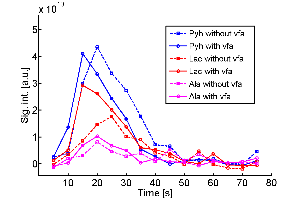
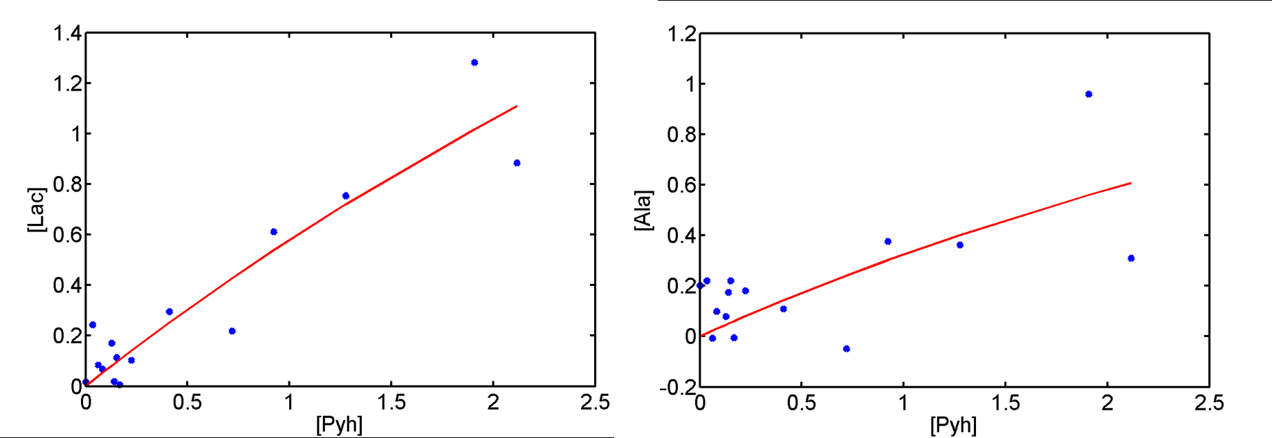
The 3D-spCSI data were acquired from the section of the abdomen centered on the liver of the rat. Pyh, Lac, and Ala peak integrals were computed from the region of interest (ROIs) in the rat liver. Time courses for the individual metabolites from the Pyh-band (Fig. 1) show higher signal for product resonances with variable flip angle as compared to constant flip angle excitation. Fig. 2 shows the representative saturable kinetics between the apparent reaction velocities of the 13C label and the Pyh concentration, and the best-fit curves for both Lac and Ala. Apparent from Fig. 2, the given dose of 0.6 mM/kg of Pyr did not saturate the enzyme ADH and LDH activities. To better estimate the apparent maximum reaction velocity Vmax and apparent Michaelis constant Km, the dose of Pyr to be injected needs to be increased in order to saturate the enzymes.Acknowledgements
NIH grants R01 DK106395, R21 CA202694, R21 NS096575, and R21 CA213020References
1. Eldirdiri A, Clemmensen A, Bowen S, Kjær A, Ardenkjær‐Larsen JH. Simultaneous imaging of hyperpolarized [1,4‐13C2]fumarate, [1‐13C]pyruvate and 18F–FDG in a rat model of necrosis in a clinical PET/MR scanner. NMR in Biomedicine. 2017;30:e3803.
2. Singh M, Josan S, Zhu M, Mayer D. Dynamic Metabolic Imaging of Co-Polarized [2-13C]Pyruvate and [1,4-13C2]Fumarate Using 3D-Spiral CSI with Alternate Spectral Band Excitation. In Proceedings of the 26th Annual Meeting of ISMRM, Paris, France, 2018. P. 3054.
3. Zhao L, Mulkern R, Tseng C, Williamson D, Patz S, Kraft R, et al. Gradient-Echo Imaging Considerations for Hyperpolarized 129Xe MR. Journal of Magnetic Resonance Series B, 1996;113(2), 179–183.
4. Xu T, Mayer D, Gu M, et al. Quantification of in vivo metabolic kinetics of hyperpolarized pyruvate in rat kidneys using dynamic 13C MRSI. NMR Biomed., 2011;24:997-1005.
Figures