4313
Effect of Opening the Blood-Brain Barrier with Focused Ultrasound on 13C Hyperpolarized Imaging in Rat Brain1Advanced Imaging Research Center, University of Texas Southwestern Medical Center, Dallas, TX, United States, 2Radiology, University of Texas Southwestern Medical Center, Dallas, TX, United States, 3Radiology, Stanford University, Stanford, CA, United States, 4Chemistry and Biochemistry, California State University Fullerton, Fullerton, CA, United States, 5Electrical and Computer Engineering, University of Texas Dallas, Richardson, TX, United States
Synopsis
We examined how opening the blood-brain barrier (BBB) with focused ultrasound (FUS) pulses changes the uptake and metabolism of hyperpolarized [1-13C]pyruvate and [1-13C]glycerate in the brain. We observed larger amounts of metabolic products, lactate and bicarbonate, from hyperpolarized [1-13C]pyruvate in the brain region where FUS was applied. Moreover, increased [1-13C]glycerate uptake in the brain was detected after the FUS-induced BBB opening.
Background
New innovations using hyperpolarized (HP) 13C labeled chemicals have allowed researchers to follow metabolic pathways in live subjects real-time using MR. This development of non-invasive metabolic mapping has granted insight into various systemic and neurological functions such as cardiac performance, cancer development, traumatic brain injury progression1-3. However, the application of HP imaging in the brain is particularly limited to small and non-polar molecules that will pass the blood-brain barrier (BBB) quickly due to the rapid relaxation of the hyperpolarized signals. In order to paint a more complete picture of energy metabolomics it is important to trace regional metabolism of other substrates involved in energetic metabolomics in the brain. Previous research from Mazuel et al. tried to overcome this issue by tracing the metabolism of the hyperpolarized [1-13C]glutamate, a polar amino acid, in the brain after temporarily opening the BBB via chemical induction (mannitol)4. Focused ultrasound (FUS) has been developed as a safe, less invasive and clinically available technique that transiently disrupts BBB5-7,11. Peeters et al. investigated the feasibility of combining FUS and HP imaging but no significant metabolic alteration or notable pyruvate uptake was detected primarily due to the low polarization and the poor signal sensitivities8. In this study, we further investigated the effects of FUS in HP imaging by opening the BBB and examining metabolic changes using two HP agents; One agent believed to readily cross the BBB, [1-13C]pyruvate9, and one polar substance unlikely to cross the BBB quickly, [1-13C]glycerate10.Methods
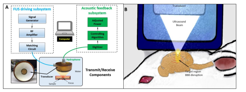
To open the BBB, the FUS exposures (f0 = 0.5MHz, pulse length = 10ms, pulse repetition frequency = 1Hz) were delivered using a custom stereotactic system (Figure 1) in three healthy Fischer rats. The exposures were delivered in a 2x4 grid manner with 2-mm spacing to cover an area of 4×8 mm2 in the left hemisphere. At each target (duration = 50s), stimulated acoustic emissions were monitored and controlled to a target threshold at an ultra-harmonic frequency (0.75MHz). The infusion rate of nanobubbles during FUS was 0.3 ml/min. All the imaging data were collected using on a GE 3T 750W wide-bore scanner and a 13C/1H dual-tuned birdcage radiofrequency (RF) coil. The rats were imaged immediately after FUS exposure (20-30mins post-exposure). T2-weighted fast spin echo (FSE) images (TE/TR=11.3ms/5000ms, thickness=2.0mm, matrix=256x256, FOV= 9.6mm2) were acquired to identify location and volume of the opened area. A 35-μL sample of 14-M [1-13C]-pyruvate mixed with OX063 trityl (15mM) was prepared for each dissolution and polarized using a SPINlab clinical DNP polarizer (GE Healthcare). After 3-4hrs of polarization, pyruvate samples were dissolved, mixed with pH-neutralization media (NaOH), and immediately injected over 10-12s intravenously (70-mM pyruvate, ~7.5 of pH). [1-13C]glycerate samples were prepared as previous described10. For both HP agents, 13C data was acquired ~30min after FUS exposure using a single timepoint 2D free-induction decay chemical shift imaging (FID CSI; spectral width=5000Hz; spectral points=256; FOV=50x50mm2; matrix=16x16, thickness=7.7mm) sequence 25s after the start of the injection. Contrast-enhanced T1 images were taken immediately after the HP injection to confirm BBB opening in the target area. Each metabolite was normalized by the total 13C (TC) signal detected within the ROI.Results and Discussion
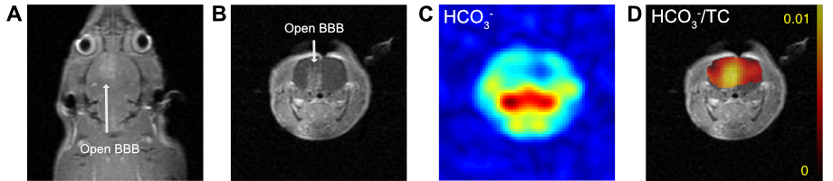
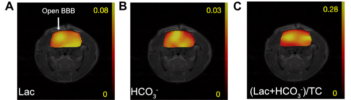
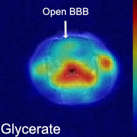
Gd-enhanced T1 images confirmed regionally targeted BBB opening immediately (30-40 mins) after FUS exposure. HP imaging of [1-13C]pyruvate showed increased 13C-products in the FUS targeted area with increased [1-13C]lactate and [13C]bicarbonate signal. Though the lactate signal between target region and contralateral side of the brain was under the detection limit in animal 1 (Figure 2), the second animal (Figure 3) showed slightly elevated lactate levels in the focal opened BBB area. Increases in bicarbonate in the focally opened BBB area were consistent in both animals, but was especially apparent in animal 1 (Figure 2). We also qualitatively observed an increase in glycerate in the BBB targeted area of the brain, but the signal-to-noise ratio was not high enough to detect any metabolic products of [1-13C]glycerate (Figure 4). We anticipate that the combination of FUS and HP imaging could potentially facilitate a wide range of feasible studies examining the diverse amount of chemicals involved in energetics metabolism in vivo. Future work is aimed to repeat the experiments with [1-13C]pyruvate and [1-13C]glycerate to confirm findings and to examine the dynamic pharmacokinetics changes of FUS delivery and metabolism of 13C-labeled compounds overtime.Acknowledgements
Funding: The Mobility Foundation; The Texas Institute of Brain Injury and Repair; National Institutes of Health of the United States (P41 EB015908, S10 OD018468, SC1 GM127213, R01 EB0901802, P41 EB01589121); GE Healthcare.References
1. Nelson, S.J., Kurhanewicz, J., Vigneron, D.B., Larson, P.E., Harzstark, A.L., Ferrone, M., van Criekinge, M., Chang, J.W., Bok, R., Park, I., Reed, G., Carvajal, L., Small, E.J., Munster, P., Weinberg, V.K., Ardenkjaer-Larsen, J.H., Chen, A.P., Hurd, R.E., Odegardstuen, L.I., Robb, F.J., Tropp, J. and Murray, J.A., 2013. Metabolic imaging of patients with prostate cancer using hyperpolarized [1-13C] pyruvate. Science translational medicine, 5(198), pp.198ra108-198ra108.
2. DeVience, S.J., Lu, X., Proctor, J., Rangghran, P., Melhem, E.R., Gullapalli, R., Fiskum, G.M. and Mayer, D., 2017. Metabolic imaging of energy metabolism in traumatic brain injury using hyperpolarized [1-13 C] pyruvate. Scientific reports, 7(1), p.1907.
3. Golman, K. and Petersson, J.S., 2006. Metabolic imaging and other applications of hyperpolarized 13C1. Academic radiology, 13(8), pp.932-942.
4. Mazuel, L., Schulte, R.F., Cladière, A., Spéziale, C., Lagrée, M., Leremboure, M., Jean, B., Durif, F. and Chassain, C., 2017. Intracerebral synthesis of glutamine from hyperpolarized glutamate. Magnetic resonance in medicine, 78(4), pp.1296-1305.
5. Hynynen, K., McDannold, N., Vykhodtseva, N. and Jolesz, F.A., 2001. Noninvasive MR imaging–guided focal opening of the blood-brain barrier in rabbits. Radiology, 220(3), pp.640-646.
6. Choi, J.J., Pernot, M., Small, S.A. and Konofagou, E.E., 2007. Noninvasive, transcranial and localized opening of the blood-brain barrier using focused ultrasound in mice. Ultrasound in medicine & biology, 33(1), pp.95-104.
7. Sheikov, N., McDannold, N., Vykhodtseva, N., Jolesz, F. and Hynynen, K., 2004. Cellular mechanisms of the blood-brain barrier opening induced by ultrasound in presence of microbubbles. Ultrasound in medicine & biology, 30(7), pp.979-989.
8. Peeters T.H., Kobus, T., Veltien, A., Heerschap, A., and Scheenen, T.W.J., 2017. Dynamic Nuclear Polarization across the barrier: a Focused Ultrasound approach. International Society of Magnetic Resonance in Medicine, #3694.
9. Hurd, R.E., Yen, Y.F., Mayer, A., Chen, A., Wilson, D., Kohler, S., Bok, R., Vigneron, D., Kurhanewicz, J., Tropp, J., Spielman, D., and Pfefferbaum, A., 2010. Metabolic imaging in the anesthetized rat brain using hyperpolarized [1-13C] pyruvate and [1-13C] ethyl pyruvate. Magnetic resonance in medicine, 63(5), pp.1137-1143.
10. Park, J.M., Wu, M., Datta, K., Liu, S.C., Castillo, A., Lough, H., Spielman, D.M. and Billingsley, K.L., 2017. Hyperpolarized Sodium [1-13C]-Glycerate as a Probe for Assessing Glycolysis In Vivo. Journal of the American Chemical Society, 139(19), pp.6629-6634.
11. Bing, C., Hong, Y., Hernandez, C., Rich, M., Cheng, B., Munaweera, I., Szczepanski, D., Xi, Y., Bolding, M., Exner, A. and Chopra, R., 2018. Characterization of different bubble formulations for blood-brain barrier opening using a focused ultrasound system with acoustic feedback control. Scientific reports, 8(1), p.7986.
Figures