4312
Cardiac metabolic imaging using hyperpolarized [1-13C]lactate as a substrate1Medical Biophysics, University of Toronto, Toronto, ON, Canada, 2Physical Sciences Platform, Sunnybrook Research Institute, Toronto, ON, Canada, 3GE Healthcare, Toronto, ON, Canada
Synopsis
The use of hyperpolarized [1-13C]lactic acid as a substrate for studying cardiac metabolism is attractive due to its high safety profile. 13C metabolite images were acquired following injection of hyperpolarized [1-13C]lactate in the porcine heart. The appearance of 13C-bicarbonate and absence of 13C-pyruvate in these images demonstrates rapid decarboxylation of pyruvate within the cardiac tissue.
Introduction
Hyperpolarized (HP) 13C MRI is a promising tool for non-invasive characterization of in vivo metabolism [1,2,3]. [1-13C]pyruvate is the most commonly used substrate for these experiments due to the high achievable polarization, long T1 relaxation time, and the important role of pyruvate in cellular metabolism. In these experiments, pyruvate is typically injected in supraphysiological concentrations, potentially altering the metabolic system that is being interrogated. [1-13C]lactate is an attractive alternative; it can be given safely at high millimolar physiological concentrations[4-6]. Potentially PDH flux can be more directly interrogated by measuring HP pyruvate and bicarbonate signal within the cardiac muscle produced from the HP lactate substrate. In this abstract, we investigate using hyperpolarized [1-13C]lactic acid as a probe of cardiac metabolism in pigs as a large animal model of the human heart.Methods
All scans were performed at 3T (GE MR750, GE Healthcare, Waukesha, MI). A Yorkshire pig was used in this experiment (weight 30 kg, HR 90 bpm). Two infusions (1 lactate, 1 pyruvate) were performed. [1-13C]lactic acid and [1-13C]pyruvic acid (Isotec) were mixed with 15 mM AH111501 and polarized in a GE SpinLab DNP polarizer. Following dissolution and neutralization, either substrate (125 mM / 15 mL) were injected at a rate of 1 mL/s, followed by 5 mL saline flush. 13C imaging was started at the end of the saline flush.
Short-axis images of 13C-bicarbonate, [1-13C]lactate, and [1-13C]pyruvate were obtained using a multi-slice, spectrally-selective sequence covering the left ventricle (1x1x1 cm3 resolution, 6 slices, scan duration 18 cardiac cycles, volume transmit coil, 2x4 channel 13C receive coil, placed on anterior chest wall)[7]. For a heart rate of 90 bpm, the total breath-held imaging time was 12 seconds. The metabolite acquisition order was modified to preserve the magnetization of the substrate (Figure 1). Following 13C imaging, dynamic 13C MRS was used to monitor residual metabolic signals from the entire heart.
Results and Discussion
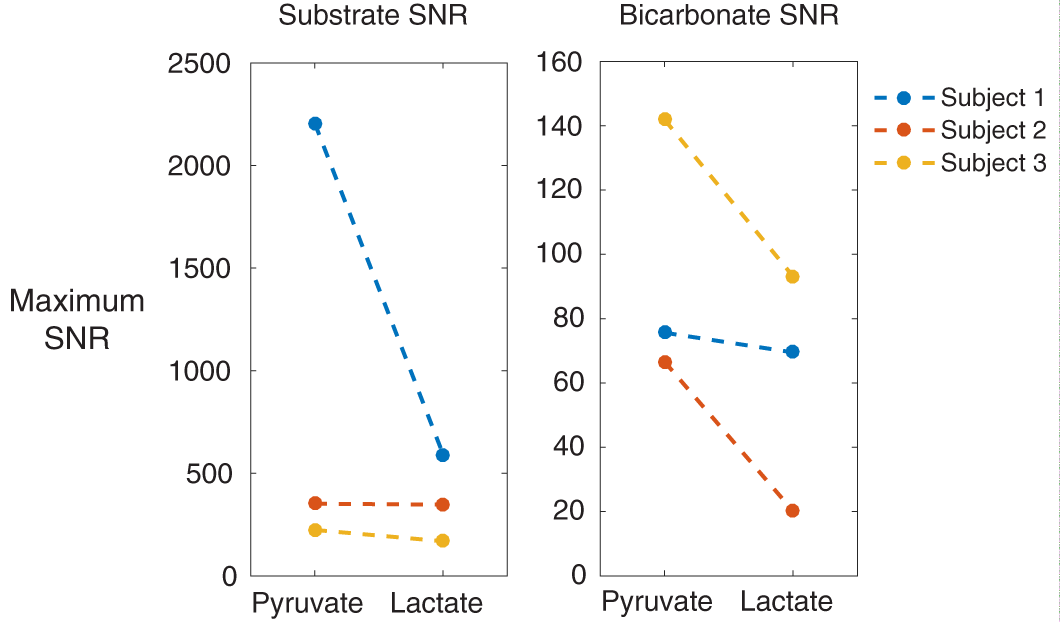
In separate experiments, the [1-13C]lactic acid liquid state polarization was found to be similar to that of [1-13C]pyruvic acid (~50%). Figures 2 and 3 show metabolite images from lactate and pyruvate injections. In the lactic acid injection, bicarbonate signal was observed spatially localized to the myocardium. No pyruvate was observed in the blood pool, which is consistent with the lactate-pyruvate redox balance. This also demonstrates that PDH-mediated irreversible decarboxylation of pyruvate occurs rapidly inside the cardiac muscle. Figure 4 shows the maximum image SNR for the respective substrate, and the corresponding maximum bicarbonate SNR in each subject. Generally, the bicarbonate SNR is observed to be lower, presumably because of the additional oxidation of lactate to pyruvate prior to PDH decarboxylation.Conclusions
13C metabolite images were acquired following injection of hyperpolarized [1-13C]lactate in the porcine heart. The appearance of 13C-bicarbonate and absence of 13C-pyruvate in these images demonstrates rapid decarboxylation of pyruvate within the cardiac tissue.Acknowledgements
The authors acknowledge funding support from NSERC and NVIDIA.References
- Ardenkjaer-Larsen JH et al. Increase in signal-to-noise ratio of > 10,000 times in liquid-state NMR. PNAS 2003 Sep 2;100(18):10158-63.
- Nelson SJ et al. Metabolic imaging of patients with prostate cancer using hyperpolarized [1-¹³C]pyruvate. Sci Transl Med. 2013 Aug 14;5(198):198ra108.
- Cunningham CH et al. Hyperpolarized 13C Metabolic MRI of the Human Heart: Initial Experience. Circ Res. 2016 Sep 15.
- Chen AP et al. Feasibility of Using Hyperpolarized [l-13C]Lactate as a Substrate for In Vivo Metabolic 13C MRSI Studies. Magn Reson Imaging. 2008 Jul; 26(6): 721–726.
- Mayer D et al. Application of Hyperpolarized [1-13C]Lactate for the In Vivo Investigation of Cardiac Metabolism. NMR Biomed. 2012 Oct; 25(10): 1119–1124.
- Chen AP et al. Using [1-13C]lactic acid for hyperpolarized 13C MR cardiac studies. Magn Reson Med. 2015 Jun;73(6):2087-93.
- Lau AZ et al. Rapid multislice imaging of hyperpolarized 13C pyruvate and bicarbonate in the heart. Magn Reson Med. 2010 Nov;64(5):1323-31.
Figures