4309
Detecting early response to PD-L1 blockade by 13C MRIYu Saida1, Tomohiro Seki1, Kazu Yamamoto1, Jeffery R. Brender1, James B. Mitchell1, Murali C. Krishna1, and Shun Kishimoto1
1Radiation Biology Branch, National Cancer Institute, Bethesda, MD, United States
Synopsis
This goal of this study is to detect metabolic changes using hyperpolarized 13C-MRI with [1-13C] pyruvate to evaluate the response to PD-L1 blockade treatment. Lactate/Pyruvate ratio tended to decrease in αPD-L1 antibody treated tumor in mouse model although not statistically significant. We will investigate capability of metabolic imaging to early response to immune checkpoint inhibitor.
Purpose
Immune checkpoint inhibitors have now become a standard therapy for several cancers. This therapy is known to show highly durable treatment response, characterized by a plateau in the tail of the survival curve. However, there are some clinical problems. First, immune checkpoint inhibitors are effective only in a limited number of patients. Secondly, although several biomarkers have been proposed for this purpose, none of them have become a gold standard for treatment assessment to date. Thirdly, the treatment response in immunotherapy is often delayed; the disease may stall or tumors may transiently enlarge even when the treatment is eventually effective1. The delayed response can pose a potential problem in designing treatment plans. It is reported that metabolic competition between tumor cells and immune cells is one of the reason that immune system can’t eradicate tumor cells. PD-1/PD-L1 blockade can restore glucose in tumor microenvironment, permitting T cell glycolysis and IFN-γ production and dampen tumor glycolysis by inhibiting mTOR activity2. In addition to this, physiologic changes by immunotherapy such as inflammation, edema, and necrosis can modulate the metabolic profile in tumor. Therefore, we employed hyperpolarized 13C MRI using [1-13C] pyruvate for detecting early changes in tumor glycolysis after PD-L1 blockade. The aim of this study is to investigate the capability of metabolic imaging to evaluate the treatment response to PD-L1 blockade.Methods
MC38 colon adenocarcinoma and B16.F10 melanoma were used as sensitive or less sensitive model to PD-L1 blockade. For in vitro antibody treatment assay, tumor cells were treated with 100 U/ml of recombinant murine IFN-γ for 24h followed by 10 μg/ml αPD-L1 antibody treatment for an additional 24h before assaying. Real-time extracellular acidification rates (ECAR) were analyzed on an XF96 Extracellular Flux Analyzer (Seahorse). C57BL/6 mice were used for all in vivo experiments. 1-10x105 tumor cells were inoculated s.c. into the right leg of mice. For in vivo antibody treatments, tumor bearing mice were injected i.p. with 200 μg of αPD-L1 antibody on days 7, 10, and 13 post tumor inoculation. Tumor bearing mice in the control group were injected with 200 μg each of IgG isotype antibody. Isolation of Tumor-Infiltrating Lymphocytes (TIL) for Flow Cytometry: The s.c. tumors were digested with collagenase, hyaluronidase and DNase I. TIL were isolated with Percoll gradient. Flow cytometry: αCD3, αCD8 and αCD4 antibody were used to identify tumor infiltrating lymphocytes. Data were collected on FACS Calibur, and analyzed by Cell Quest Pro software. Hyperpolarized 13C-MRI studies: [1-13C] pyruvic acid (30 uL), containing 15 mmol/L OX063 and 2.5 mmol/L gadolinium, was hyperpolarized using the Hypersense DNP polarizer (Oxford Instruments). After 30 to 60 minutes, the hyperpolarized sample was rapidly dissolved in 4.5 mL of a superheated alkaline buffer. A hyperpolarized [1-13C] pyruvate solution (96 mmol/L) was intravenously injected through a catheter placed in the tail vein of the mouse (12mL/g body weight). Hyperpolarized 13C MRI studies were performed on a 3T scanner using a 17mmhome-built 13C solenoid coil placed inside of a saddle coil for 1H. 13C spectra were acquired every 1 second for 240 seconds.Results
Tumor growth of both MC38 and B16F10. were delayed with in vivo αPD-L1 antibody treatment (Fig. 1A, 1B). CD4 T cells and CD8 T cells increased in αPD-L1 antibody treated MC38 bearing mice in both tumor and tumor draining lymph node by flow cytometry analysis (Fig.2). These results confirmed the therapeutic effect of αPD-L1 antibody treatment in mouse model. Further, in in vitro assay, ECAR, an indicator of aerobic glycolysis, was similar between αPD-L1 antibody treated cells and control IgG treated cells in both MC38 and B16.F10. by Seahorse assay (Fig. 3A, 3B), suggesting that the effect of αPD-L1 antibody therapy on glycolysis of tumor cells was minor. Hyperpolarized 13C-MRI with [1-13C] pyruvate showed that Lactate/Pyruvate ratio (L/P) tended to decrease in αPD-L1 antibody treated MC38 tumor compared to control IgG treated tumor, but the difference was not significant (Fig. 4)(B16.F10 unexamined).Conclusion
We showed that L/P tends to decrease in αPD-L1 antibody treated tumor compared to non-treated tumor. The change in glycolytic profile is thought to be the result of immune cell infiltration or resultant necrosis. Since treatment response vary individually even in an antibody treated group. We will further investigate time dependent L/P change in individual mouse. We also plan to evaluate the treatment efficacy using necrosis probe [1.4]-13C2 fumarate.Acknowledgements
No acknowledgement found.References
1. Hodi FS, et al. Evaluation of Immune-Related Response Criteria and RECIST v1.1 in Patients With Advanced Melanoma Treated With Pembrolizumab. J Clin Oncol 2016 1;34(13):1510-7.
2. Chang C-H, et al. Metabolic Competition in the Tumor Microenvironment Is a Driver of Cancer Progression. Cell. 2015;162(6):1229-41.
Figures
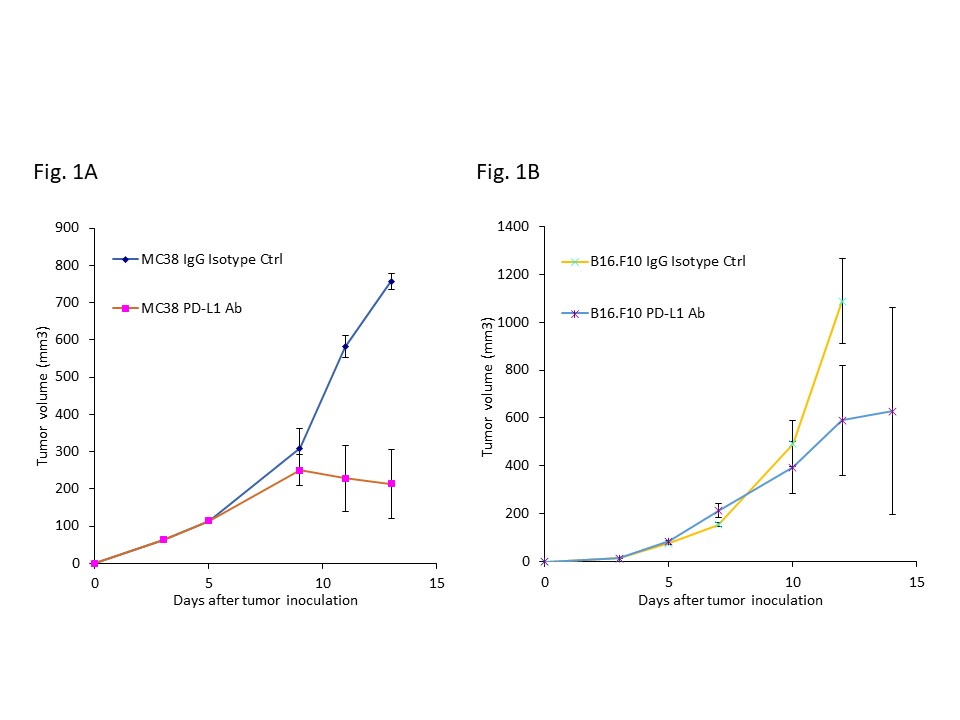
Tumor growth in skin tumor model of MC38 (A) and B16.F10 (B).
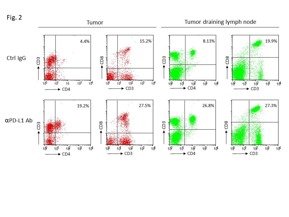
Flow cytometry analysis of tumor infiltrating lymphocytes and tumor
draining lymphocytes in MC38 tumor.
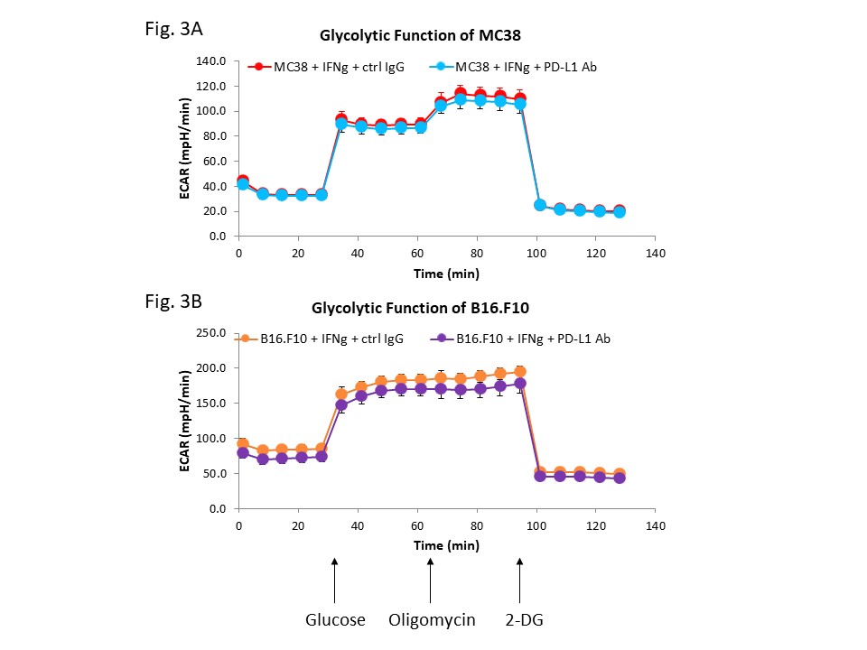
Seahorse assay evaluating glycolytic function in MC38 tumor (A) and B16.F10
(B) tumor.
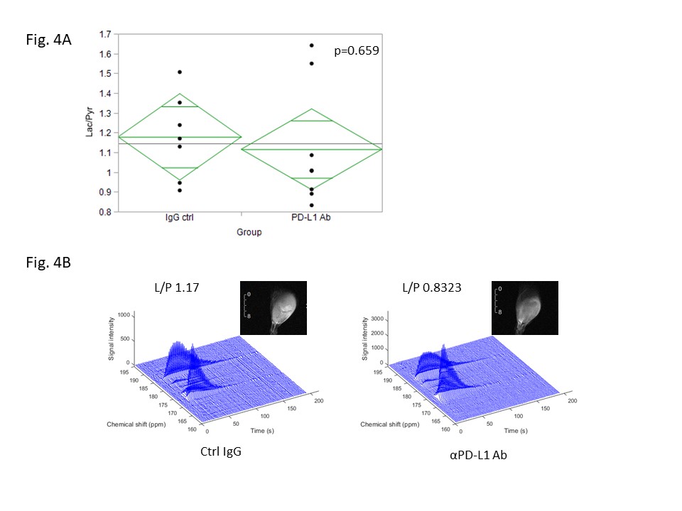
Hyperpolarized [1-13C] Pyr. (A) Comparison of Lactate/Pyruvate ratio
with hyperpolarized [1-13C] Pyr between αPDL-1 antibody treated and control IgG
treated MC38 tumor. (B) Representative dynamic spectra of hyperpolarized
[1-13C] Pyr in MC38, and its anatomical 1H image.