4301
Detection of pyruvate carboxylation and age related changes in glycolysis in human brain using hyperpoplarized 13C MR spectroscopy1GE Healthcare, Toronto, ON, Canada, 2Physical Sciences, Sunnybrook Research Institute, Toronto, ON, Canada, 3Medical Biophysics, University of Toronto, Toronto, ON, Canada, 4Pharmacy, Sunnybrook Health Sciences Centre, Toronto, ON, Canada, 5Sunnybrook Health Sciences Centre, Toronto, ON, Canada
Synopsis
The feasibility of acquiring hyperpolarized 13C images from human brains following injections of HP [1-13C]pyruvate solution has been recently demonstrated. In this study, following each dynamic 3D volume 13C imaging acquisition (5 s temporal resolution), 13C spectroscopic data were acquired from a large slab through the brain. 13C pyruvate, lactate, alanine and bicarbonate were observed in all volunteer subjects. 13C aspartate was also detect in some of the subjects. Good inter-subject consistency in metabolite ratios as well as potential age related trend were observed in these data.
Introduction
Hyperpolarized 13C MR spectroscopy has been used to investigate metabolic changes in brain tumor patients1-2. While direct MR imaging of hyperpolarized 13C substrate and its metabolites following frequency selective excitation3 can improve the coverage, temporal and spatial resolutions in this application, the spectroscopic data may still provide useful information. In this study, an MRS acquisition was interleaved with a 3D time-resolved EPI sequence4 for 13C MRI following injections of hyperpolarized [1-13C]pyruvate in healthy volunteers. The spectroscopic data obtained from this acquisition provided complementary information that may help to interpret the imaging results and to design acquisition methods in future studies.Methods
Subjects: Healthy subjects (N=9) were recruited and gave written informed consent under a protocol approved by the institutional Research Ethics Board and approved by Health Canada as a Clinical Trial Application. Hardware and sample preparation: Each subject was positioned supine and head first in a MR750 3.0 Tesla MRI scanner (GE Healthcare). A home-built 13C birdcage T/R coil was used for 13C imaging and spectroscopy acquisition. Hyperpolarized [1-13C] pyruvate solution was prepared using a GE SPINLabTM system equipped with the Quality Control (QC) module. After sample dissolution, the QC parameters were evaluated by the study pharmacist to ensure the parameters were within specifications. Upon release, the dose syringe was rapidly loaded onto a Spectris Solaris power injector (Medrad) and a 0.43 cc/kg dose of the ~250mM pyruvate solution was injected at 4mL/s followed by a 25mL saline flush at 5mL/s. 13C MRS acquisitions: The 13C imaging data acquisition was initiated at the end of the saline flush. 13C images of bicarbonate [1-13C]pyruvate and [1-13C]lactate were acquired using a dual-echo EPI sequence with a spectral-spatial excitation pulse4. Temporally resolved, 3D images from the whole brain for these 3 metabolites were acquired every 5s for a total of 60s. Following each set of imaging acquisitions, one 13C spectrum was acquired from a 4 cm axial slab in the center of the FOV using the simple pulse-acquire method (10° flip angle, 62.5 kHz/16384 pts), for a total of 12 spectra at the same temporal resolution as the imaging data (5s).Results and Discussions
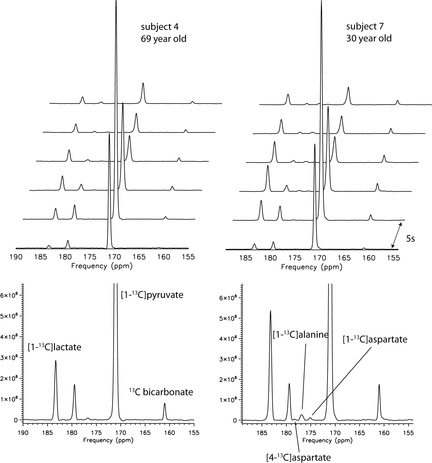
Representative spectra from two volunteers are shown in Figure 1. Besides the metabolites that are typically observed in the brain in studies utilizing [1-13C]pyruvate, [1-13C]asparate was also detected in five of the subjects, and [4-13C]asparate was detected in two of those subjects (when summing the spectra from all time points). To our knowledge, these data represent the first instance of the observation of pyruvate carboxylation product in human brain using hyperpolarized 13C MR. Unlike prior data from mouse liver where pyruvate carboxylation products were observed5-6 using hyperpolarized [1-13C]pyruvate, no 13C malate resonances were detected in these experiments.
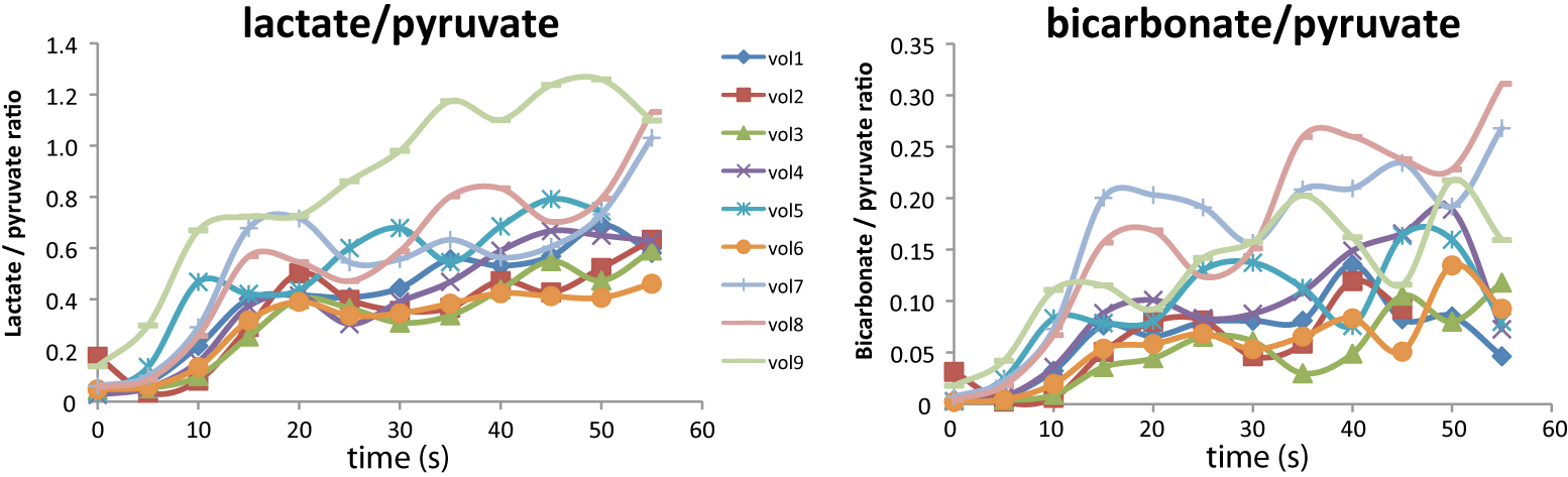
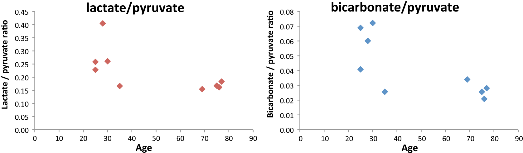
Lactate and bicarbonate to substrate ratios from all volunteers from the time-resolved 13C MRS data are plotted in Figure 2. The metabolite to substrate ratios increased sharply in the first 15 to 20 s of the data due to the bolus arrival in the brain (high substrate and low metabolite signals), then they level off to a much slower but still upward trend during the remainder of the data acquisition window. It also appears that the lactate to pyruvate ratio is less variable than bicarbonate to pyruvate ratio in these datasets (F-test of equality of variances, p < 0.05). When metabolite ratios were calculated from the summed signal from all spectra for each volunteer and then plotted against age of the volunteers (figure 3.), there is a strong trend that the metabolite to substrate ratios decreased with age, and the correlation for bicarbonate to pyruvate ratio and age is significant (Pearson correlation, two-tailed student t-test, p = 0.02). The data suggested that the lower aerobic glycolysis known to occur with aging7 can be detected using hyperpolarized 13C MRS.
Conclusions
13C MRS data acquired from the brain in normal human subjects after injection of hyperpolarized [1-13C]pyruvate provided additional information not available in the imaging data. Product of pyruvate carboxylation was observed for the first time in human brain using hyperpolarized 13C MR. Age dependent changes in brain glycolytic metabolism were also demonstrated. These initial results may provide useful information for investigation of metabolic perturbations in human brain due to disease or neuronal activities.Acknowledgements
The authors are grateful to Sumeet Sachdeva for coordinating the studies and to Ruby Endre and Garry Detzler for MR technician support.References
1. Park I et al. MRM. 2018;80(3):864-873.
2. Miloushev VZ et al. Cancer Res. 2018;78(14)3755-3760.
3. Lau AZ et al. NMR in Biomed. 2011;24:988-996.
4. Geraghty BJ et al. MRM. 2018;79(2)643-653.
5. Merritt ME et al. PNAS. 2011;108(47):19084-9.
6. Lee P. et al. Hepatology. 2013;57(2):515-24.
Figures