4300
Spiral-IDEAL for Time-Resolved Imaging of Hyperpolarized 129Xe Kinetics in the Rat Brain1Translational Medicine, Hospital for Sick Children, Toronto, ON, Canada, 2Department of Medical Biophysics, University of Toronto, Toronto, ON, Canada, 3Department of Medical Imaging, University of Toronto, Toronto, ON, Canada
Synopsis
Rapid, spectrally resolved imaging of 129Xe in the rat brain may allow for measurement of cerebral blood flow, as well as tissue characteristics such as relaxation rates and partition coefficient which may be useful for characterizing neurological disease or injury such as stroke. In this work, the feasibility of spiral-IDEAL for spectrally resolved 129Xe in the rat brain was demonstrated during steady state ventilation of a rat. With future improvements to the signal-to-noise ratio, it is proposed that spiral-IDEAL will allow for rapid, spectrally resolved imaging of the brain.
Introduction
Analogous to O2 delivery, inhaled hyperpolarized (HP) 129Xe dissolves through the lungs and into blood where it transits to the brain via the vascular system, crosses the blood brain barrier, and dissolves into the brain tissue. Furthermore, signal from dissolved 129Xe in the brain is spectrally resolvable into several peaks including signal originating from 129Xe in the red blood cells (RBCs) and 129Xe in the brain tissues1. Therefore, imaging the kinetics (i.e. wash-in/wash-out timecourse) of 129Xe dissolved in the brain may allow measurement of cerebral blood flow, as well as tissue characteristics such as relaxation rates and partition coefficient2–4. For example, regional imaging of 129Xe kinetics in the brain could be useful for characterizing neurological disease or injury expected to alter the partition coefficient of brain tissue such as infarcted brain tissue following a stroke5. Example applications of 129Xe MRI of the brain include the imaging of perfusion in healthy humans6, spectroscopy of 129Xe kinetics in the brain in Alzheimer’s disease7, and multi-voxel chemical shift imaging of 129Xe8. However, due to lack of spectral resolution, spatial resolution, and/or imaging speed none of these reported methods have explored the kinetics of spatially and spectrally resolved 129Xe signals in the brain. Spiral-IDEAL imaging is a technique that can overcome the spatial, spectral, or temporal limitations of the aforementioned imaging methods9–11. Here, we demonstrate the feasibility of spiral-IDEAL imaging of HP 129Xe dissolved in the RBCs and tissues in the brain of ventilated rats.Methods
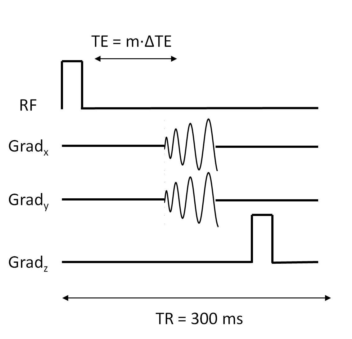
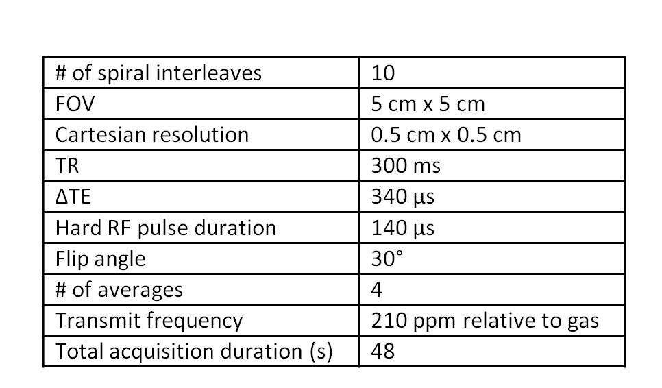
Healthy Sprague Dawley rats were mechanically ventilated12 with alternating breaths of HP (12-15% polarization, model 9800, Polarean, Durham, NH) xenon gas (86% 129Xe) and O2. Imaging was performed using a 3T MRI Scanner (Prisma, Siemens, Germany) and a quadrature transmit/receive birdcage coil (Morris Instruments, Canada) placed over the head of the rat. After waiting 15s of ventilation for 129Xe concentration in the brain to reach physiological steady state2, spiral-IDEAL images were acquired10,11. A pulse sequence timing diagram of the spiral-IDEAL sequence is shown in Fig. 1. The spiral-IDEAL echo time shift was chosen so that spectral separation was optimized for the chemical shift of RBCs (210ppm), brain tissue(s) (195ppm), and gas (0ppm) (requiring a maximum TE of 1.2ms). The spectral separation was restricted to 3 components as prohibitively long echo times would otherwise be required to resolve addition dissolved signal components, given the short T2* of 129Xe dissolved in RBCs (empirically measured to be 2-3ms). Furthermore, the spiral acquisition of k-space was interleaved (Nint=10) to reduce the read-out time from 4.64ms to 0.61ms. The imaging parameters, (summarized in Table 1), were empirically chosen to provide sufficient signal at a resolution of 0.5cm x 0.5cm. A 3D GRE 1H image was also acquired of the rat brain to co-localize the 129Xe MR images to anatomical reference points.Results
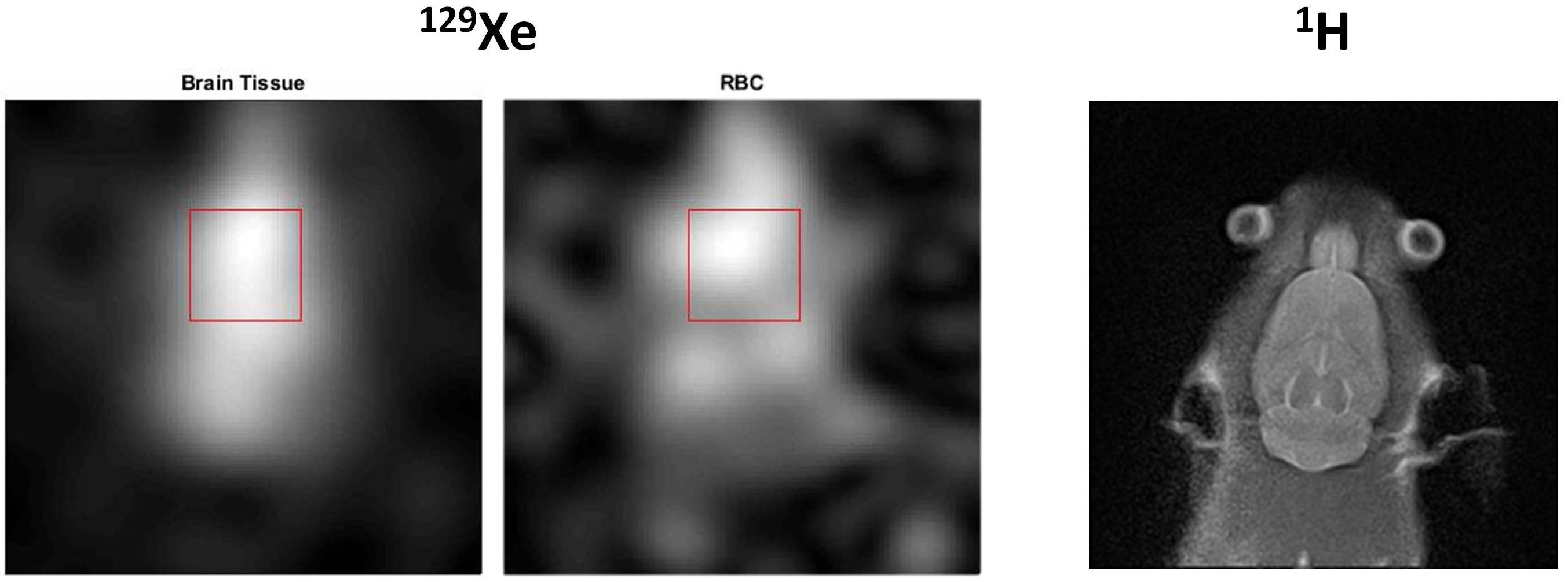
Representative coronal spiral-IDEAL and 1H images are shown in Fig. 2. The signal-to-noise ratio (SNR) in the indicated region of interest is 16±2 for the RBC image and 31±4 for the brain tissue image. Although of limited resolution, the dissolved 129Xe images demonstrate reasonable spatial agreement with the structural 1H image.Discussion
This study demonstrates the feasibility of spiral-IDEAL for spectrally resolved imaging of 129Xe in the rat brain during steady state ventilation. This is an important step towards rapid, spectrally resolved imaging of 129Xe kinetics in the brain. At this time, the imaging sequence (Fig. 1) requires a TR of 300ms during each arm of the interleaved spiral acquisition. Thus, the imaging time per average was 12s, prohibiting the investigation of 129Xe kinetics. The long TR was necessary to allow more 129Xe to wash in between acquisitions, improving the SNR4. However, past studies have shown that a temporal resolution of approximately 1s is required to measure 129Xe kinetics3,4,7. Although, the SNR in this study was insufficient to acquire images with sufficient temporal resolution, it is expected that improvements in optimization of the pulse sequence (e.g. gradient trajectories and interleaves) and hardware (e.g. polarization, reduced ventilator noise) will improve SNR in the future and allow for a temporal resolution of <1s.Conclusion
The feasibility of spiral-IDEAL of 129Xe in the rat brain during steady state ventilation was demonstrated. This is an important step towards rapid, spectrally resolved imaging of 129Xe kinetics in the brain and translation to imaging of humans.Acknowledgements
Thanks to A. Lindenmaier and D. Li for their assistance with imaging experiments. This work was supported by funding from CIHR. Y.F. was financially supported by a Queen Elizabeth II Graduate Scholarship in Science and Technology.References
1. Antonacci MA, Zhang L, Burant A, McCallister D, Branca RT. Simple and robust referencing system enables identification of dissolved-phase xenon spectral frequencies. Magn Reson Med. 2017;00(October). doi:10.1002/mrm.27042.
2. Peled S, Jolesz FA, Tseng C-H, Nascimben L, Albert MS, Walsworth RL. Determinants of tissue delivery for 129Xe magnetic resonance in humans. Magn Reson Med. 1996;36(3):340-344. doi:10.1002/mrm.1910360303.
3. Kilian W, Seifert F, Rinneberg H. Dynamic NMR spectroscopy of hyperpolarized (129)Xe in human brain analyzed by an uptake model. Magn Reson Med. 2004;51(4):843-847. doi:10.1002/mrm.10726.
4. Imai H, Kimura A, Akiyama K, Ota C, Okimoto K, Fujiwara H. Development of a fast method for quantitative measurement of hyperpolarized 129Xe dynamics in mouse brain. NMR Biomed. 2012;25(2):210-217. doi:10.1002/nbm.1733.
5. Pereira RS, Prato FS, Wisenberg G, Sykes J, Yvorchuk KJ. The use of Gd-DTPA as a marker of myocardial viability in reperfused acute myocardial infarction. Int J Cardiovasc Imaging. 2001;17(5):395-404. doi:10.1023/A:1011989626052.
6. Rao M, Stewart NJ, Griffiths PD, Norquay G, Wild JM. Imaging Human Brain Perfusion with Inhaled Hyperpolarized 129Xe MR Imaging. Radiology. 2018;000(0):1-7. doi:10.1148/radiol.2017162881. [Epub ahead of print].
7. Hane F, Li T, Plata J-A, Hassan A, Granberg K, Albert M. Inhaled Xenon Washout as a Biomarker of Alzheimer’s Disease. Diagnostics. 2018;8(2):41. doi:10.3390/diagnostics8020041.
8. Rao M, Stewart NJ, Norquay G, Griffiths PD, Wild JM. High resolution spectroscopy and chemical shift imaging of hyperpolarized 129Xe dissolved in the human brain in vivo at 1.5 tesla. Magn Reson Med. 2016;75(6):2227-2234. doi:10.1002/mrm.26241.
9. Wiesinger F, Weidl E, Menzel MI, et al. IDEAL spiral CSI for dynamic metabolic MR imaging of hyperpolarized [1-13C]pyruvate. Magn Reson Med. 2012;68(1):8-16. doi:10.1002/mrm.23212.
10. Doganay O, Wade T, Hegarty E, McKenzie C, Schulte RF, Santyr GE. Hyperpolarized 129Xe imaging of the rat lung using spiral IDEAL. Magn Reson Med. 2016;76(2):566-576. doi:10.1002/mrm.25911.
11. Zanette B, Stirrat E, Jelveh S, Hope A, Santyr G. Physiological gas exchange mapping of hyperpolarized 129 Xe using spiral-IDEAL and MOXE in a model of regional radiation-induced lung injury. Med Phys. 2018;45(2):803-816. doi:10.1002/mp.12730.
12. Fox MS, Ouriadov A, Thind K, et al. Detection of radiation induced lung injury in rats using dynamic hyperpolarized 129Xe magnetic resonance spectroscopy. Med Phys. 2014;41(7):072302. doi:10.1118/1.4881523.
Figures