4294
Improving Hyperpolarized 13C Spiral Chemical Shift Imaging Using Model-Based Iterative Reconstruction and Prior Knowledge1Diagnostic Radiology & Nuclear Medicine, University of Maryland Baltimore, Baltimore, MD, United States
Synopsis
A model-based iterative reconstruction algorithm for spiral chemical shift imaging (spCSI) of hyperpolarized 13C metabolites is implemented and compared to conventional CSI reconstruction. The iterative reconstruction utilizes the prior knowledge of 1) the off-resonance frequencies for each metabolite and 2) the common spatial distribution for pyruvate and pyruvate hydrate. The proposed method improves quantification of bicarbonate and reduces artifacts for pyruvate maps caused by
Introduction
Fast spiral chemical shift imaging (spCSI) acquires 2D spatial and 1D spectral information simultaneously. It can be accelerated by performing spectral under-sampling at a cost of aliased spectral peaks1. In conventional CSI reconstruction, the raw k-space data are first transformed in image and spectral domain. This includes the correction of the phase evolution of off-resonance components during the spiral read-out. Then metabolic maps are reconstructed through quantification, e.g., line integral, of the respective peak in the map of spectra. Due to B0 inhomogeneity, $$$T_{2}^{\ast}$$$ decay and limited number of echoes, spectra from spCSI suffer from broad line width and added phase noise. Consequently, it is difficult to quantify low SNR metabolites whose resonance peak is close to that of a high SNR metabolite, such as bicarbonate with respect to pyruvate. Furthermore, in the case where two spectral peaks partially overlap due to aliasing, such as [1-13C] pyruvate and [1-13C] pyruvate hydrate (~270Hz difference at 3T with a commonly used spectral width of ~280Hz), CSI reconstruction can only correct the phase evolution along spiral read-out for one spectral peak, while signal from the aliased peak will contribute to a blurred superpositioned image as a result of the chemical shift artifact. In this study, we implement a model-based iterative reconstruction scheme using the prior knowledge of the known metabolite spectrum as well as the pyruvate - pyruvate hydrate chemical balance to improve the image quality.Method
The iterative reconstruction generates the metabolic maps through solving the minimization problem $$$\hat{\rho} = \underset{\rho}{\operatorname{argmin}} \| s - E\cdot \rho\| $$$, where $$$E$$$ represents the encoding matrix relating the metabolic map $$$ \rho $$$ and acquired signal $$$S$$$. For an M-metabolite system, the encoding matrix describes the following forward signal model2:
$$ S\left(t\right)=\int\limits_{V} e^{i\vec{k}\left(t\right)\cdot\vec{r}} \displaystyle\sum_{\text{i}}^{M}\rho_{\text{i}}\left(\vec{r}\right)e^{i2\pi\Delta f_{\text{i}}\cdot t} d\vec{r}$$
$$$\Delta f_{\text{i}}$$$ is the off-resonance frequency for metabolite $$$\text{i}$$$ and $$$\vec{k}\left(t\right)$$$ is the spiral readout trajectory in k-space, both known priorily.
To resolve the overlapped peaks of pyruvate and pyruvate hydrate, we utilize the prior knowledge that they have the same spatial distribution with a fixed ratio $$$k_{p} ={}^{\rho_{pyrh}}/_{\rho_{pyr}}$$$due to their chemical balance. The signal equation can be expanded as:
$$ S\left(t\right)=\int\limits_{V} e^{i\vec{k}\left(t\right)\cdot\vec{r}} \rho_{Pyr}(\vec{r})\left(e^{i2\pi\Delta f_{pyr}\cdot t} + k e^{i2\pi\Delta f_{pyrh}\cdot t}\right)d\vec{r}$$
After contructing the encoding matrix $$$E$$$ properly, the solution $$$\rho$$$ can be derived using conjugate gradient algorithm with fast convergence3.
Results and Discussion
Digital simulations performed in Matlab (R2018a, The MathWorks, Natick, MA) evaluated the effectiveness of iterative reconstruction in removing the chemical shift artifact from spectrally overlapped pyruvate hydrate. Signal acquisition is simulated according to a 2D spiral CSI sequence with FOV = 80mm, matrix size 16×16, 3 spatial interleaves, 32 echoes and 276Hz spectral width (ΔTE=3.62ms). Conventional CSI reconstruction for a 4mm-radius circular disc phantom containing pure [1-13C] pyruvate (Δfpyr=0Hz, relative amplitude (RA)=1.00) is set as ground truth, shown in Figure 1a. Conventional CSI reconstruction for a 4mm-radius circular disc phantom containing pyruvate and pyruvate hydrate mixture (Δfpyrh=269Hz, RA=0.11) is shown in Figure 1b. Iterative reconstruction using the given prior knowledge for the same mixture phantom is shown in Figure 1c. The result from the iterative reconstruction demonstrates the effect of de-blurring at the side lobes.
Two in vivo experiments were conducted on a 3T MR scanner on healthy rats with intravenous injections of hyperpolarized [1-13C] pyruvate. In the first experiment, image data were acquired at a 10mm slice through the kidneys by the same spCSI sequence as in the digital simulation. Metabolic maps for four metabolites were derived: lactate (Δflac=195Hz), alanine (Δfala=-16Hz), pyruvate (Δfpyr=-195Hz) and bicarbonate (Δfbic=-516Hz). Results from both CSI reconstruction and iterative reconstruction are shown in Figure 2. By using the prior knowledge of the spectrum and $$$k_{p}=0.08$$$, the metabolic maps using iterative reconstruction gives better quantification for bicarbonate. Results for iterative reconstruction are prone to error if pyruvate hydrate is treated as an independent metabolite.
In the second experiment, a dynamic 3D spCSI sequence (FOV=80mm×80mm×60mm, matrix size 16×16×12, 4 spatial interleaves, 24 echoes, 274Hz spectral width, 12 time points with 6 seconds temporal resolution) was applied. A time-averaged signal between 48 sec and 72 seconds after injection was used to reconstruct the metabolic maps. Figure 3 shows the results at the slice through the liver, where a urea syringe phantom was placed on top of the rat. Since urea peak (Δfurea=-242Hz) and pyruvate peak (Δfpyr=-482Hz) partially overlap in the under-sampled spectrum, severe artifacts exist for the CSI reconstruction, while the iterative reconstruction can resolve the two metabolites successfully.
Conclusion
The model-based iterative reconstruction method improves quantification of low SNR metabolites such as bicarbonate. It also helps to remove the blurring/artifact from two partially overlapped spectral peaks on the under-sampled spectrum, such as pyruvate hydrate and urea.Acknowledgements
This work was supported by NIH grants R01 DK106395, R21 CA202694, R21 NS096575, and R21 CA213020References
- Mayer, Dirk, et al. "Fast metabolic imaging of systems with sparse spectra: application for hyperpolarized 13C imaging." Magnetic resonance in medicine 56.4 (2006): 932-937.
- Gordon, J. W., Niles, D. J., Fain, S. B., & Johnson, K. M. Joint spatial‐spectral reconstruction and k‐t spirals for accelerated 2D spatial/1D spectral imaging of 13C dynamics. Magnetic resonance in medicine 71.4 (2014): 1435-1445.
- Sutton, B. P., Noll, D. C., & Fessler, J. A. Fast, iterative image reconstruction for MRI in the presence of field inhomogeneities. IEEE transactions on medical imaging 22.2 (2003): 178-188.
Figures
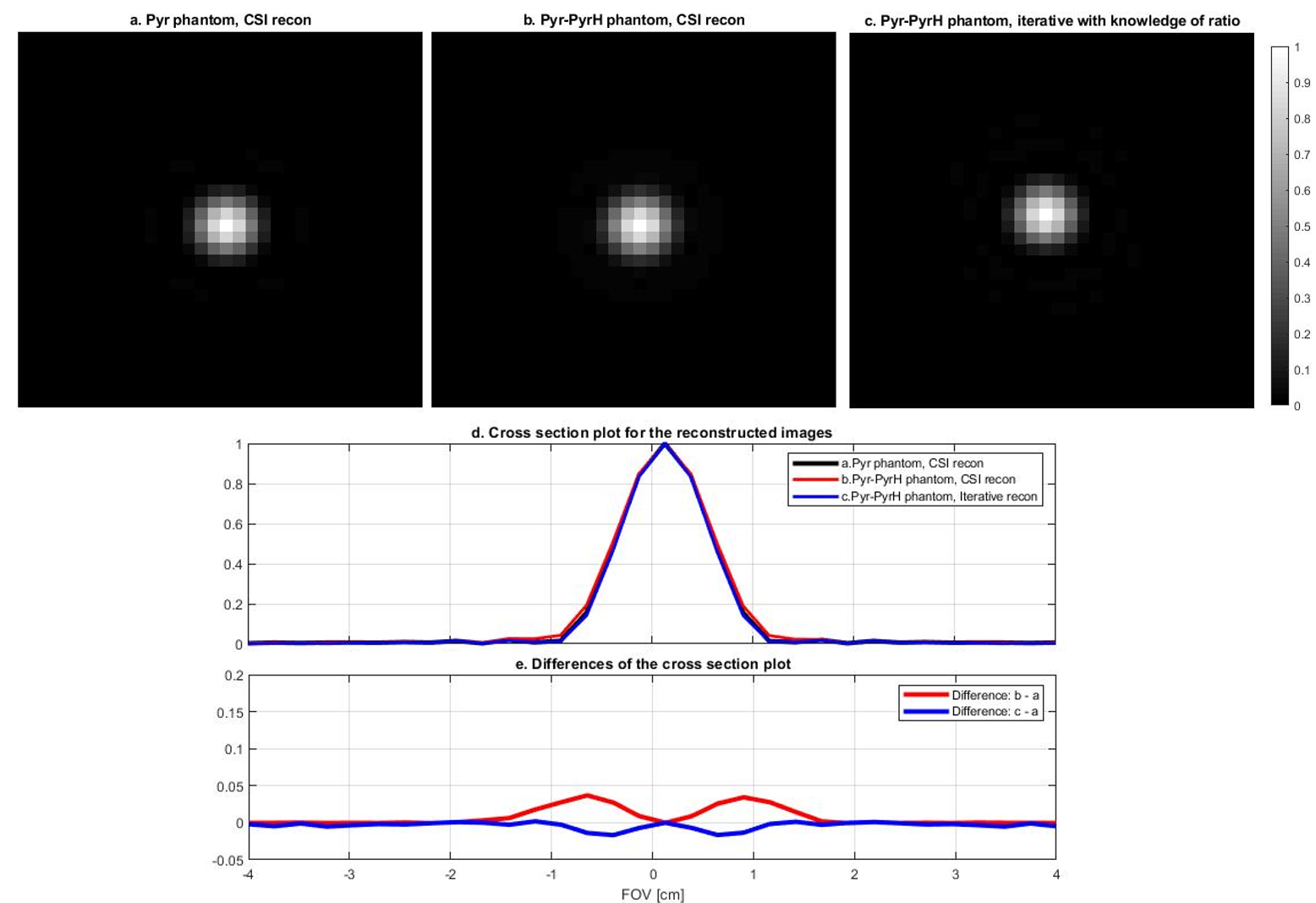
Figure 1: Simulation results for 4mm radius disc phantoms, all acquired with the same 2D spCSI sequence.
(a) pure pyruvate phantom reconstructed by CSI reconstruction
(b) pyruvate - pyruvate hydrate mixture phantom reconstructed by CSI reconstruction
(c) pyruvate - pyruvate hydrate mixture phantom reconstructed by iterative reconstruction.
Cross section of the reconstructed images are shown in (d), and the differences between the results from (b) and (c) relative to (a) are shown in (e).
The difference plot illustrates the effect of de-blurring at the side lobes through iterative reconstruction.
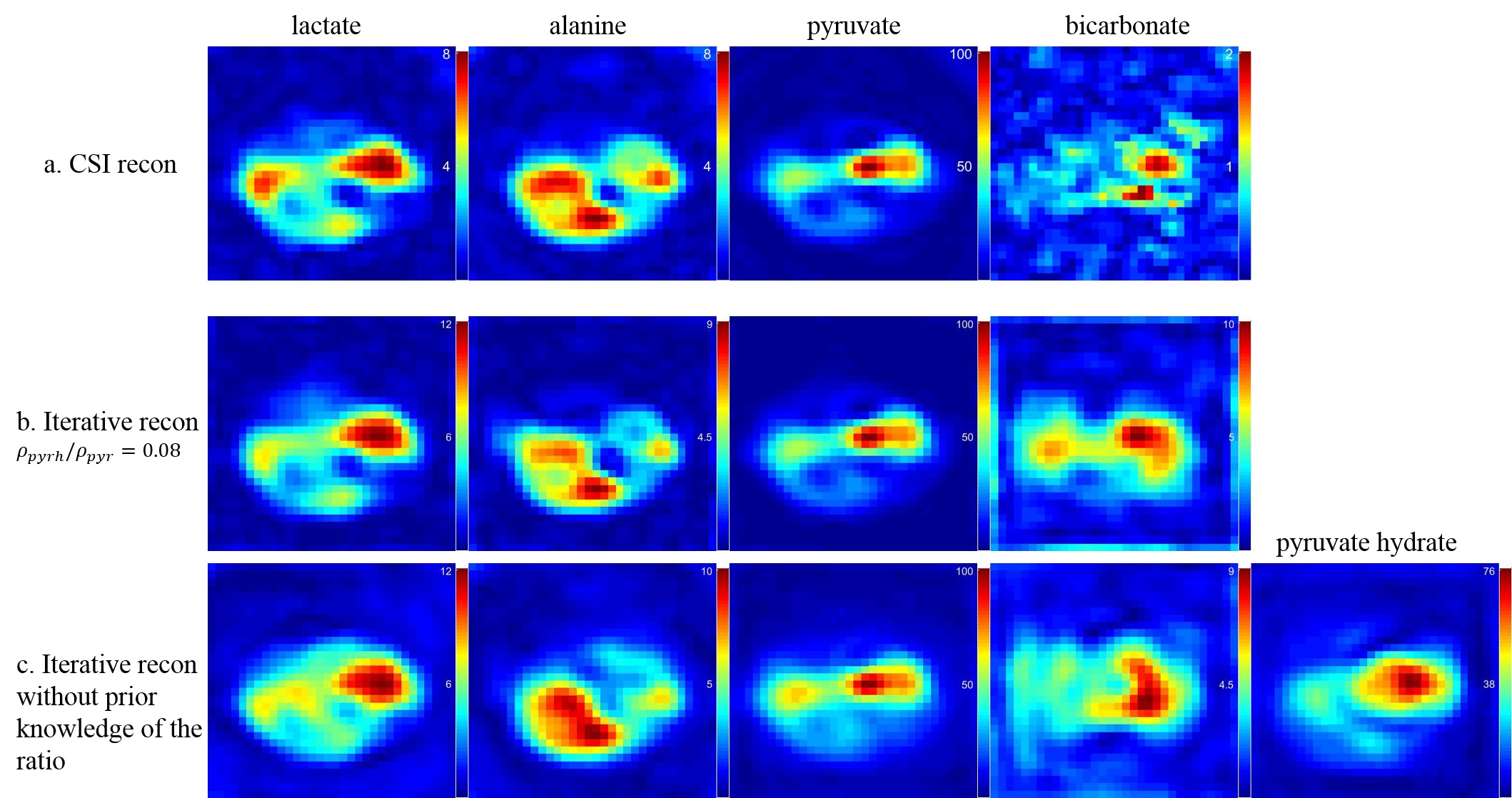
Figure 2: Reconstruction results for in vivo rat kidney with 2D spCSI sequence.
Top row: CSI reconstruction
Middle row: iterative reconstruction with fixed ratio ρpyrh / ρpyr = 0.08
Bottom row: Iterative reconstruction without using the fixed ratio (treated as independent metabolites)
Iterative reconstruction gives better quantification for bicarbonate images with prior knowledge of the pyruvate - pyruvate hydrate ratio. The reconstruction is prone to error if the pyruvate hydrate is treated as an independent metabolite.
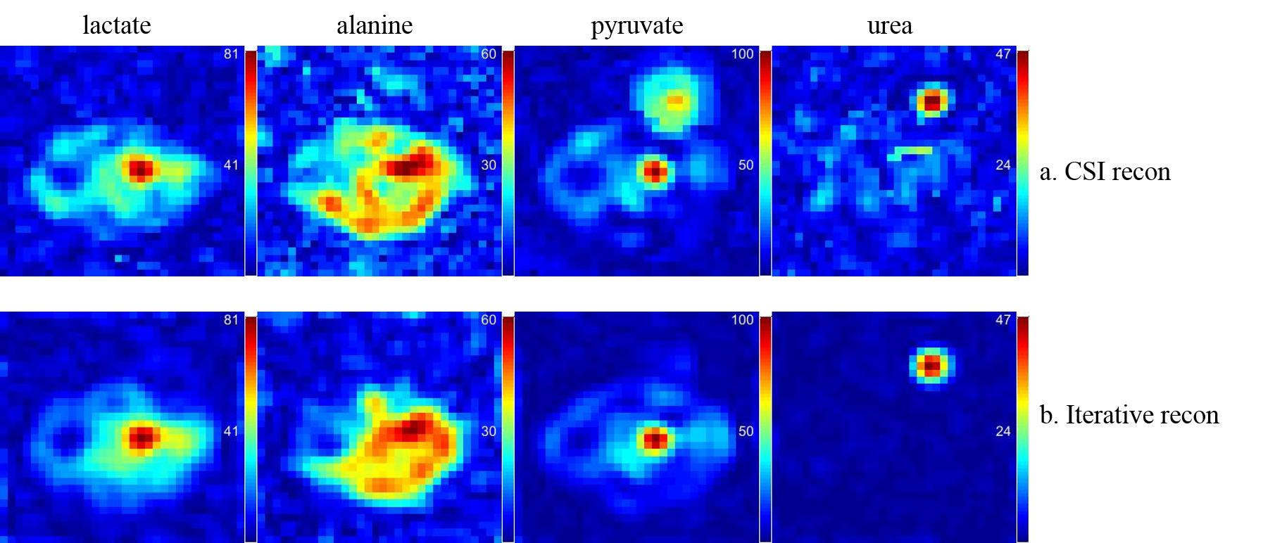
Figure 3: Reconstruction results for in vivo rat liver with 3D spCSI sequence, urea reference phantom exists on top of the rat.
Top row: CSI reconstruction
Bottom row: Iterative reconstruction
Artifacts from the urea phantom signal are seen in CSI reconstruction and removed in iterative reconstruction.