4290
Partial hyperpolarization transfer to protons in [15N2]urea1Cancer Research UK Cambridge Institute, University of Cambridge, Cambridge, United Kingdom, 2Department of Radiology, Hebrew University of Jerusalem, Jerusalem, Israel, 3Department of Biochemistry, University of Cambridge, Cambridge, United Kingdom
Synopsis
Direct observation of 15N hyperpolarization is limited by nitrogen’s low gyromagnetic ratio. We overcome this limitation here by transferring 15N hyperpolarization in [15N2]urea to spin coupled protons. The larger gyromagnetic ratio of the protons will increase the sensitivity of detection provided that there is not a significant loss of polarization during the transfer. We show that our polarization transfer pulse sequence, which has been modified for partial polarization transfer, is tolerant of B1 and centre frequency variations and can be used for dynamic spectroscopy and imaging measurements.
Introduction
Hyperpolarized 15N labelled substrates have been suggested as contrast agents for investigating perfusion (urea, glutamine)1and as pH probes (pyridine derivatives)2. Recently it has been shown that substrates labelled with this low gamma nucleus can be polarized to levels of around 5% relatively easy with equipment widely used for 13C dissolution DNP3. The advantage of 15N labelled substrates over 13C labelled substrates is a very long T1 when kept in D2O 3. However, the 2.5-fold lower gamma compared to 13C (10-fold lower when compared to 1H) results in lower magnetization and precession frequency, and therefore lower sensitivity, and also requires larger gradients for imaging. One way of overcoming this while still benefiting from the long T1 times is to transfer the hyperpolarization, piecemeal, from 15N to spin-coupled protons.Methods
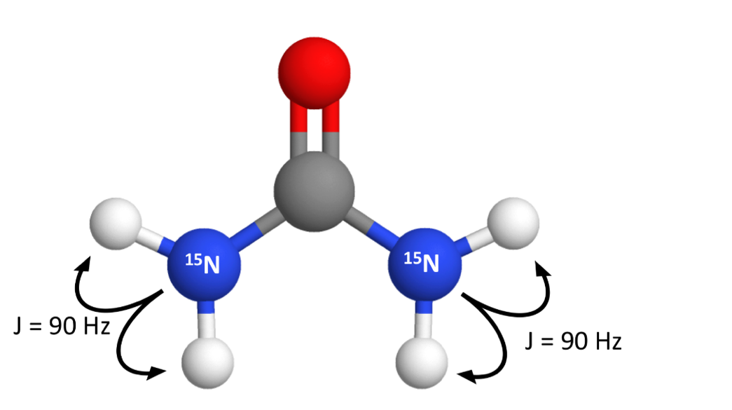
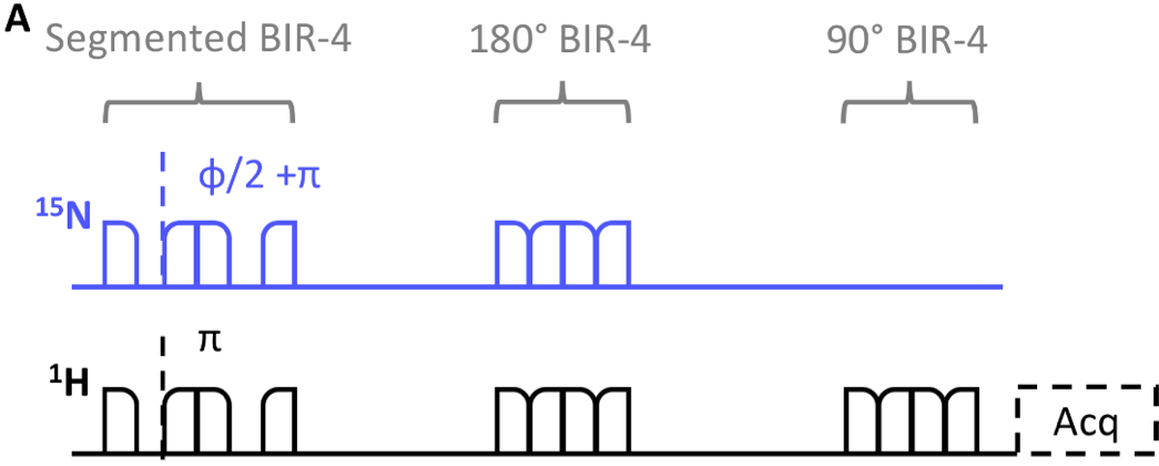
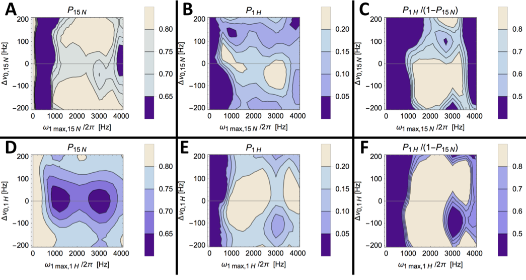
Pulse Sequence: The polarization transfer sequence (Fig. 2) is based on the BINEPT sequence4 modified for partial transfer5. In one acquisition ~20% of the 15N hyperpolarization is transferred to coupled (JNH=90 Hz) protons. The last adiabatic BIR4 pulse in the sequence, shown in Fig.2, flips the proton polarization onto the z-axis. Then, either a non-localized excitation pulse or a 2D-single-shot imaging sequence can be used for proton acquisition (detailed below). The proton polarization P1H divided by the depleted 15N polarization (1-P15N) at the end of the transfer block describes the efficiency of polarization transfer. This parameter was calculated from simulations in SpinDynamica6 for a range of 15N and 1H excitation frequency offsets and transmitter powers.
Dynamic Nuclear Polarization: HyperSense polarizer (Oxford Instruments, Abingdon, United Kingdom) was used for dynamic nuclear polarization. [15N2]urea was prepared as described in Harris et al.3. Polarization time was three hours or more.
Phantom measurements: Experiments were performed on an Agilent 7 T spectrometer using a home-made 1H/15N transmit/receive surface coil7. A spherical flask filled with 3 ml water was positioned in the isocentre of the scanner. Shimming and reference imaging were performed. For the hyperpolarized acquisitions, 1.5 ml of the water were removed and replaced with hyperpolarized [15N2]urea solution. For the spectroscopic and imaging experiments six water presaturation pulses with crusher gradients were added prior to the polarization transfer block. Spectra were acquired with a 90° BIR4 excitation pulse after the polarization transfer block. Parameters were TR = 2 s, sweep width = 10000 Hz, number of points = 4096. Images were acquired with a 2D EPI sequence after the polarization transfer block. Imaging parameters were: FOV = 32x32x1 mm, TR = 1 s, bandwidth = 250 kHz, matrix size = 32x32.
Results
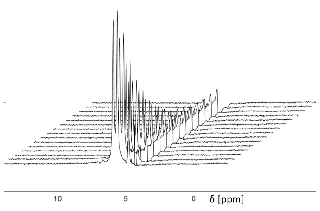
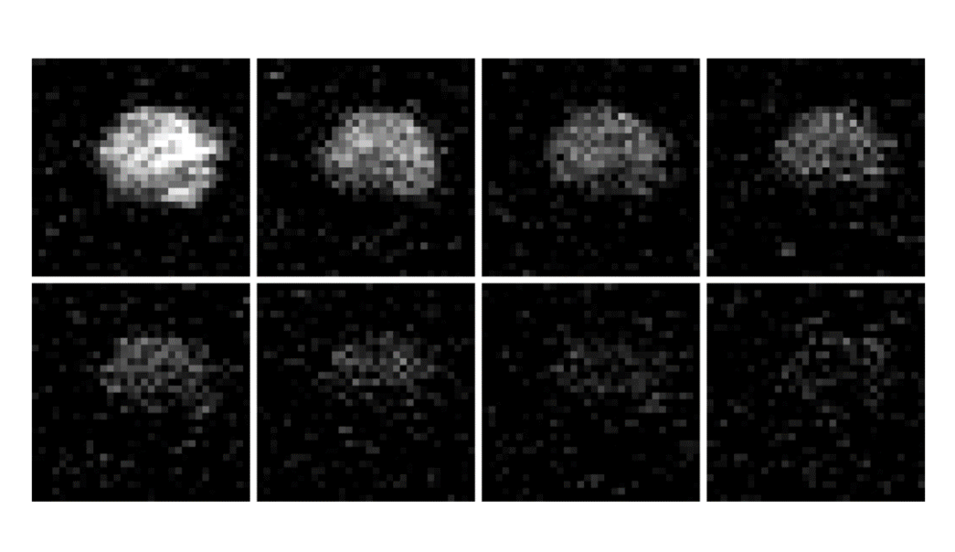
The transfer efficiency is preserved for large regions of parameter space (Fig.3). Partial transfer of polarization was successful. The proton polarization could be used for dynamic spectral acquisition (Fig. 4) and imaging (Fig.5). In the spectra the splitting due to the 90 Hz coupling is visible.Discussion
Simulations (Fig.3) show that the B1 insensitivity of the BINEPT sequence is preserved when the sequence is adapted for partial transfer. We did not include polarization loss due to relaxation in the simulations, because unlike in previous 13C to 1H polarization transfer experiments8, the polarization transfer via the strong coupling is fast (~12ms vs 278ms).Conclusion
We showed that partial polarization transfer followed by proton detection is an alternative to direct detection of hyperpolarized very low gamma nuclei. Potential applications should be evaluated with regards to signal to noise ratio, when compared to Carbon-13 labelled molecules, with the consideration given that typical initial 15N hyperpolarization is lower than 13C hyperpolarization.Acknowledgements
FK received founding from the European Union’s Horizon 2020 research and innovation program under the Marie Sklodowska-Curie grant agreement No 642773 (EUROPOL).References
1. Durst M, Chiavazza E, Haase A, Aime S, Schwaiger M, Schulte RF. α-trideuteromethyl[15N]glutamine: A long-lived hyperpolarized perfusion marker. Magn Reson Med. 2016;76(6):1900-1904.
2. Jiang W, Lumata L, Chen W, et al. Hyperpolarized15N-pyridine Derivatives as pH-Sensitive MRI Agents. Sci Rep. 2015;5:1-6.
3. Harris T, Gamliel A, Uppala S, et al. Long-lived 15N Hyperpolarization and Rapid Relaxation as a Potential Basis for Repeated First Pass Perfusion Imaging - Marked Effects of Deuteration and Temperature. ChemPhysChem. 2018;32(1):60-73.
4. Merkle H, Wei H, Garwood M, Ugurbil K. B1-insensitive heteronuclear adiabatic polarization transfer for signal enhancement. J Magn Reson. 1992;99(3):480-494.
5. Norton VA, Weitekamp DP. Communication: Partial polarization transfer for single-scan spectroscopy and imaging. J Chem Phys. 2011;135(14).
6. Bengs C, Levitt MH. SpinDynamica: Symbolic and numerical magnetic resonance in a Mathematica environment. Magn Reson Chem. 2018;56(6):374-414.
7. Wetterling F, Högler M, Molkenthin U, et al. The design of a double-tuned two-port surface resonator and its application to in vivo Hydrogen- and Sodium-MRI. J Magn Reson. 2012;217:10-18.
Figures