4287
A 3D HYBRID-SHOT SPIRAL FOR HYPERPOLARIZED 13 C IMAGING (3D-HYSS)1Department of Physiology, Anatomy and Genetics, University of Oxford, Oxford, United Kingdom, 2Oxford Centre for Clinical Magnetic Resonance Research, John Radcliffe Hospital, Oxford, United Kingdom, 3Department of Physics, University of Oxford, Oxford, United Kingdom
Synopsis
Spiral k-space trajectories are a powerful tool for in vivo 13C hyperpolarized cardiac MRI, however short readout durations limit the maximum resolution that single-shot spiral trajectories can achieve for a given field of view. This leaves the researcher with a choice at acquisition time, use a single-shot trajectory with poor spatial resolution or switch to a multiple-shot scheme with improved spatial resolution at the cost of degraded temporal resolution. We present a hybrid-shot spiral trajectory, which by compounding a single-shot and multiple-shot spiral, shifts this decision to analysis time.
Introduction
Hyperpolarized 13C cardiac MRI has the potential to greatly improve patient care by providing metabolic imaging which could help clinicians make better decisions when providing targeted treatments1. The first hyperpolarized 13C images of the human heart were acquired using a single-shot spiral k-space trajectory2. Spiral trajectories are signal efficient, robust to subject motion (particularly important for cardiac imaging) and minimize eddy currents. However, for a given field of view, the maximum spatial resolution that can be achieved with a single-shot spiral is limited by the readout duration allowed by the T2* of the hyperpolarized nuclei.
Spatial resolution can be improved at the expense of temporal resolution by utilizing a multiple-shot spiral trajectory. Multiple rotated spiral readouts, each of which has a smaller field of view than the required image, are interleaved together to allow a full field of view high resolution image to be reconstructed. This leaves the user with a choice at acquisition time between spatial and temporal resolution. In this abstract we present a new hybrid-shot spiral pulse sequence which shifts this decision to reconstruction time so that both cases can be analyzed from a single experiment.
Sequence Design
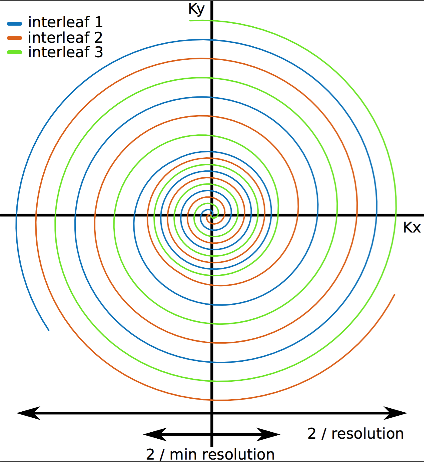
We propose a hybrid-shot spiral k-space encoding trajectory. A single-shot and N-shot spiral are compounded together so that the central region of k-space is fully sampled every shot and the outer region is fully sampled every N shots. This allows for a low spatial resolution image to be reconstructed every shot, giving maximum temporal resolution or for N consecutive shots to be combined giving a higher spatial resolution, albeit at the cost of temporal resolution. The sequence is implemented using a modified version of the Hargreaves variable density spiral algorithm3 to generate the spirals which are compounded together, for example, the N = 3 case is shown in figure 1. A schematic representation of the pulse sequence is shown in figure 2.1H Imaging
For proof of concept, a lemon was imaged using a 72 mm birdcage coil in a Varian 7T preclinical scanner (G max = 1000 mT/m, S max = 5000 mT/m/ms) with an N = 3 hybrid-shot sequence. Figure 3 shows the central slice of the lemon, as reconstructed from the full extent of sampled k-space by combining 3 interleaves (2), from only the single-shot portion of a single interleaf (3) and from a conventional 3D gradient echo acquisition of the same slice (1) for comparison. Scan times are indicated in the lower right corner. The difference in resolution, 1 mm against 8 mm, for the 3 interleaves and single interleaf reconstruction is visible in the additional internal structure resolved in the 3 interleaves reconstruction.13C Imaging
Two male Wistar rats (600 & 540 g) were placed in a Varian 7T preclinical scanner with a 2-channel 40mm 13C receive array and 72mm 1H/13C birdcage transmit coil (Rapid Biomedical) and 2 mL of 80 mM hyperpolarized [1-13C]pyruvate was injected into the tail vein over 12 s. Pyruvate, bicarbonate and lactate were imaged using a cardiac gated, N = 3 hybrid-shot sequence (one TR per R-R interval, minimum TR 150 ms) with an interleaved spectral-spatial excitation scheme. Axial images of the rat hearts are shown in figure 4. Reconstruction combining 3 spiral interleaves (full res) gave a resolution of 2 mm and revealed the cardiac chambers, with clear delineation of the myocardium, in the pyruvate images. When only the inner region of one interleaf was reconstructed, the resolution dropped to 10 mm and this detail was lost. The 3-interleaf reconstruction of the bicarbonate and lactate signal was also well resolved. Approximate scan times were ~1.8 s for a single interleaf reconstruction and ~5.4 s for a 3-interleaf reconstruction.Conclusion
The experiments reported in this abstract demonstrate the applicability of 3D-HYSS to hyperpolarized 13C cardiac imaging in vivo. Greater flexibility is afforded to the researcher when planning their study by moving the choice of trade-off between spatial and temporal resolution from scan time to analysis time. Future work will translate this pulse sequence to clinical systems in order to perform hyperpolarized 13C imaging on the human heart with analysis time control of spatial/temporal resolution.Acknowledgements
This work was supported by funding from the British Heart Foundation, the Engineering and Physical Sciences Research Council (EPSRC) and Medical Research Council (MRC) [grant number EP/L016052/1]References
1. Apps A, Lau J, Peterzan M, et al Hyperpolarised magnetic resonance for in vivo real-time metabolic imaging Heart 2018;104:1484-1491.
2. C.H. Cunningham et al, Circ Res, 119, 1177 (2016).
3. J.H. Lee et al, Magn Reson Med, 50, 1276 (2003).
Figures