4284
Over 70% 13C polarization on [U-13C]glucose1Electrical Engineering, Danish Technical University, Kongens Lyngby, Denmark
Synopsis
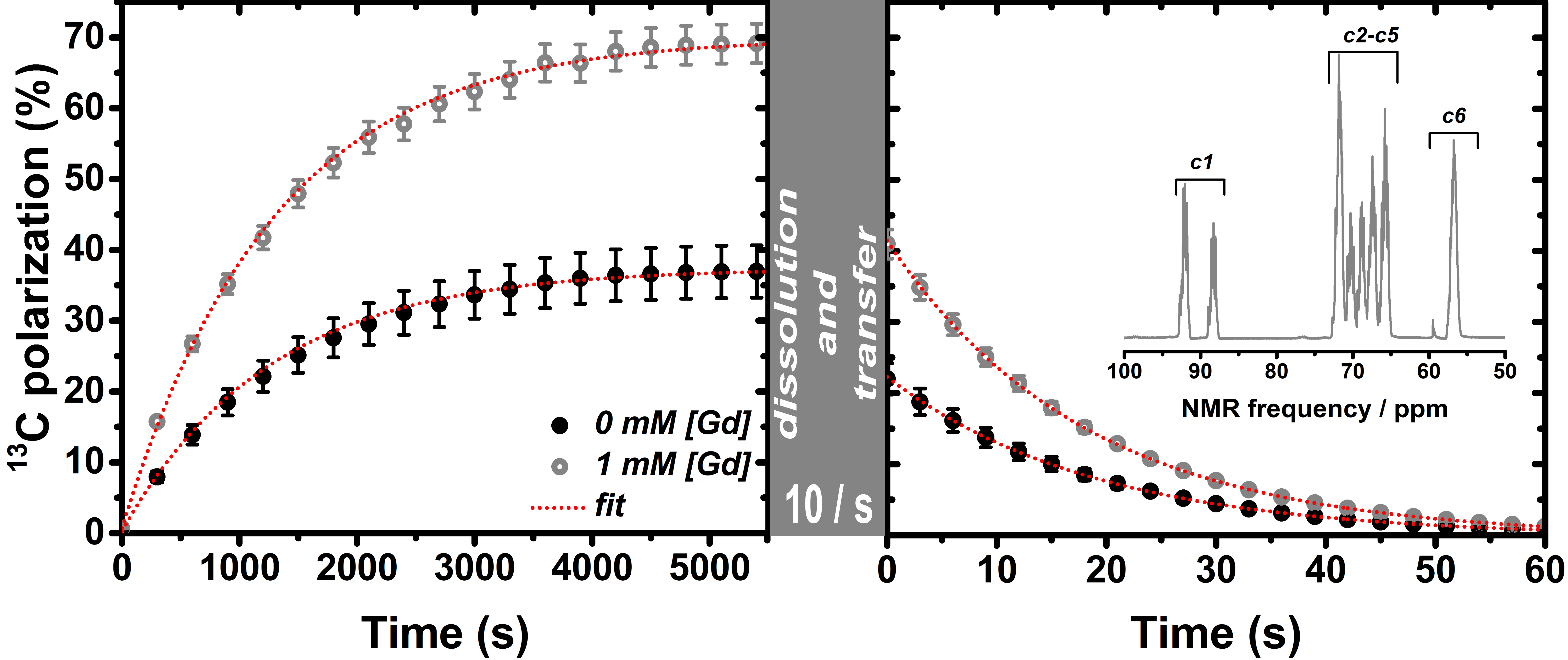
The undisputed usefulness in biomedical applications, the high carbon polarization (up to 70%) and the long relaxation time constant have made “neat [1-13C]pyruvic acid plus trityl” the most studied dissolution-DNP sample. Nevertheless, too often results valid for this particular preparation are considered to hold also for other trityl-based samples, limiting the potential of other biologically interesting substrates. In the present study we demonstrate that the sample composition and magnetic field affect the trityl ESR properties. A thorough optimization of the these parameters lead to a dramatic increase of [U-13C]glucose polarization from 37±4% to 69±3%, the highest value reported to date.
Introduction
Hyperpolarization of nuclear spins via dissolution-DNP1 has proven its great potential and versatility in enhancing the sensitivity of a broad variety of molecules, in particular for detection with 13C NMR and MRI.2-3 In this scenario trityl dissolved in neat [1-13C]pyruvic acid (PA) has become the most studied dissolution-DNP sample because of [1-13C]pyruvate’s undisputed usefulness in biomedical applications, the really high carbon polarization (up to 70%)4-5 and its long relaxation time constant (more than 60s).
Too often results valid for this particular preparation are considered to hold also for other trityl-based samples, limiting the potential of different biologically interesting substrates. A crucial example is the almost negligible “gadolinium effect” at high magnetic field.4-7 Accordingly, at these experimental conditions, no big effort has been put in investigating the effect of Gd3+ doping on trityl based non-PA sample preparations. Nevertheless, the latter benefit much less from increasing the magnetic field strength.8-9
Recently, hyperpolarized glucose has received increasing interest in the hyperpolarization community thanks to the richer metabolic pathways it can give access to.9,11 However, the lower polarization and faster relaxation time constant, compared to pyruvate, has been precluding its widespread employment.
The aim of this work was to measure the trityl main ESR-features at high field DNP conditions (6.7T) and cryogenic temperatures (1.1K) on a generally applicable sample composition, trityl in glycerol-water, to polarize [U-13C]glucose and compare this to the well-studied neat [1-13C]PA/trityl sample. We aimed at understanding why, for the same trityl radical concentration and experimental conditions, neat PA samples outperform other preparations and tried to “fill the gap” by tuning the radical properties via Gd3+ doping.
Methods
[U-13C6,1,2,3,4,5,6,6-d7]-D-glucose was dissolved in 200uL of glycerol:water 1:1 (v/v) to obtain a final glucose concentration of 2M. 30mM trityl radical AH111501 was then admixed to the final solution. Six 30uL samples were pipped out and doped with the following Gd3+ concentrations: 0mM, 0.5mM, 1mM, 2mM, 4mM, and 8mM. A control sample of neat [1-13C]PA with 30mM trityl radical AH111501 was also prepared. From this point onward the control sample will be referred to as PA-sample and the six glucose sample identified through their Gd3+ concentration only (e.g. 1mM-sample). DNP and LOD-ESR measurements were performed on a homebuilt dissolution-DNP polarizer working at 1.10±0.05K and 6.7T. All LOD-ESR acquisition were obtained by means of a homemade spectrometer.12 After dissolution and transfer (10s) the liquid state relaxation was monitored in a 9.4T high resolution vertical NMR magnet (Varian, USA).Results and Discussion
Although the radical concentration was the same for both samples reported in Figure1, the 0mM-sample showed a T1e twice as long as the PA-sample and the ESR-spectrum appeared grossly broadened. Moreover the PA-sample DNP-sweep was narrower and the carbon maximum achievable enchantment double. PA samples at 3.35T and 1.1K have a T1e of about 1s and the most commonly acknowledged explanation for the polarization improvement driven by gadolinium doping is T1e decrease.13-14 The “already short T1e” we measured for the PA-sample, without any addition of gadolinium, can justify its better DNP performance at 6.7T compared to the glucose undoped sample. Indeed, the phonons spectral distribution, responsible for the electron spin-lattice relaxation in the solid-sate, depends on the glassing matrix stiffness and density and thus from its composition.15 As reported in Figure2, the T1e value for the glucose samples could be changed by adding increasing concentrations of Gd3+. The carbon polarization was significantly improved already for the 0.5mM-sample, it reached its maximum for the 1mM-sample and slowly decreased at higher gadolinium concentrations. It is worth noticing that the 1mM-sample was the one with the most similar T1e to the PA-sample.
Besides T1e shortening, a new feature stemmed from our measurements: increasing concentrations of Gd3+ reduced the trityl ESR-linewidth in good agreement with the gradual DNP-spectrum narrowing. If this effect is a direct consequence of the T1e changing or more related to Gd3+ affecting electron spectral diffusion parameters it is not clear.16-17 Nevertheless, the values giving the best polarization enhancement were the ones close to the 13C NMR frequency at 6.7T (i.e. 71.8MHz).
Finally in Figure3 we report the comparison between the best glucose sample and the undoped one. All numerical results are summarized in Table1.
Conclusions
We have demonstrated that trityl T1e and ESR-linewidth strongly depend on sample composition and are both crucial for efficient DNP. At 6.7T the trityl T1e and FWHM values in water-glycerol matrix are too large and lead to a glucose polarization around 35%. The two crucial parameters could be tuned by Gd3+ doping of the sample. At optimal conditions, we doubled the glucose polarization matching 1-13CPA high standards.Acknowledgements
The research leading to these results has received funding from the Danish National Research Foundation (DNRF124) and the European Union's Horizon 2020 research and innovation programme under the Marie Sklodowska-Curie grant agreement no. 713683 (COFUNDfellowsDTU).References
1. Ardenkjaer-Larsen, J. H.; Fridlund, B.; Gram, A.; Hansson, G.; Hansson, L.; Lerche, M. H.; Servin, R.; Thaning, M.; Golman, K. Increase in signal-to-noise ratio of > 10,000 times in liquid-state NMR. P Natl Acad Sci USA 2003, 100 (18), 10158-10163.
2. Kurhanewicz, J.; Vigneron, D. B.; Brindle, K.; Chekmenev, E. Y.; Comment, A.; Cunningham, C. H.; DeBerardinis, R. J.; Green, G. G.; Leach, M. O.; Rajan, S. S.; Rizi, R. R.; Ross, B. D.; Warren, W. S.; Malloy, C. R. Analysis of Cancer Metabolism by Imaging Hyperpolarized Nuclei: Prospects for Translation to Clinical Research. Neoplasia 2011, 13 (2), 81-97.
3. Lerche, M. H.; Jensen, P. R.; Karlsson, M.; Meier, S. NMR Insights into the Inner Workings of Living Cells. Anal Chem 2015, 87 (1), 119-132.
4. Yoshihara, H. A. I.; Can, E.; Karlsson, M.; Lerche, M. H.; Schwitter, J.; Comment, A. High-field dissolution dynamic nuclear polarization of [1-C-13]pyruvic acid. Phys Chem Chem Phys 2016, 18 (18), 12409-12413.
5. Ardenkjaer-Larsen, J. H.; Bowen, S.; Petersen, J. R.; Rybalko, O.; Vinding, M. S.; Ullisch, M.; Nielsen, C. N. Cryogen-Free dissolution Dynamic Nuclear Polarization polarizer operating at 3.35 T, 6.70 T and 10.1 T. Magnet Reson Med 2018, accepted manuscript.
6. Meyer, W.; Heckmann, J.; Hess, C.; Radtke, E.; Reicherz, G.; Triebwasser, L.; Wang, L. Dynamic polarization of C-13 nuclei in solid C-13 labeled pyruvic acid. Nucl Instrum Meth A 2011, 631 (1), 1-5.
7. Johanneson, H.; Macholl, S.; Ardenkjaer-Larsen, J. H. Dynamic Nuclear Polarization of [1-C-13]pyruvic acid at 4.6 tesla. J Magn Reson 2009, 197 (2), 167-175.
8. Yoshihara, H. A. I.; Bastiaansen, J. A. M.; Karlsson, M.; Lerche, M. H.; Comment, A.; Schwitter, J. Myocardial fatty acid metabolism probed with hyperpolarized [1-13C]octanoate. Journal of Cardiovascular Magnetic Resonance 2015, 17.
9. Mishkovsky, M.; Anderson, B.; Karlsson, M.; Lerche, M. H.; Sherry, A. D.; Gruetter, R.; Kovacs, Z.; Comment, A. Measuring glucose cerebral metabolism in the healthy mouse using hyperpolarized C-13 magnetic resonance. Sci Rep-Uk 2017, 7.
10. Rodrigues, T. B.; Serrao, E. M.; Kennedy, B. W. C.; Hu, D. E.; Kettunen, M. I.; Brindle, K. M. Magnetic resonance imaging of tumor glycolysis using hyperpolarized C-13-labeled glucose. Nat Med 2014, 20 (1), 93-+.
11. Meier, S.; Karlsson, M.; Jensen, P. R.; Lerche, M. H.; Duus, J. O. Metabolic pathway visualization in living yeast by DNP-NMR. Mol Biosyst 2011, 7 (10), 2834-2836.
12. Capozzi, A.; Karlsson, M.; Petersen, J. R.; Lerche, M. H.; Ardenkjaer-Larsen, J. H. Liquid-State C-13 Polarization of 30% through Photoinduced Nonpersistent Radicals. J Phys Chem C 2018, 122 (13), 7432-7443.
13. Ardenkjaer-Larsen, J. H.; Macholl, S.; Johannesson, H. Dynamic nuclear polarization with trityls at 1.2 K. Appl Magn Reson 2008, 34 (3-4), 509-522.
14. Serra, S. C.; Filibian, M.; Carretta, P.; Rosso, A.; Tedoldi, F. Relevance of electron spin dissipative processes to dynamic nuclear polarization via thermal mixing. Phys Chem Chem Phys 2014, 16 (2), 753-764.
15. Abragam, A.; Goldman, M. Principles of Dynamic Nuclear-Polarization. Rep Prog Phys 1978, 41 (3), 395-467.
16. Walker, S. A.; Edwards, D. T.; Siaw, T. A.; Armstrong, B. D.; Han, S. Temperature dependence of high field C-13 dynamic nuclear polarization processes with trityl radicals below 35 Kelvin. Phys Chem Chem Phys 2013, 15 (36), 15106-15120.
17. Ravera, E.; Shimon, D.; Feintuch, A.; Goldfarb, D.; Vega, S.; Flori, A.; Luchinat, C.; Menichetti, L.; Parigi, G. The effect of Gd on trityl-based dynamic nuclear polarisation in solids. Phys Chem Chem Phys 2015, 17 (40), 26969-26978.
Figures