4283
Phenylglyoxylic acid as a generic UV-induced radical precursor to hyperpolarize 13C-labelled substrates for in vivo metabolic studies1Institute for Bioengineering of Catalonia, Barcelona, Spain, 2University of Cambridge, Cambridge, United Kingdom, 3Aix-Marseille University, CNRS, ICR, Marseille, France, 4Department of Electrical Engineering, Center for Hyperpolarization in Magnetic Resonance, Technical University of Denmark, Lyngby, Denmark, 5GE Healthcare, Cambridge, United Kingdom
Synopsis
Photo-irradiation of phenylglyoxylic acid (PhGA) using visible light produces a non-persistent radical that, in principle, can be used to hyperpolarize any molecule. We show that PhGA can be used as a radical precursor to polarize photo-sensitive metabolic substrates (such as the gluconeogenic probe 13C-dihydroxyacetone) as well as 13C-glucose. We also present our preliminary DNP data obtained in PhGA-doped samples using a novel custom-built cryogen-free 7T/1.4K polarizer.
Introduction
Free radicals are an essential component for any sample to be hyperpolarized by Dynamic Nuclear Polarization (DNP). Conventionally, these are supplied by doping the sample with stable free radicals. These radicals accelerate the polarization decay after DNP and need to be removed from the hyperpolarized solution before it is used in clinical applications.
Photo-induced radicals generated by ultraviolet-visible (UV-Vis) irradiation can be annihilated inside the polarizer (at around 200K) following DNP while preserving high polarization levels [1,2]. We have shown recently that irradiation of phenylglyoxylic acid (PhGA) with ultraviolet-visible light produces radicals suitable to polarize any substrate, even some photosensitive molecules like dihydroxyacetone (DHAc) [3].
Methods
Photo-irradiation and radical concentration measurement: Frozen 4-μl beads of the solution of interest were formed by dispensing droplets from a syringe into an ESR quartz dewar flask filled with liquid nitrogen (LN2). The beads were photo-irradiated using a narrowband 405nm light source (Dymax BlueWave LED VisiCure 405nm).
ESR spectra of single frozen beads were acquired using a benchtop X-band ESR spectrometer (MiniScope MS5000, Magnettech). Radical concentration was determined by comparing double integration of the first derivative ESR spectrum, corrected for bead volume, with a calibration curve obtained from beads of known concentrations of TEMPOL.
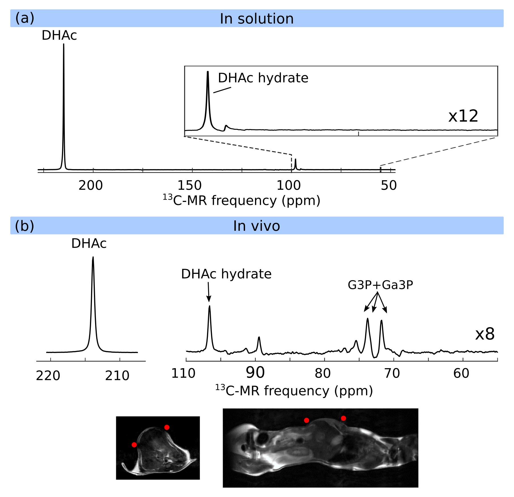
Dissolution DNP of DHAc at 3.35T: Approximately 30 frozen beads from a sample containing 8M [2-13C]DHAc, 1M PhGA and 1.2mM gadolinium in water were UV-irradiated for 200s and loaded into a 3.35T HyperSense polarizer. The sample was polarized and dissolved in 6ml of PBS. This solution (400μl) was injected into a female C57B6 mouse (bw=32.2g) via a tail-vein over a period of 3s. The mouse had been anesthetized previously with 2% isoflurane, its body temperature maintained at 37ºC, and placed inside a small-animal 7T MR system. 13C MR acquisition started 12s after the injection. The parameters used for 13C MR acquisition were: TR=0.2s, flip angle=15º, pulse width=2ms, spectral width=6kHz, total acquisition time=80s. The transmitter was centered at 72.3 ppm, and for every 10th acquisition it was switched to 214ppm for a single acquisition before returning to 72.3ppm. Therefore, the spectral region around 72.3ppm was sampled 360 times (every 0.2s) and the region around 214ppm was sampled 40 times (every 2s). A 42-mm-diameter bird-cage 1H (transmit/receive)/13C (transmit only) volume coil was used to excite signal from the whole body. A 20mm outer diameter 13C receive surface coil was placed over the liver of the mouse. Positioning of the surface coil was confirmed with sagittal, coronal and axial T2-weighted 1H images.
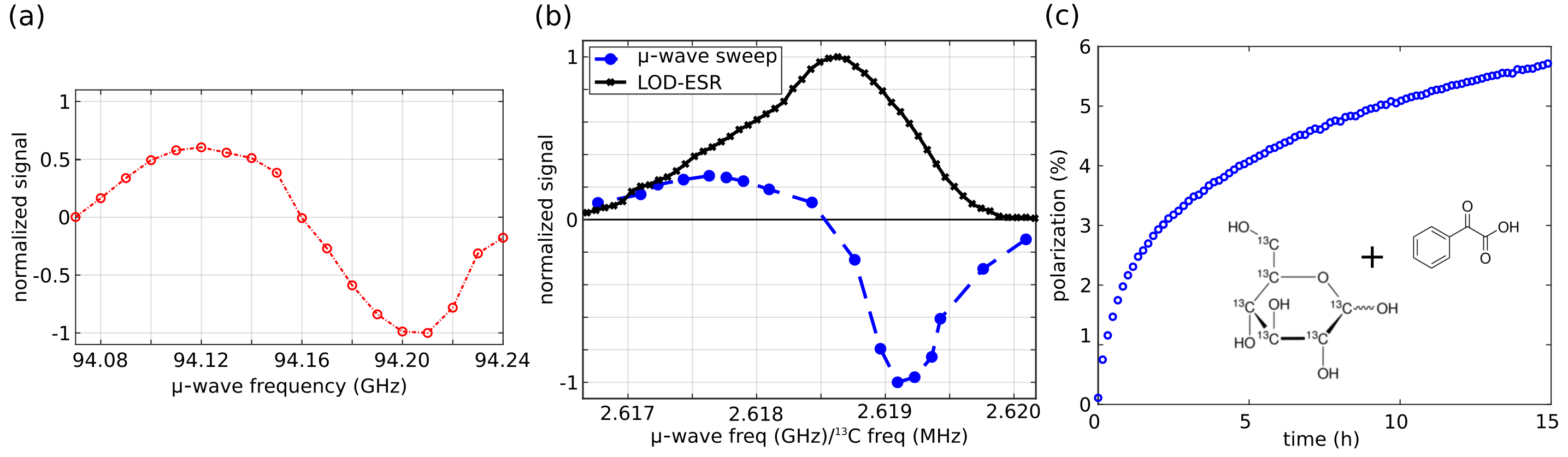
Solid-state DNP at 7.0T: Approximately 30 frozen beads of a solution containing 3.2M [U-13C]glucose and 2.3M PhGA dissolved in a mixture of glycerol/water were UV-irradiated for 100s and loaded into a custom-built, cryogen-free 7T polarizer. A microwave sweep, LOD-ESR and polarization build-up curve at the optimal microwave irradiation frequency were measured.
Results and Discussion
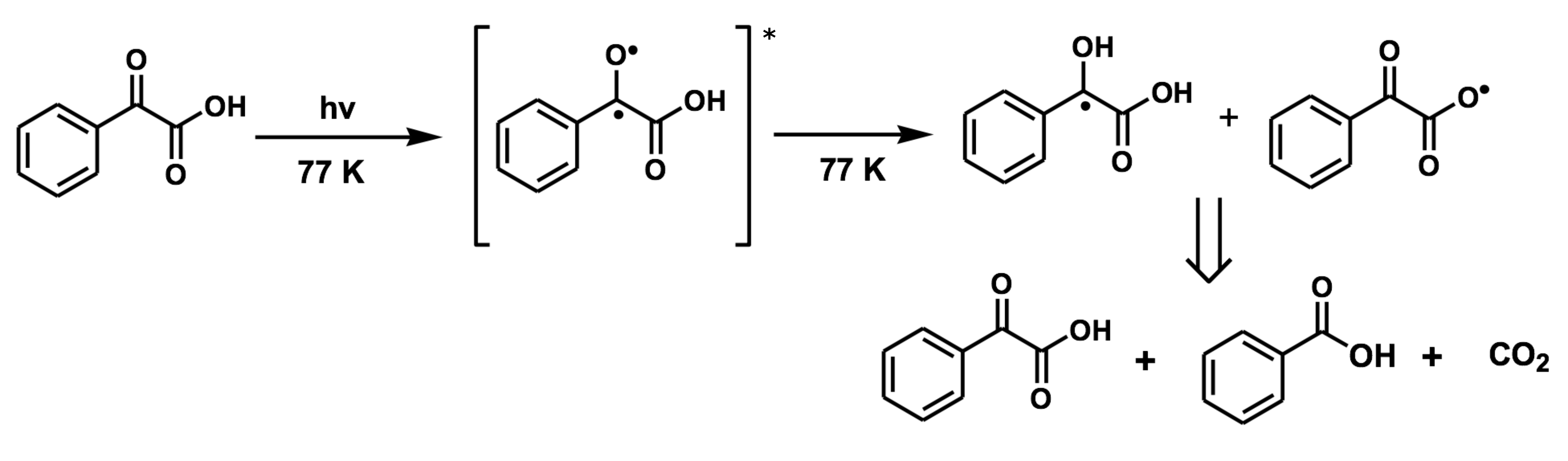
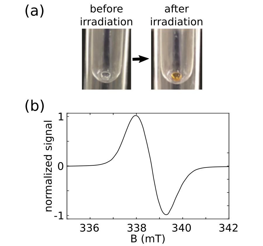
Photo-irradiation of PhGA produces radicals that are stable at 77K and are suitable for hyperpolarization of 13C-labeled substrates but quench upon dissolution. Figure 1 shows the photo-excitation products of PhGA. Upon photo-irradiation of a frozen bead of PhGA, radical generation can be confirmed by visual inspection – a change in the color of the bead from transparent to orange (Figure 2a) – and by the characteristic ESR spectrum of the radical (Figure 2b). As an example of the suitability of PhGA as a radical precursor for metabolic studies, we hyperpolarized [2-13C]DHAc with PhGA radicals using a 3.35T polarizer and injected it into a mouse in vivo. The hyperpolarized spectrum of [2-13C]DHAc in solution and in vivo are shown in Figure 3.The solid-state microwave sweep measured at 3.35T/1.2K in a PhGA-doped DHAc sample and at 7T/1.4K in a PhGA-doped [U-13C]glucose sample are shown in Figure 4. The microwave sweep at 7T is superimposed on the ESR spectrum measured inside the polarizer, which shows an asymmetric profile, characteristic of the g-anisotropy of the PhGA-derived radical. The long build-up time and polarization level of ~6% shown in Figure 4c suggest that the 25mM radical concentration chosen for polarization at 7T/1.4K was too low. Optimization of the sample and microwave irradiation strategy should speed up the process and increase the polarization levels.Conclusion
Photo-irradiated PhGA can be used as a source of unpaired electrons to hyperpolarize 13C-labeled substrates using a 3.35T or a 7T polarizer. To improve the DNP performance of PhGA radicals at 7T, we plan to increase the radical concentration and we will also implement microwave modulation. These adaptations together with the low toxicity profile of PhGA are expected to make this compound an attractive radical precursor for in vivo metabolic imaging.Acknowledgements
This work is part of a project that has received funding from the European Union’s Horizon 2020 European Research Council (ERC Consolidator Grant) under grant agreement No 682574 (ASSIMILES), a Cancer Research UK Programme grant (17242) and by the CRUK-EPSRC Imaging Centre in Cambridge and Manchester (16465). FK and SP received funding from the European Union’s Horizon 2020 research and innovation program under the Marie Sklodowska-Curie grant agreement No 642773 (EUROPOL). A. Capozzi received funding from the European Union's Horizon 2020 research and innovation program under the Marie Sklodowska-Curie grant agreement no. 713683 (COFUNDfellowsDTU). IMR received financial support through the Junior Leader Postdoctoral Fellowship Programme from “la Caixa” Banking Foundation.References
[1] Eichhorn TR, Takado Y, Salameh N, et al. Hyperpolarization without persistent radicals for in vivo real-time metabolic imaging. Proc Natl Acad Sci USA. 2013; 110, 18064
[2] Capozzi A, Cheng T, Boero G, et al. Thermal annihilation of photo-induced radicals following dynamic nuclear polarization to produce transportable frozen hyperpolarized 13C-substrates. Nat Commun. 2017; 8, 1
[3] Marco-Rius I, Cheng T, Gaunt A, et al. Photogenerated radical in phenylglyoxylic acid for in vivo hyperpolarized 13C MR with photosensitive metabolic substrates. J Am Chem Soc. 2018; 140, 14455
Figures