4282
Hyperpolarized metabolic imaging with high SNR in vivo using low volume photo-induced nonpersistent trimethylpyruvic acid radicals1Department of Radiology, University Hospital (CHUV), Lausanne, Switzerland, 2Department of Electrical Engineering Technical University of Denmark, Center for Hyperpolarization in Magnetic Resonance, Kgs Lyngby, Denmark
Synopsis
Recently it was demonstrated that UV irradiation of trimethylpyruvic acid (TMP) generates nonpersistent radicals with narrow electron spin resonance linewidth. TMP however has never been used to hyperpolarize 13C substrates to study in vivo metabolic processes, and relatively large volumes of TMP were needed to generate high nonpersistent radical concentrations. The aim of this study was to increase the nonpersistent radical yield in TMP-doped [1-13C]lactic acid, [1-13C]butyric acid and 13C glucose preparations to achieve high polarization levels. In vivo metabolism was measured with signal levels similar to that of persistent radical preparations, demonstrating TMP as a promising nonpersistent radical for dissolution DNP and potential clinical translation.
Introduction
Dissolution dynamic nuclear polarization (DNP) enhances the MR signal of 13C-labelled metabolites by several thousand folds allowing the noninvasive measurement of metabolic reactions within minutes [1], and has been successfully applied in patients [2,3]. The solid-state DNP process requires the addition of free radicals to the 13C metabolite preparation. However, the presence of free radicals currently imposes signal losses because the radical filtration step is time consuming, and the presence of radicals shorten the relaxation time of 13C nuclei [4], leading to a decrease in polarization level at the time of the acquisition. Recent work [5,6,7] demonstrated that alpha-keto acids, such as pyruvate or trimethyl pyruvate (TMP), form radicals following irradiation with ultraviolet (UV) light. These radicals annihilate at temperatures above 190 K [5], yielding hyperpolarized solutions that are radical free and transportable. The aim of this study was to increase and quantify the formation of photo-induced radicals in TMP to enable in vivo metabolic studies using [1-13C]lactic acid, [1-13C]butyric acid and [U2H, U13C]glucose with signal levels similar to that of persistent radical preparations.Methods
Results
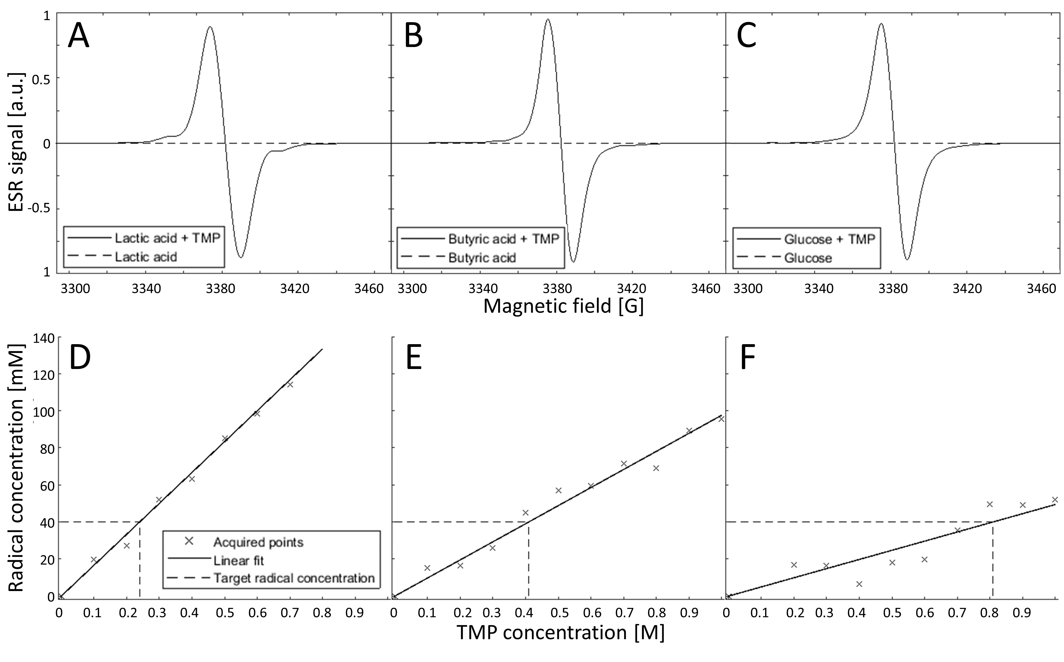
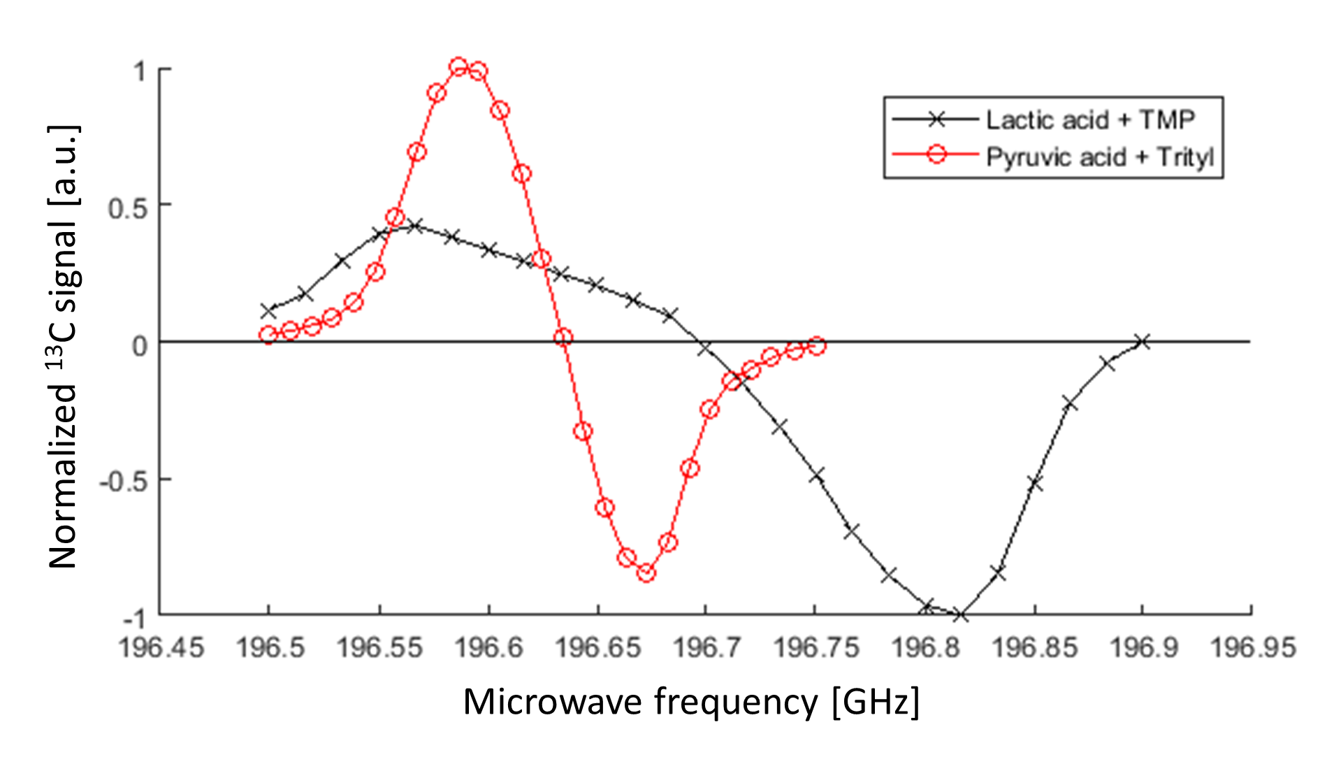
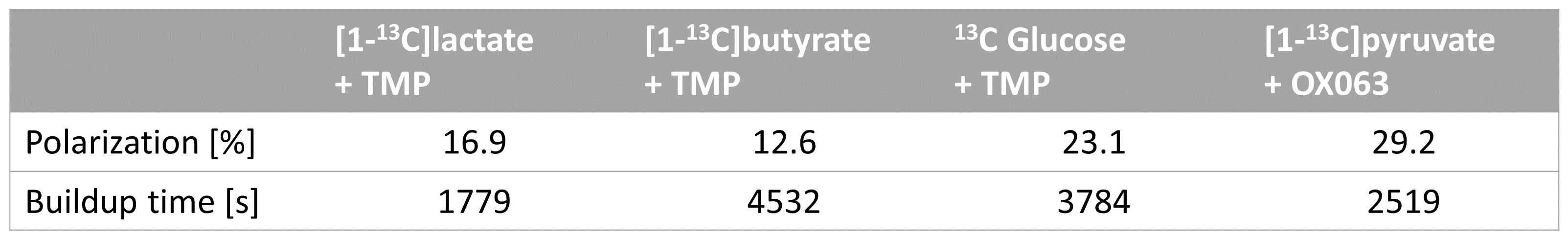
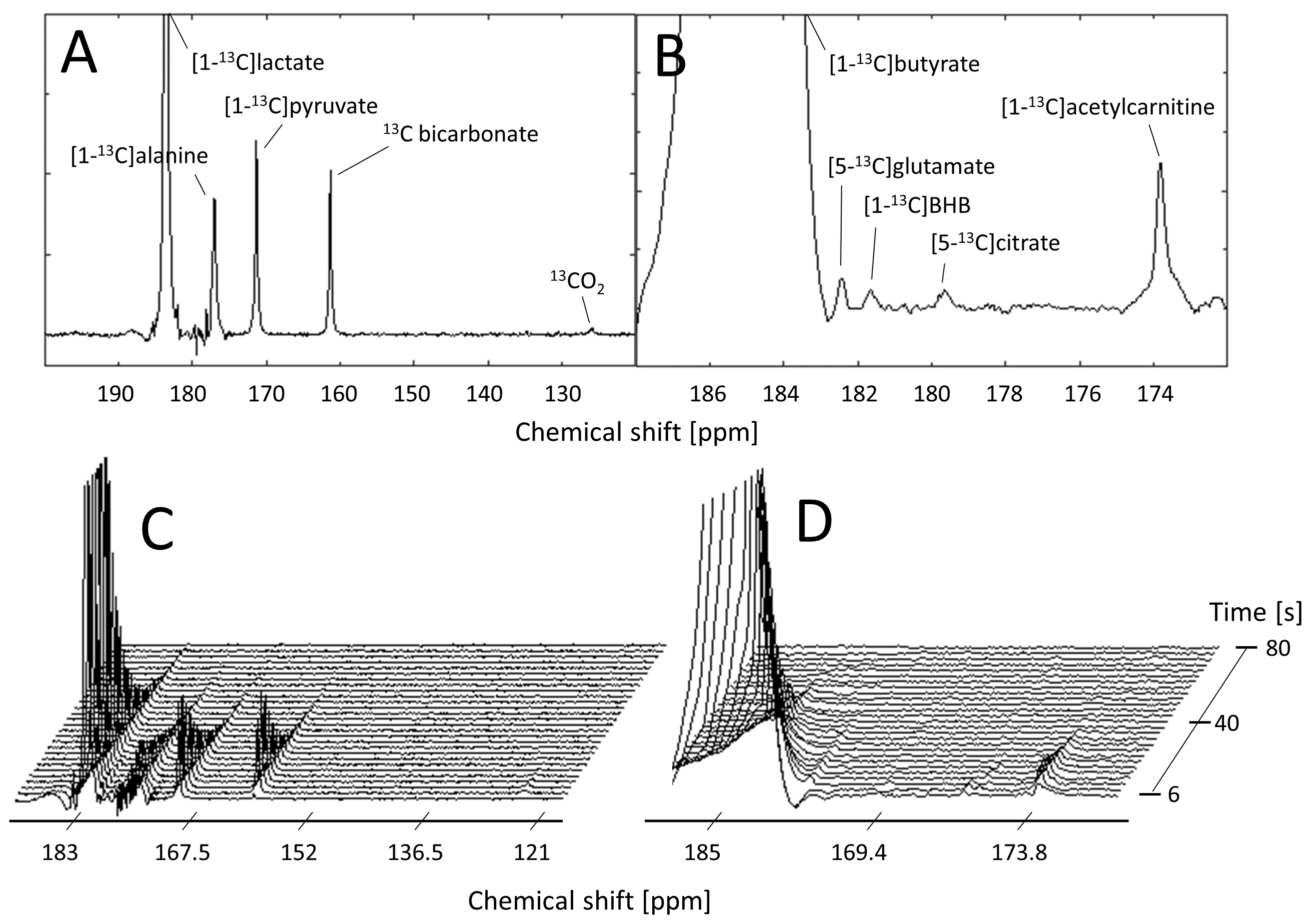
After 60 s of UV irradiation, a 40 mM radical concentration was obtained in lactic acid, butyric acid, and glucose samples doped with TMP concentrations of 0.24 M, 0.41 M, and 0.81 M respectively (Fig. 1). The MW sweep on the [1-13C]lactic acid sample doped with TMP indicated a negative maximum at 196.79 GHz compared to 196.59 GHz for [1-13C]pyruvate + OX063 (Fig. 2). The SS polarization are reported in Table 1. In vivo metabolism of hyperpolarized [1-13C]lactate and [1-13C]butyrate show the presence of lactate (peak SNR=5070±3200), bicarbonate (122±92), alanine (121±77), pyruvate (160±133), CO2(7.7±1.5), butyrate (5300±3200), acetylcarnitine (19.2±8.6), glutamate (8.2±3.6), β-hydroxybutyrate (7.3±3.2) and citrate (7.9±4.8) (Fig. 3 A-D). No myocardial metabolism of glucose was detected. The resonance frequency of natural abundance [1-13C]TMP was observed at 174.5 ppm in glucose experiments.Discussion
In our glucose preparation, UV irradiation generated 50 mM of nonpersistent radicals in a solution containing 1.0 M of TMP, compared with 1.6 M of TMP in [6], a significant reduction in TMP addition. In comparison with the generation of 18.1 mM nonpersistent radicals in 1M of phenylglyoxylic acid [8], radical formation in UV-irradiated TMP with a high power UV source is significantly higher. This enables the reduction of the amount of non-endogenous TMP needed to hyperpolarize 13C substrates. TMP was not observable in the lactic acid and butyric acid experiments because the TMP concentration was below detection limits. However, the TMP resonance could be observed in the glucose experiments, where the amount of TMP was 15x higher than lactate. Therefore it is suggested that the natural abundance carboxylic TMP resonance will not pose any limitations on metabolite quantification.Conclusion
High concentrations of non-persistent radicals were obtained following UV irradiation in 13C metabolite solutions containing TMP at significantly lower volumes. The use of nonpersistent TMP radicals led to high polarization levels of [1-13C] lactic acid and [1-13C] butyric acid, allowing the measurement of metabolic processes in vivo of the same quality as using persistent radicals.Acknowledgements
SNF Ambizione PZ00P3_167871
R'Equip SNF grant 326030_150828
References
[1] Ardenkjaer-Larsen JH et al. Increase in signal-to-noise ratio of > 10,000 times in liquid-state NMR. Proc Natl Acad Sci U S A 2003;100:10158–63.
[2] Nelson SJ et al. DNP-Hyperpolarized 13C Magnetic Resonance Metabolic Imaging for Cancer Applications. Appl Magn Reson 2008; 34(3-4): 533–544.
[3] Cunningham CH et al. Hyperpolarized 13C Metabolic MRI of the Human Heart: Initial Experience. Circ Res. 2016;119(11):1177-1182.
[4] Lumata L et al. Influence of Deuteration in the Glassing Matrix on C-13 Dynamic Nuclear Polarization. Phys Chem Chem Phys 2013; 15, 7032−7035.
[5] Capozzi A et al. Thermal annihilation of photo-induced radicals following dynamic nuclear polarization to produce transportable frozen hyperpolarized 13C-substrates. Nature Communication 2017; 8, 15757
[6] Capozzi A et al. A narrow line UV-induced non-persistent radical to generate highly polarized transportable glucose solid samples (2018). Abstract from 59th Experimental Nuclear Magnetic Resonance Conference, Orlando, United States.
[7] Bastiaansen JAM et al. Probing cardiac metabolism by hyperpolarized 13C MR using an exclusively endogenous substrate mixture and photo-induced non-persistent radicals. Magn Reson Med 2018; 79(5): 2451–2459.
[8] Marco-Rius I et al. Photogenerated Radical in Phenylglyoxylic Acid for in Vivo Hyperpolarized 13C MR with Photosensitive Metabolic Substrates. J. Am. Chem. Soc. 2018; 140, 14455−14463
Figures