4280
Partial Fourier Reconstruction of Hyperpolarized 13C EPI in the Human Brain1Medical Biophysics, University of Toronto, Toronto, ON, Canada, 2Physical Sciences, Sunnybrook Research Institute, Toronto, ON, Canada, 3GE Healthcare, Toronto, ON, Canada, 4Pharmacy, Sunnybrook Health Sciences Centre, Toronto, ON, Canada, 5Radiation Oncology, Sunnybrook Health Sciences Centre, Toronto, ON, Canada
Synopsis
Asymmetric in-plane k-space sampling of EPI can reduce the minimum achievable TE in hyperpolarized 13C with spectral-spatial radio frequency pulses, thereby reducing T2* weighting and signal-losses. Partial Fourier image reconstruction exploits the approximate Hermitian symmetry of k-space data and can be applied to asymmetric data sets to synthesize unmeasured data. Here we present the results of applying partial Fourier image reconstruction from hyperpolarized [1-13C]pyruvate scans in human brain. A quantitative evaluation of image sharpness using no-reference image quality assessment agreed with a perceived improvement in contrast and resolution.
Introduction
Recent studies have demonstrated the ability of hyperpolarized HP [1-13C]pyruvate to cross the blood-brain barrier in humans and become metabolized by cells, as evidenced by the appearance of 13C-labelled bicarbonate in the brain1-3. Thus, HP [1-13C]pyruvate MR spectroscopy and imaging hold tremendous potential for studying neurophysiological metabolism the brain.
While numerous acquisition methods exist for HP 13C MRI with [1-13C]pyruvate, time-resolved volumetric imaging is now most commonly achieved with spectrally- and spatially-selective radio frequency (ssRF) excitation followed by rapid echo-planar spatial encoding4-7. In this work, 3D dual-echo echo-planar imaging8 (DE-EPI) readouts (2D DE-EPI + phase encoding along the “slice” direction) were used.
Compared to non-spectrally selective RF excitation, the long TE associated with ssRF pulses increases T2* weighting and can reduce SNR. Previously9, we showed that for a fixed readout length, asymmetric sampling along the blipped direction in k-space improved SNR without artefacts in preclinical 13C images, thereby motivating the adoption of asymmetrically sampled DE-EPI for our ongoing HP [1-13C]pyruvate brain imaging study in healthy volunteers.
Asymmetrically acquired k-space data can be used to synthesize “missing” lines through partial Fourier (PF) reconstruction10,11. Here we present PF reconstruction of HP 13C images obtained in the human brain. A no-reference image quality assessment12,13 metric was used for quantitative comparisons.
Methods
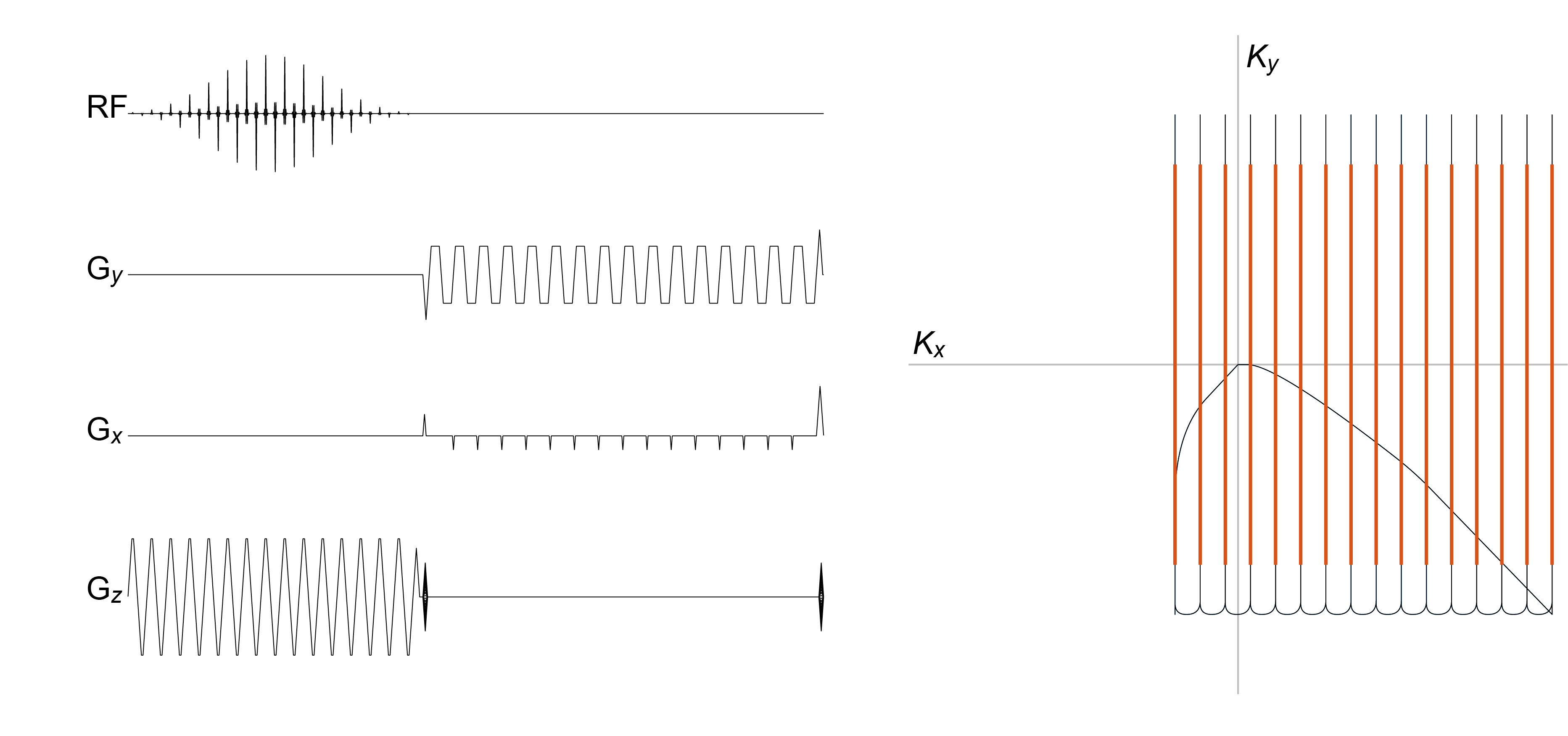
DE-EPI gradient waveforms were designed for 24×24 cm2 FOV at 15 mm in-plane resolution. The ±125 KHz readout bandwidth allowed 8-fold oversampling in the readout-direction. Dephasing and rephasing trapezoids for the blipped waveform were scaled to shift the sampling window in k-space by the equivalent of 5 lines, reducing TE by 7.6 ms (TE = 13.5 ms). Through-plane coverage was performed with centre-out phase encoding across 36 cm with 15 mm slice thickness. The ssRF pulse4 had a duration of 18.4 ms. Waveforms were incorporated into a 3D gradient-echo sequence (figure 1).
Subjects (N=9) were positioned in a General Electric MR750 3T MRI scanner. [1-13C]pyruvate doses were polarized using a SPINLab polarizer (Research Circle Technologies Inc.), dissolved and neutralized to final concentration of 250 mM, and 0.43 mL/kg (0.01 mmol/kg) was injected at 4.0 mL/s via a 20 gauge intravenous catheter. At the end of a 25 mL saline flush at 5.0 mL/s, the 13C image acquisition was initiated and lasted 60 seconds. The centre frequency was toggled to excite in sequence one of [1-13C]lactate, 13C-bicarbonate or [1-13C]pyruvate, resulting in separate, time-resolved volumes for each metabolite at a temporal resolution of 5 s. Afterwards, the 13C head coil was replaced with an 8-channel neurovascular array (Invivo Inc.) and T2wFLAIR ( TR/TE 8000/120 ms, 22×22 cm2, 0.6875×0.982 mm2, 3 mm slice thickness, 111°) and T1wFSPGR images (TR/TE 7.6/2.9 ms, 25.6×25.6×16.6 cm3, 1 mm-isotropic resolution, 11°) were acquired for anatomical reference.
Image reconstruction was performed offline with MATLAB R2018a (The Mathworks Inc.). Off-resonance correction was carried out as described in8 and the Projection On Convex Sets (POCS) algorithm10,11 was used for PF reconstruction.
Results & Discussion
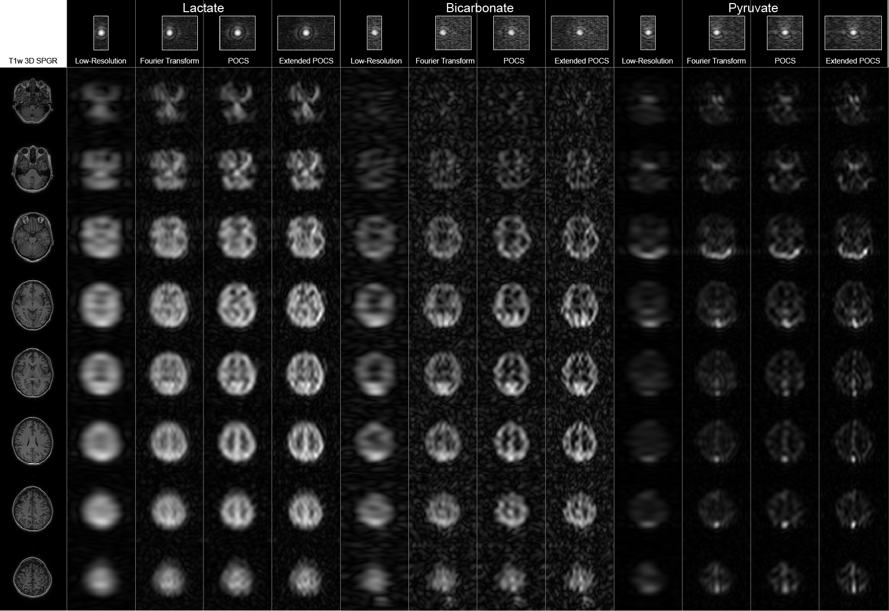
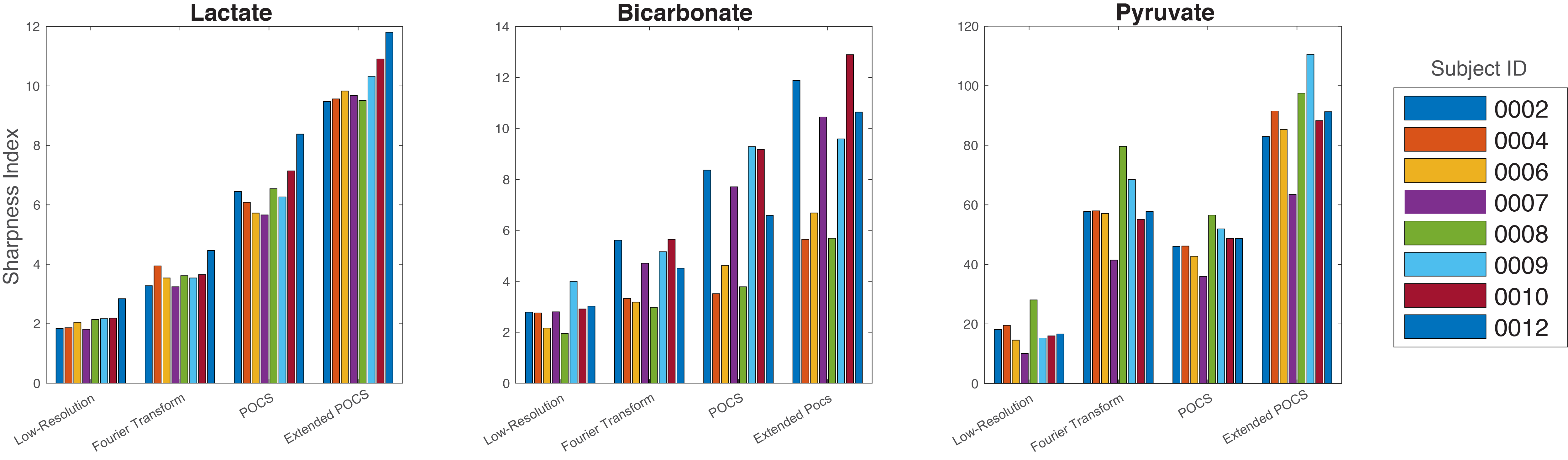
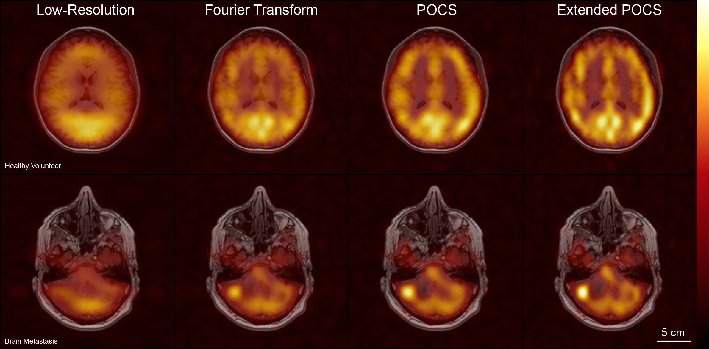
Three variants of the reconstruction were evaluated: direct Fourier transform (15 mm resolution); POCS (15 mm) and “Extended POCS” (9.2 mm). Additionally, a low-resolution reconstruction was performed by keeping the lowest 6 lines of k-space. Time-integrated 13C images for each variant were resampled to match the FOV and resolution of the T1-weighted anatomicals. To compare between reconstructions, a subset of resampled slices for each subject, metabolite and reconstruction variant was extracted and vertically concatenated into single images (figure 2). Sharpness Index12,13 (SI) was used as a no-reference image quality assessment for each image subset (figure 3). Image overlays from a normal volunteer and from a renal cell carcinoma patient with a brain metastasis are shown for each reconstruction in figure 4.
The most striking difference between the POCS and Fourier transform (FT) reconstruction is the apparent improvement in contrast, seen especially in the ventricular regions of the lactate images. The extended POCS reconstruction represents a 62.5% increase in non-padded matrix size, which provides a clear improvement in image sharpness. In all cases, extended POCS had a Sharpness Index (SI) greater than symmetric POCS. Interestingly, SI was increased in POCS over FT except for pyruvate, where it was slightly reduced.
Conclusion
Although SNR is always a limiting factor, metabolic images at spatial resolution that approaches the anatomical imaging resolution would be highly desirable. Partial Fourier imaging and reconstruction were shown to improve the image quality and contrast of HP 13C DE-EPI. A quantitative evaluation of image quality using sharpness index, a no-reference image quality assessment, agreed with the perceived improvements, with the extended POCS reconstruction improving the sharpness index by approximately a factor of 2.Acknowledgements
The authors thank Julie Green and Sumeet Sachdeva for coordinating the study and Ruby Endre for MR technical support. Funding support from the Brain Canada and Canadian Institutes for Health Research.References
- Park, Ilwoo, Peder EZ Larson, Jeremy W. Gordon, Lucas Carvajal, Hsin‐Yu Chen, Robert Bok, Mark Van Criekinge et al. "Development of methods and feasibility of using hyperpolarized carbon‐13 imaging data for evaluating brain metabolism in patient studies." Magnetic resonance in medicine 80, no. 3 (2018): 864-873.
- Miloushev, Vesselin Z., Kristin L. Granlund, Rostislav Boltyanskiy, Serge K. Lyashchenko, Lisa M. DeAngelis, Ingo K. Mellinghoff, Cameron W. Brennan et al. "Metabolic Imaging of the Human Brain with Hyperpolarized 13C Pyruvate Demonstrates 13C Lactate Production in Brain Tumor Patients." Cancer research (2018): canres-0221.
- Gordon, Jeremy W., Hsin‐Yu Chen, Adam Autry, Ilwoo Park, Mark Van Criekinge, Daniele Mammoli, Eugene Milshteyn et al. "Translation of Carbon‐13 EPI for hyperpolarized MR molecular imaging of prostate and brain cancer patients." Magnetic resonance in medicine (2018).
- Cunningham, Charles H., Albert P. Chen, Michael Lustig, Brian A. Hargreaves, Janine Lupo, Duan Xu, John Kurhanewicz et al. "Pulse sequence for dynamic volumetric imaging of hyperpolarized metabolic products." Journal of magnetic resonance 193, no. 1 (2008): 139-146.
- Lau, Angus Z., Albert P. Chen, Nilesh R. Ghugre, Venkat Ramanan, Wilfred W. Lam, Kim A. Connelly, Graham A. Wright, and Charles H. Cunningham. "Rapid multislice imaging of hyperpolarized 13C pyruvate and bicarbonate in the heart." Magnetic resonance in medicine 64, no. 5 (2010): 1323-1331.
- Miller, Jack J., Angus Z. Lau, Irvin Teh, Jürgen E. Schneider, Paul Kinchesh, Sean Smart, Vicky Ball, Nicola R. Sibson, and Damian J. Tyler. "Robust and high resolution hyperpolarized metabolic imaging of the rat heart at 7 T with 3D spectral‐spatial EPI." Magnetic resonance in medicine 75, no. 4 (2016): 1515-1524.
- Gordon, Jeremy W., Daniel B. Vigneron, and Peder EZ Larson. "Development of a symmetric echo planar imaging framework for clinical translation of rapid dynamic hyperpolarized 13C imaging." Magnetic resonance in medicine 77, no. 2 (2017): 826-832.
- Geraghty, Benjamin J., Justin YC Lau, Albert P. Chen, and Charles H. Cunningham. "Dual‐Echo EPI sequence for integrated distortion correction in 3D time‐resolved hyperpolarized 13C MRI." Magnetic resonance in medicine 79, no. 2 (2018): 643-653.
- Geraghty, Benjamin J., Justin Y.C. Lau, Albert P. Chen, and Charles H. Cunningham. "Comparison of Asymmetric and Symmetric K-space Sampling in EPI for 3D Time-Resolved Hyperpolarized 13C MRI with [1-13C]Pyruvate." In Proceedings of the 26th Annual Meeting of ISMRM; Paris, France, abstract #3071
- Haacke, E. M., E. D. Lindskogj, and W. Lin. "A fast, iterative, partial-fourier technique capable of local phase recovery." Journal of Magnetic Resonance 92 (1991): 126-145.
- McGibney, G., M. R. Smith, S. T. Nichols, and A. Crawley. "Quantitative evaluation of several partial Fourier reconstruction algorithms used in MRI." Magnetic resonance in medicine 30, no. 1 (1993): 51-59.
- Blanchet, Gwendoline, Lionel Moisan, and Bernard Rougé. "Measuring the global phase coherence of an image." In Image Processing, 2008. ICIP 2008. 15th IEEE International Conference on, pp. 1176-1179. IEEE, 2008.
- Blanchet, Gwendoline, and Lionel Moisan. "An explicit sharpness index related to global phase coherence." In Acoustics, Speech and Signal Processing (ICASSP), 2012 IEEE International Conference on, pp. 1065-1068. IEEE, 2012.
Figures