4271
Micelles-like Biodegradable conjugates as Biocompatible MRI Contrast Agents for Advanced Imaging of Tumor and Angiography1Huaxi MR Research Center, Department of Radiology, West China Hospital, Sichuan Unversity, Chengdu, China
Synopsis
To improve the contrast enhancement of solid tumor, we prepared enzymes responsive biodegradable micelles-like agents (MpH-DOTA-Gd), and the HMPA based micelle-like polymeric MR contrast agents provided a possibility of selectively great contrast in MR imaging of tumor and angiography.
Propose: To improve the contrast enhancement of solid tumor in CE-MRI and get more clear images of tumor-around vessels in CE-MRA by use of macromolecular MRI contrast agents rather than clinical used small molecular agents.
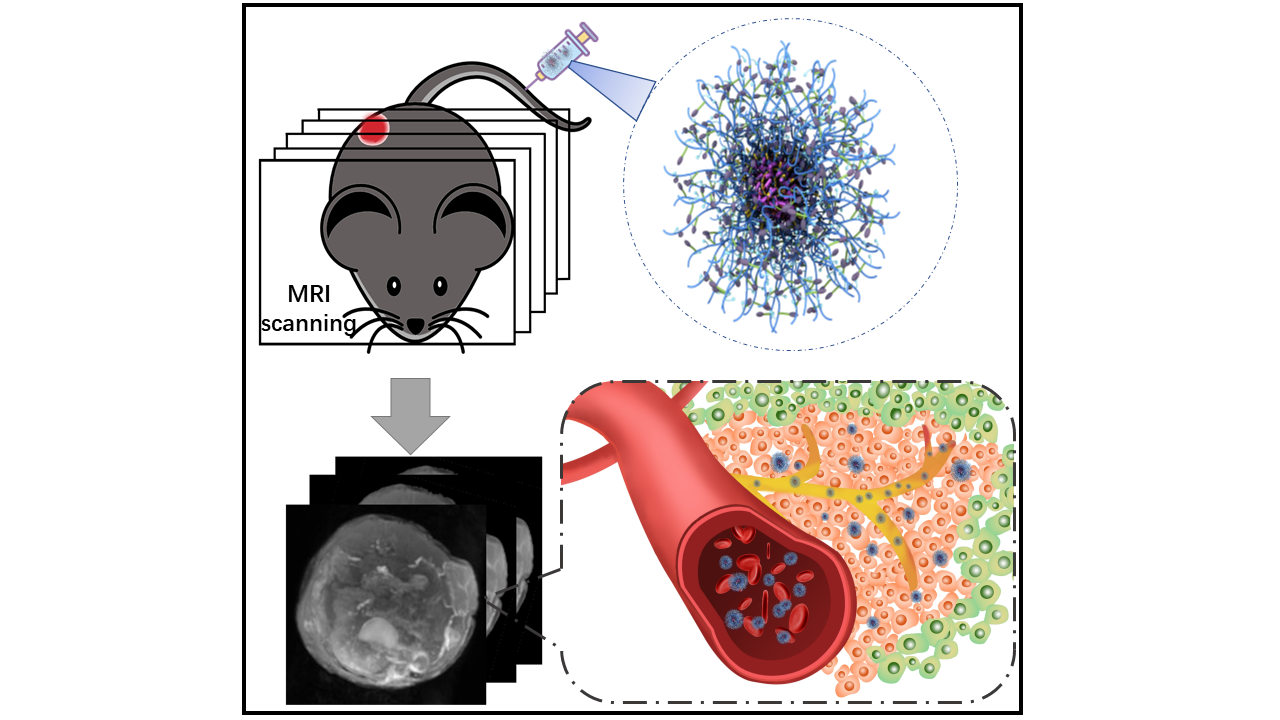
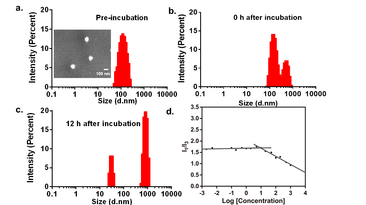
Methods and Materials: In this study, we prepared enzymes responsive biodegradable micelles-like agents (MpH-DOTA-Gd) by use of chemical synthesis. We tested and verified the macromolecular agents via 1H NMR spectroscopy, size exclusion chromatography (SEC) and scanning electron microscope (SEM). The contrast MR images of tumor and vessels were studies in 3T MRI scanner. Then biocompatibility, biodegradability and biosafety of the agents were carried out.
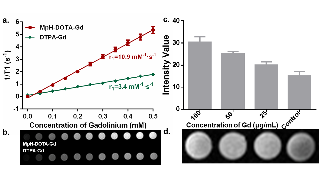
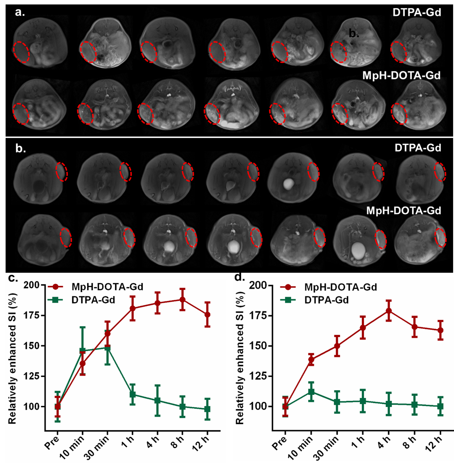
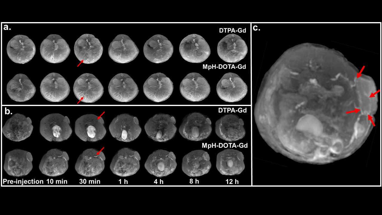
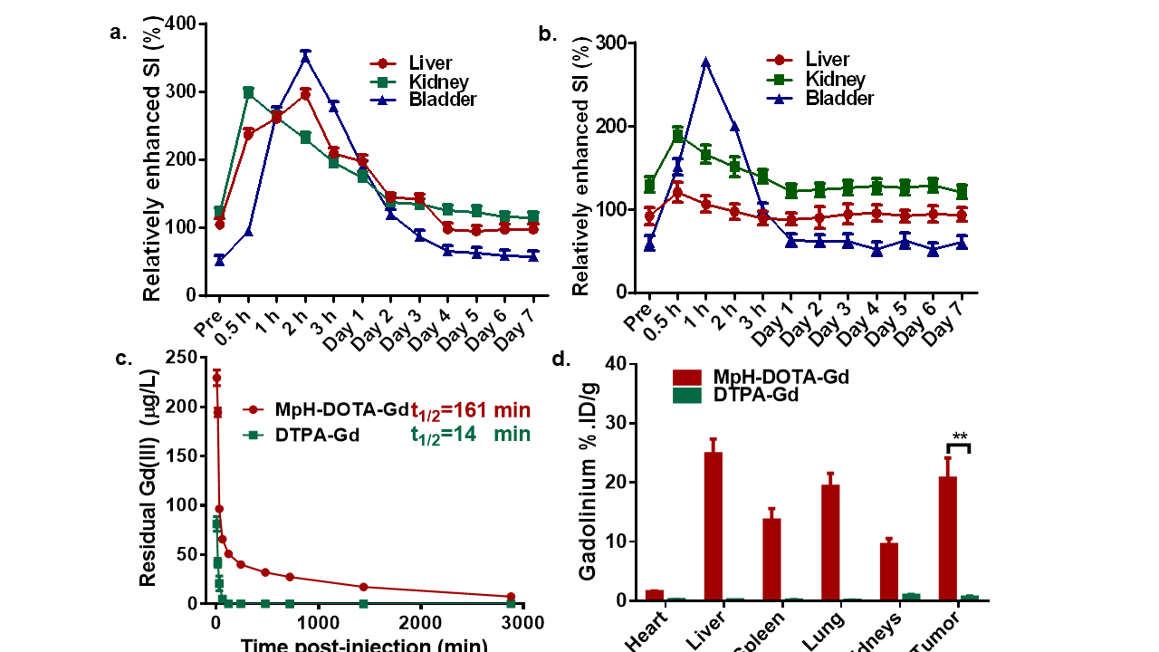
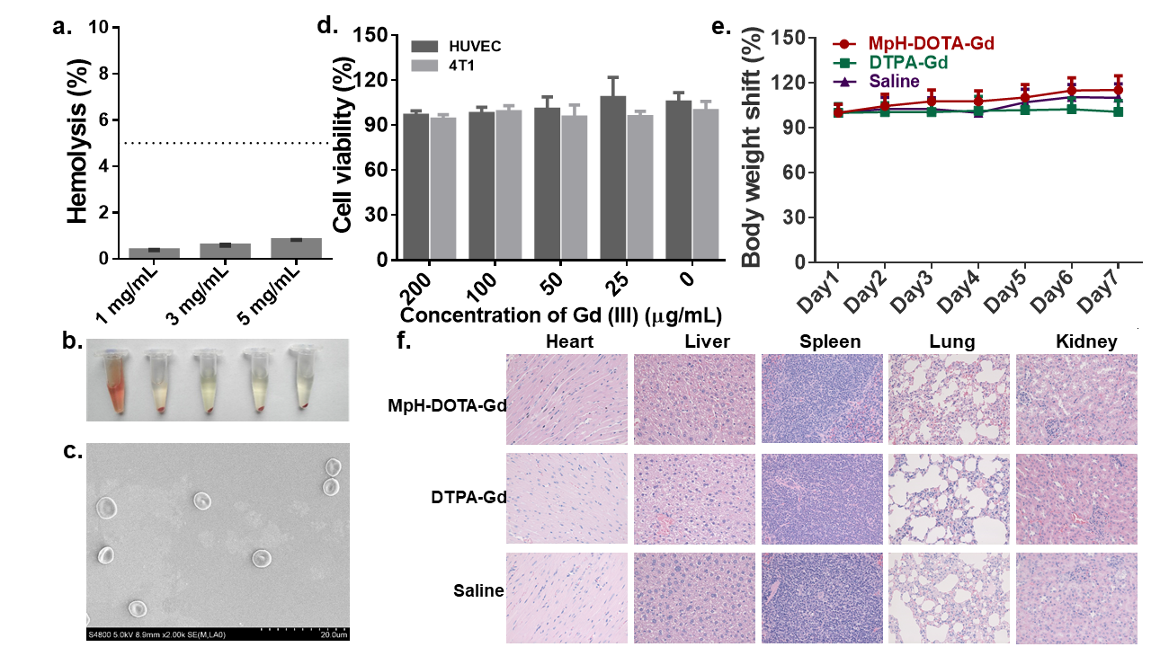
Results: The relaxivity of MpH-DOTA-Gd is 10.9 mM-1·s-1, which is three-fold higher than clinical used DTPA-Gd. In vivo tumor imaging in both orthotopic and subcutaneous tumor model showed great contrast enhancement due to selectively accumulate and high relaxivity of the MpH-DOTA-Gd in tumor. Then, MR angiography of vessels was studied. The small vessels and microvessels in liver and tumor around vessels could be clearly imaged, and the time-window of imaging was larger than DTPA-Gd. In addition, the details of small supply vessels in tumor could also be shown after administration of MpH-DOTA-Gd, instead of DTPA-Gd. Moreover, biodegradable linker in the structure help the agents be degraded into small segments and excreted out from body. Good biosafety and hemocompatibility promote the application of agents in the future.
Conclusion: In summary, the HMPA based micelle-like polymeric MR contrast agents provided a possibility of selectively great contrast in MR imaging of tumor and angiography.
Acknowledgements
The authors declare no competing financial interest.References
(1) Zhou, Z.; Lu, Z.-R. Gadolinium-Based Contrast Agents for MR Cancer Imaging. Wires. Nanomed. Nanobi. 2013, 5, 1-18.
(2) Bogdanov, A.; Mazzanti, M. L. Molecular MR Contrast Agents for the Detection of Cancer: Past and Present. Semin. Oncol. 2011, 38, 42-54.
(3) Zhou, S.; Wu, Z.; Chen, X.; Jia, L.; Zhu, W. PEGylated Polyethylenimine as Enhanced T1 Contrast Agent for Efficient Magnetic Resonance Imaging. ACS Appl. Mater. Interfaces 2014, 6, 11459-11469.
(4) Luk, B. T.; Zhang, L. Current Advances in Polymer-Based Nanotheranostics for Cancer Treatment and Diagnosis. ACS Appl. Mater. Interfaces 2014, 6, 21859-21873.
Figures