4270
Liver Distribution and Delayed Phase Liver Enhancement using Mn-PyC3A1Surgical Oncology, Massachusetts General Hospital/ Harvard Medical School, Boston, MA, United States, 2Radiology/ Martinos Center, Massachusetts General Hospital, Charlestown, MA, United States
Synopsis
Mn-PyC3A was rationally designed as a Gd-free alternative to commercially available MRI contrast agents. Quantification of Mn excretion using rats shows that the agent is efficiently eliminated with a fractional clearance that is 85% renal and 15% hepatobiliary. Based on the substantial fractional hepatobiliary excretion, we hypothesized that Mn-PyC3A would provide delayed, liver specific contrast enhancement that can be used to identify liver tumors. Mn-PyC3A was demonstrated to provide delayed phase liver enhancement that rendered liver tumors conspicuously hypointense in a murine model of colorectal metastasis.
Purpose
Mn-PyC3A was developed as a gadolinium free alternative to commercial MRI contrast agents, which pose a safety risk to renally impaired patients and are currently receiving intense regulatory scrutiny due to concerns over Gd retention and delayed toxicity.1-3 Mn-PyC3A was rationally designed with modest degree of lipophilicity to promote more efficient elimination via participation of the hepatobiliary system.4 The purpose of this study is to quantify fractional clearance of Mn-PyC3A via the hepatobiliary path in rats, to quantify liver distribution in mice, and to compare the efficacy of Mn-PyC3A to visualize liver tumors vs. the liver specific Gd contrast agent Gd-EOB-DTPA.Methods
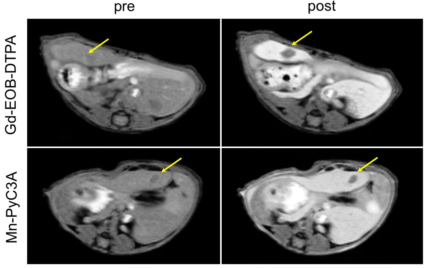
To quantify fractional excretion of Mn-PyC3A in rats (N = 10), a 2.0 mmol/kg dose of [52Mn]Mn-PyC3A was administered via tail vein injection. The rats were housed in metabolic cages for 24h after injection. Total activity recovered in the urine and feces were quantified using a gamma counter and reported as percentage injected dose (%ID). To quantify liver distribution of Mn-PyC3A in mice, the mice were simultaneously injected with 0.1 mmol/kg Mn-PyC3A and 0.025 mmol/kg Gd-EOB-DTPA, euthanized, and the livers harvested for quantification of Mn and Gd content by ICP-MS (N = 8). The efficacy of Mn-PyC3A vs Gd-EOB-DTPA to visualize liver lesions were compared using a mouse model of colorectal liver metastasis (implantation of mc26 cells). Mice were imaged at 4.7T using a 2D T1-weighted FLASH sequence prior to and 10 min following following injections of 0.1 mmol/kg Mn-PyC3A and 0.025 mmol/kg Gd-EOB-DTPA administered to the same mouse 24h apart (N = 8). The increase in liver vs. tumor contrast-to-noise ratio (DCNR) generated using Mn-PyC3A and Gd-EOB-DPTA were quantified and compared via a paired t-test.Results
A total of 82.26±3.501 %ID and 14.74±5.260 %ID of the injected [52Mn]Mn-PyC3A were recovered in the urine and feces, respectively, 24 h after RVP-001 injection. This corresponds to a fractional excretion in the urine and feces of 84.92±4.919 % and 15.08±4.919 %, respectively. A total of 97.47±3.995 %ID was recovered in excreta 24h after injection. A total of 0.11±0.022 µmol/g Mn and 0.15±0.022 µmol/g Gd were recovered from liver tissue 10 min after injection of 0.1 mmol/kg Mn-PyC3A or 0.025 mmol/kg Gd-EOB-DTPA to mice, respectively, corresponding to 2.72±0.554 %ID and 15.1±2.22 %ID. Mn-PyC3A was shown to render liver lesions conspicuously hypointense 10 min after injection, Fig 1. Liver vs. tumor DCNR generated 10 min after injection of Mn-PyC3A and Gd-EOB-DTPA were 30.0±12.3 and 46.6±14.3, respectively, P = 0.0316.Discussion
Mn-PyC3A is very efficiently excreted from rats with a fractional hepatobiliary clearance of 15.08±4.919%. Mn-PyC3A was not designed as a liver specific agent but we hypothesized that the substantial hepatobiliary excretion could enable visualization of liver lesions during the delayed phase after contrast injection. Delayed phase Mn-PyC3A enhancement enables conspicuous contrast enhanced visualization of liver tumors in mice. Liver vs. tumor CNR was significantly greater following Gd-EOB-DTPA injection, consistent with greater liver accumulation of Gd-EOB-DTPA.Conclusion
Mn-PyC3A is very effectively eliminated from rats with a fractional hepatobiliary clearance of 15.08±4.919%. Mn-PyC3A provided strong delayed phase liver enhancement that rendered liver tumors conspicuously hypointense in a murine model of colorectal metastasis.Acknowledgements
This work was supported by grants from the National Institutes of Health: HL128899, HL119145, EB022804, EB009062, RR014075, RR023385, and OD010650.References
1. Grobner T, Prischl FC. Gadolinium and Nephrogenic Systemic Fibrosis. Kidney. Int. 2007;72(3):260-264.
2. Kanda T, Fukusato T, Matsuda M, Toyoda K, Oba H, Kotoku J, Haruyama T, Kitajima K, Furui S. Gadolinium-Based Contrast Agent Accumulates in the Brain Even in Subjects Without Severe Renal Dysfunction: Evaluation of Autopsy Brain Specimens with Inductively Coupled Plasma Mass Spectroscopy. Radiology 2015;276(1):228−232.
3. Semelka RC, Ramalho J, Vakharia A, AlObaidy M, Burke LM, Jay M, Ramalho M. Gadolinium Deposition Disease: Initial Description of a Disease That has been Around for a While. Magn. Reson. Imaging 2016;34(10):1383−1390.
4. Gale, E. M.; Atanasova, I.; Blasi, F.; Ay, I.; Caravan, P. A Manganese Alternative to Gadolinium for MRI Contrast. J. Am. Chem. Soc. 2015;137(49):15548−15557.
Figures