4269
Dysprosium complexes encapsulated apoferritin T2 MRI contrast agent for ultra-high field MRIHee-Kyung Kim1,2, Ahrum Baek3, Sunghwan Hwang3, Soyeon Kim3, Garam Choi3, Bo Kyung Sung3, Byeong Woo Yang3, Heejin Seo3, MD. Kamrul Islam3, Hoesu Jung4, Taekwan Lee4, and Yongmin Chang5
1Department of Molecular Medicine & BK21 Plus KNU Biomedical Convergence Program, Kyungpook National University, Daegu, Korea, Republic of, 2Institute of Biomedical Engineering Research, Daegu, Korea, Republic of, 3Department of Medical &Biological Engineering, Kyungpook National University, Daegu, Korea, Republic of, 4Laboratory Animal Center, Daegu-Gyeongbuk Medical Innovation Foundation, Daegu, Korea, Republic of, 5Department of Radiology, Kyungpook National University, Daegu, Korea, Republic of
Synopsis
Apo[Dy(DO3A-ph-NH2)] is synthesized according to the precede literature. The absorbance is changed at 300-400 nm after encapsulation of Dy(DO3A-ph-NH2), and we confirmed the morphology of Dy(DO3A-ph-NH2) encapsulated protein with TEM image. The final concentration of paramagnetic composition is calculated by ICP. The relaxivities of Dy(DO3A-ph-NH2) encapsulated protein at various external fields (3 - 15.2 T) are considerably increased, and also r2/r1 ratios are increased up to 55 at 15.2 T.
Introduction
Lanthanide metals except gadolinium (Gd) have highly anisotropic electron spin structure, their electron relaxation rates are on the order of ~10-13 s-1. This anisotropic electron structure results in the short electron relaxation time (T1e), molecular tumbling correlation time (τR) is always larger than T1e. The curie relaxation mechanism becomes important on this account, it cause to increase transverse relaxation shortening effect especially with a high magnetic moment (μeff) and external field. Dysprosium (Dy) has the largest μeff (10.6 μB) among lanthanides, therefore, it can be used a useful low molecular T2 MRI contrast agent (CA) in ultra-high field. We synthesized Dy complex (Dy(DO3A-ph-NH2)) encapsulated apoferritin for use as a T2 MRI CA for ultra-high field MRI.Materials and Methods
All reagents were purchased from commercial sources and used as received. Characterization of the synthesized compounds have been determined by analytical spectroscopic methods.1, 2 The final paramagnetic composition concentration of Apo[Dy(DO3A-ph-NH2)] was measured by ICP mass. T1 measurements were carried out using an RARE-inversion recovery method with variable inversion times (TI) in various external fields (3 - 15.2 T). For T2, the multi-slice multi-echo (MSME) sequence was adapted in same the external fields. R1 and R2 relaxation times were obtained from the nonlinear least-squares fit of the mean pixel values at variable TI and TE, respectively. The relaxivities (r1 and r2) were then calculated as a slope of linear fit of relaxation rate along with concentrations. The in vivo T2 weighted MR image was obtained at 9.4 T animal MRI equipment (Agilent, U.S.). In these studies, the mice (25 ~ 27 g) were anesthetized by 1.5% isoflurane in oxygen. MR images were acquired before and after intravascular injection of Apo[Dy(DO3A-ph-NH2)] (dose: 0.3 mmol Dy/kg). Also image parameters of axial images for fast spin-echo are as follows: Repetition time (TR) = 4000 msec; echo time (TE) = 12 msec; 30 30 mm field of view (FOV); 128×128 matrix size; 1.0 mm slice thickness; echo train length = 4; scan time of each image = 8 min 40 sec.Results and Discussion
Low molecular Dy complex was synthesized according to precede literature1, the purity was characterized with FAB mass spectroscopy and HPLC analysis. Encapsulation of apoferritin was proceeded carefully by adjusting pH (pH 2.0-7.4)2. After encapsulation, morphology of protein was well maintained in TEM images (figure 1). The absorbance change of Apo[Dy(DO3A-ph-NH2)] was observed at 300-400 nm because of encapsulated Dy(DO3A-ph-NH2) (figure 1). The r2 relaxivities were considerably increased up to 69.63 mM-1s-1 at 15.2 T. And the r2/r1 ratio per Dy increased from 3.82 at 3 T to 55.26 at 15.2 T. (figure 2). The curie relaxation is affected by molecular tumbling correlation time (τR) and water residence time (τM). The preceding results from large molecular weight (~480 kDa) and restricted water access by protein out-sphere that respectively increase τR and τM. The concentration of Apo[Dy(DO3A-ph-NH2)] for in vivo test were prepared to 44 mM per Dy from ICP-spectrometer measurement. The MR images of Apo[Dy(DO3A-ph-NH2)] were acquired at 9.4T with liver tumor mice (figure 3). This senseful negative enhancement in liver shows the effect of T2 MRI contrast agent at ultra-high field.Conclusion
In conclusion, we have successfully synthesized Apo[Dy(DO3A-ph-NH2)] as a T2 MRI CA for ultra-high field MRI. Due to the high increment of r2/r1 ratio of Apo[Dy(DO3A-ph-NH2)] above 9.4 T, we expected that it can be used as biocompatible negative-enhancing CA replacing metal-oxide based T2 MRI CAs.Acknowledgements
We gratefully acknowledge financial support from the Basic Science Research Program (NRF-2017R1D1A1B03031640) and BK21 Plus KNU Biomedical Convergence Program.References
(1) Iman Daryaei, “Study, evaluation, and applications of MRI contrast agents that work based on CEST and T2-EX mechanism”, Dr diss., University of Arizona, 2017.
(2) Akira M. Nanomedicine: NBM. 2011, 7, 638-646
Figures
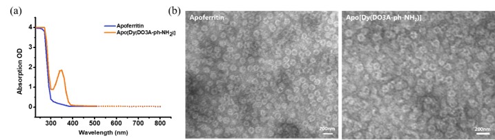
Figure 1. (a)
UV-absorbance spectrum and (b) morphology TEM images of Apo[Dy(DO3A-ph-NH2)].
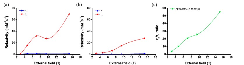
Figure 2. (a)
Apo[Dy(DO3A-ph-NH2)] and (b) Dy(DO3A-ph-NH2) relaxivities
at various external fields. (c) r2/r1 ratios per Dy of
Apo[Dy(DO3A-ph-NH2)].
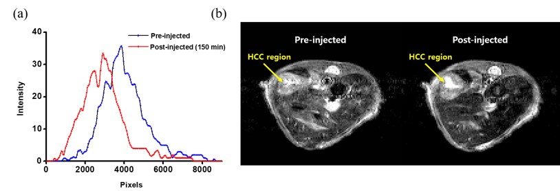
Figure 3. (a) Intensity histogram in liver and (b) in-vivo T2-weighted MR images (nude
mouse, male, 14 weeks-old, 25 g) of mice bearing Huh-7 tumor obtained with
Apo[Dy(DO3A-ph-NH2)], measured 150 min after injection. Dose: 0.3
mmol/kg.