4263
Macrocyclic Manganese (II) Complex of Mefenamic acid (MA) Conjugate as an Inflammation Targeting MRI Contrast Agent1Medical and Biological Engineering, Kyungpook National University, Daegu, Korea, Republic of, 2Molecular Medicine, Kyungpook National University, Daegu, Korea, Republic of, 33Institute of Biomedical Engineering Research, Kyungpook National University, Daegu, Korea, Republic of, 4Laboratory Animal Center, Daegu-Gyeongbuk Medical Innovation Foundation, Daegu, Korea, Republic of, 5Radiology, Kyungpook National University, Daegu, Korea, Republic of
Synopsis
The purpose of the present study is to design and synthesis of novel macrocyclic Mn2+ complex as an alternative to the well-established Gd3+ based chelates for use as an inflammation targeting MR imaging agent. This new complex exhibits higher r1 relaxivity (2.32 mM−1 s−1) than clinically approved Mn-DPDP® (1.6 mM−1 s−1) at 4.7 T. T1-weighted MR coronal images display strong signal enhancement in liver, heart, gallbladder and kidney. In an inflammatory mouse model, it shows greater inflammatory tissues detection with in a clinical dose.
Introduction
Inflammation is an adaptive response that arises in the initial stages of some disease, which involve the formation of different inflammatory mediators. Aiming these mediators by using the nonsteroidal anti-inflammatory drugs (NSAIDs) can be a promising approach to target the site of inflammation. Gadolinium (Gd3+) is the popular choice among the paramagnetic metals due to its suitable magnetic properties. However, Gd3+ based CAs are associated to the nephrogenic systemic fibrosis (NSF), a rare but potential fibrosis of the skins and internal organs that can arise in renally compromised patients.1 Moreover, a number of lessons confirmed that intravenously administered Gd3+ deposit in the brains and bones of the patients with normal renal function.2 In this context, several approaches have been made based on the non-lanthanide metals, particularly less toxic manganese (Mn) are getting special attention owing to the necessary physical properties desired for MRI. Though, no relationship has been found between Mn and NSF, but the potential neurotoxicity of free Mn2+ ions remain as key safety concern. Herein, we report the design of a novel Mn2+ complex based on 1,4,7-triaazacyclononane1,4,7-triacetic acid (NOTA) chelating ligand by conjugating a mefenamic acid (MA) moiety with high chelation stability for use as an inflammation targeting agent.3Materials and Methods
All reagents were purchased from commercial sources and used without further purification unless otherwise stated. Solvents were purified and dried using standard procedures. Deionized water was used for all experimental procedures. T1 measurements were carried out using an inversion recovery method with a variable inversion time (TI) at 4.7 T (Bruker BIOSPEC 47/40, Ettlingen, Germany). T1 and T2 relaxation times were obtained from the non-linear least squares fit of the mean pixel values for the multiple spin-echo measurements at each TI value and echo time. Relaxivities (r1 and r2) were then calculated as an inverse of relaxation time per mM. MR images of anaesthetized six weeks inflammatory ICR mice were obtained pre- and post-injection of MnL1 (0.05 mmol Mn/kg) by tail vein with a 4.7 T MR unit (Bruker BIOSPEC 47/40, Ettlingen, Germany) using home-made small animal RF coil. The imaging parameters were as follows: repetition time (TR) = 400 ms; echo time (TE) = 7.5 ms; FOV (for coronal images 50 mm and for axial images 30 mm); 128×128 matrix size; 1.0 mm slice thickness; number of acquisition (NEX) = 2.Results and Discussion
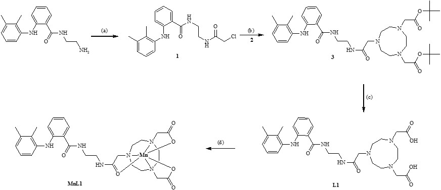
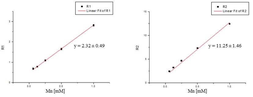
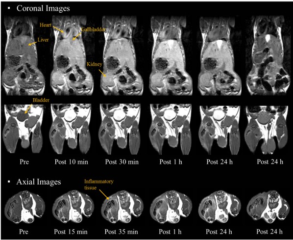
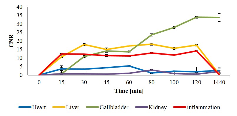
The synthesis of MnL1 complex are described in Scheme 1. The formation of new compounds and metal complex were confirmed by spectroscopic and microanalysis methods such as, 1H NMR, HR-FAB-MS and elemental analysis (EA). MnL1 shows high r1 relaxivity (2.32 mM-1s-1) than that of clinically approved Mn2+ based MRI contrast agent Mn-DPDP® (1.6 mM-1s-1) (Figure 1).4 T1-weighted MR coronal images of inflammatory ICR mice before and after intravenous injection of MnL1 ((0.05 mmol Mn/kg) are shown in Figure 2. It shows strong signal enhancement in liver, gallbladder, kidney and heart. Time-dependent MR signal intensity enhancements of the gallbladder, inflammation, liver and kidney are shown in Figure 3. Both the kidney and liver showed significant signal enhancements suggesting that both organs are responsible for the elimination of MnL1. In the axial image, the inflammatory tissues showed significant signal enhancement suggesting that this new complex can be a prominent MR imaging agent for inflammation (Figure 2).Conclusions
In the current study, we successfully designed, synthesized and T1 weighted image evaluation of MnL1 chelate as a new family of stable inflammation MRI contrast agent. This new complex can be suitable for MR imaging of inflammation-related diseases.Acknowledgements
No acknowledgement found.References
1. Grobner, T. Gadolinium a specific trigger for the development of nephrogenic fibrosing dermopathy and nephrogenic systemic fibrosis? Nephrol. Dial. Transplant. 2006, 21, 1104–1108.
2. Kanal, E.; Tweedle, M. F. Residual or Retained Gadolinium: Practical Implications for Radiologists and Our Patients. Radiology, 2015, 275, 630–634.
3. Wong, W. T. et al. Inflammation Targeted Gd3+-Based MRI Contrast Agents Imaging Tumor and Rheumatoid Arthritis Models. Bioconjugate Chem. 2014, 25, 1112−1123
4. Rohrer, M and Weinmann, H. J. et al. Comparison of Magnetic Properties of MRI Contrast Media Solutions at Different Magnetic Field Strengths. Investigative Radiology. 2005, 40(11), 715−724.
Figures