4262
Quantitative Time-dependent Mapping of Liver Cirrhosis Heterogeneity with A Protein-based MRI Contrast Agent1Biomedical Engineering, Georgia Institute of Technology / Emory University, Atlanta, GA, United States, 2Department of Chemistry, Georgia State University, Atlanta, GA, United States
Synopsis
This work is focused on quantitative and qualitative analysis of the spatial and time-dependant heterogeneity of liver cirrhosis using a novel protein-based MRI contrast agent which targets collagen type I.
Introduction
Spatial heterogeneity in fibrosis is one of the major limitations and challenges of liver biopsy and elastography-based techniques, as the current inability to provide comprehensive imaging of the entire liver volume cannot accurately reflect the overall extent and stage of fibrosis [1, 2]. Liver fibrosis heterogeneity has differential special distribution and is also dependent on animal models and causes [3]. Liver fibrosis heterogeneity and the process for the formation of liver fibrosis currently cannot be accurately detected by any methods. Collagen type I is heterogeneously accumulated in liver fibrosis, depending on the etiology which leads to fibrosis that disrupts liver cytoarchitecture and function [4]. Biopsy has many limitations such as sampling errors, high inter-observer variability with 33-50% error rate even for diagnosis of advanced stages of liver fibrosis such as cirrhosis likely due to heterogeneity. MRI offers several unique advantages compared to other clinical imaging modalities with its deep tissue penetration, high spatial resolution, and coverage of the entire liver, however, MRI cannot detect liver fibrosis with regional heterogeneity due to limitations of currently available MRI contrast agents [5, 6]. We have developed a collagen I targeted protein-based MR contrast agent, ProCA32.collagen1 capable of detecting cirrhosis heterogeneity in a time-dependent manner with dynamic molecular imaging (DMI) property.Methods
In order to induce liver cirrhosis, 14-day-old male mice with C57BL/6 genetic background were treated with a single dose of DEN (Sigma–Aldrich # N0756) given dissolved in saline at a dose of 25 mg/kg body weight by i.p. injection on day 12. Mice were sacrificed 10 months after DEN administration for histological and biochemical analyses. ProCA32.collagen1 was administered via intravenous injection. All mice were imaged on a 7-T Agilent MRI scanner at the University of Georgia. Animals were anesthetized using isoflurane and their respiration rate was monitored with a small animal physiological monitoring system. Anesthesia was adjusted to maintain a respiration rate of 65 ± 5 breaths per minute. T1-weighted MRI images were collected before and after intravenous (I.V.) administration of 5 mmol/L of ProCA32.collagen1. T1 weighted images acquired with TR=500 ms and TE=14.89 ms. Other acquisition parameters include: field of view FOV = 35×35 mm, matrix = 256×256, slice thickness = 1.0 mm, and 12 image slices with no gap. The images were collected before and after the contrast agent I.V. injection of 5 mM ProCA32.collagen1 with different time points. The voxel based relative enhancement analysis using the pre-injection T1 weighted image as the baseline was used to determine the spatial and the time dependent effectiveness of the contrast agent. All the analysis was done using MATLAB 2018 (MathWorks, Natick, MA) custom designed codes.Results and Discussion
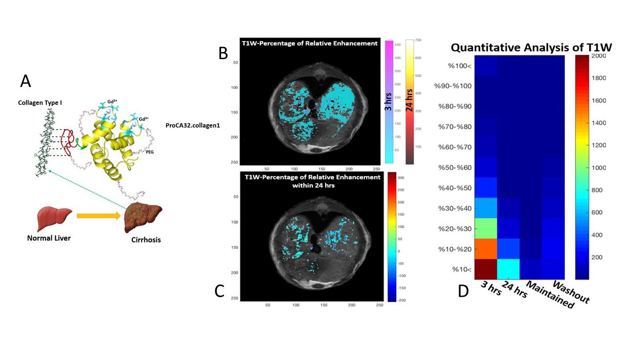
We have addressed liver fibrosis heterogeneity induced by DEN with ProCA32.collagen1-enabled molecular imaging and DMI. ProCA32.collagen1 possesses high relaxivities per particle (r1 and r2) at both 1.4 and 7.0 T which enables the detection of cirrhosis heterogeneity via dual contrast modes. ProCA32.collagen1 also mitigates metal toxicity due to lower dosage and strong resistance to transmetallation (104 and 1012-fold higher metal selectivity for Gd3+ over Ca2+ and Zn2+, respectively) compared to clinical contrast agents. This DEN-induced late-stage cirrhosis mouse model exhibited strong liver heterogeneity mimicking patient cirrhosis. The T1-weighted image before injection of contrast agent used as the gray scale base map, the T1-wighted image after 3 h (cold color map) and the T1-weighted image after 24 h (hot color map) were superimposed upon it to demonstrate the time-dependent enhancement of cirrhotic liver at 3 h and 24 h post-injection of ProCA32.collagen1 with the maximum enhancement at 3 h post-injection, specifically at the right segment of the liver (Figure 1-B). The voxels with positive value represent the regions that have an increased enhancement after 24 h in comparison to 3 h and voxels where the contrast agent washed out have the negative values (Figure 1-C). The quantitative analysis matrix (Figure 1-D) verifies the qualitative maps, showing that the most significant enhancement happens after 3 h from the injection time.Conclusion
Overall, the results demonstrate that enhancement characteristics after administration of ProCA32.collagen1 reflect liver cirrhosis heterogeneity. The development of the contrast agent is expected to overcome the major clinical barriers in early diagnosis, noninvasive detection and staging of chronic liver diseases, and have strong translational potential in facilitating effective treatment and image-guided biopsy.Acknowledgements
This work was supported by National Institute of Health (NIH) Research Grants EB007268, R41CA183376 and R41AA025863 (to Jenny J.Yang).References
1- Dodd GD, 3rd, Baron RL, Oliver JH, 3rd, Federle MP. Spectrum of imaging findings of the liver in end-stage cirrhosis: Part II, focal abnormalities. AJR American journal of roentgenology 173, 1185-1192 (1999).
2 - Dodd GD, 3rd, Baron RL, Oliver JH, 3rd, Federle MP. Spectrum of imaging findings of the liver in end-stage cirrhosis: part I, gross morphology and diffuse abnormalities. AJR American journal of roentgenology 173, 1031-1036 (1999).
3 - Fuchs BC, et al. Molecular MRI of collagen to diagnose and stage liver fibrosis. J Hepatol 59, 992-998 (2013).
4 - Chen BB, et al. Dynamic contrast-enhanced magnetic resonance imaging with Gd-EOB-DTPA for the evaluation of liver fibrosis in chronic hepatitis patients. Eur Radiol 22, 171-180 (2012).
5 - Tschirch FT, Struwe A, Petrowsky H, Kakales I, Marincek B, Weishaupt D. Contrast-enhanced MR cholangiography with Gd-EOB-DTPA in patients with liver cirrhosis: visualization of the biliary ducts in comparison with patients with normal liver parenchyma. Eur Radiol 18, 1577-1586 (2008).
6 - Tamada T, et al. Gd-EOB-DTPA-enhanced MR imaging: evaluation of hepatic enhancement effects in normal and cirrhotic livers. Eur J Radiol 80, e311-316 (2011).
Figures