4261
A noninvasive T1-weighted MR tool for tracking of vicinal thiols motif of thioredoxin protein in live cells1Bio-imaging Research Team, Korea Basic Science Institute, Chungcheongbuk-do, Korea, Republic of, 2Chungnam National University, Daejeon, Korea, Republic of, 3Amrita School of Engineering, Ettimadai, India
Synopsis
We have synthesized a couple of new MRI contrast agent for recognition of cellular vicinal thiols motif protein thioredoxin.
Introduction
The vicinal thiols-motif protein namely thioredoxin a class of small redox protein known to be present in all organisms. It plays a significant role in many important biological processes, including redox signaling. Also it plays a central role in reducing process of disulfide bond in various enzymes such as ribonucleotide reductase, methionine sulfoxide reductases, and peroxiredoxins. and etc. Dysfunction of this class of protein responsible for many diseases such as cancer, diabetes, human immunodeficiency virus type 1 (HIV-1), and neurodegenerative diseases. Thus, noninvasive tracking of vicinal thiols-motif protein thioredoxin in cellular level is critical.Methods
Preparation of Gd3+ Complex. Ligand (80 µm) was in ultrapure water (10 mL) and the solution was adjusted to ~pH 7 with sodium bicarbonate. Gadolinium chloride hexahydrate (78 µm) was dissolved in 3.0 mL of ultrapure water and added to the solution of ligand in three separate aliquots. After the addition of each aliquot, the pH was adjusted back to a pH between 6.5–7.0 using 0.1 M potassium carbonate solutions. The solution was allowed to stir for 30 min to allow for Gd3+ chelation to occur, dialyzed against ultrapure water for overnight, and lyophilized to yield respective complex. Gd3+ complex .ESI- m/z (M +1) for CA1: calcd. 789.96, found 789.98; ESI- m/z (M +K+) for CA2: calcd. 943.96, found 943.06.
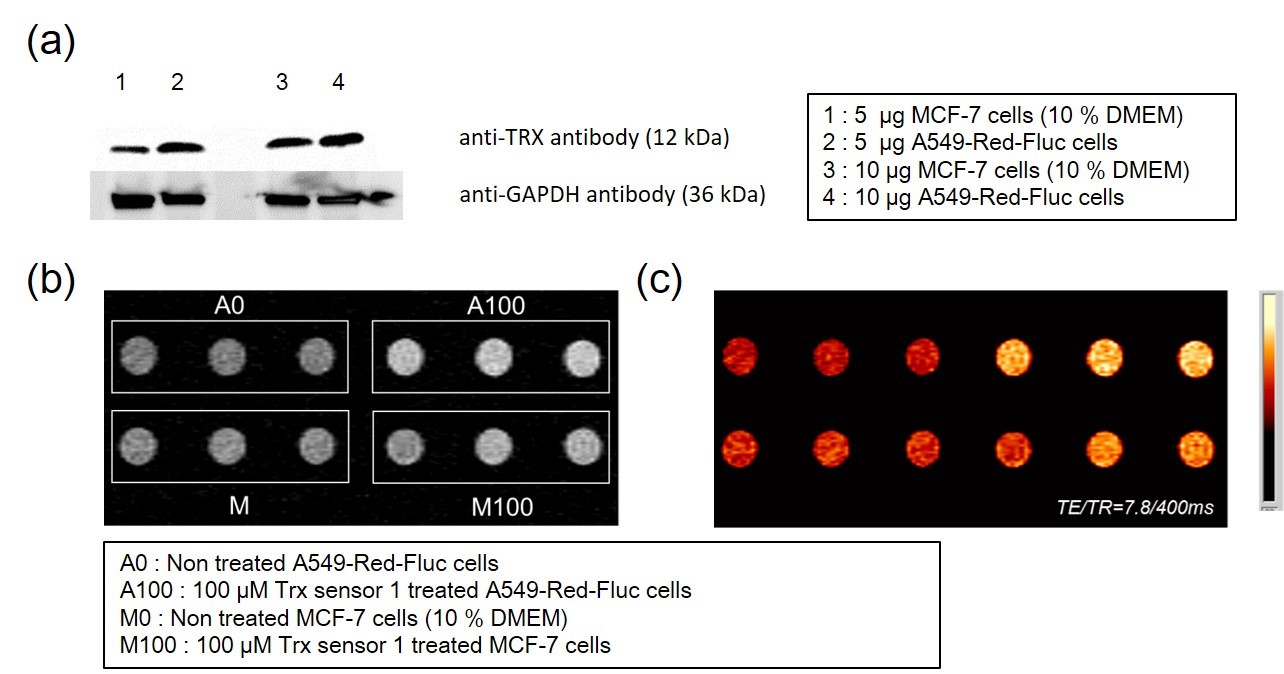
MRI phantom image. MRI phantom images were acquired in 4.7 T MRI instrument (Biospec 47/40, Bruker, Germany). T1 weighted MR image obtained using following scanning parameters : MSME (Multi-slice multi-echo) pulse sequence, TE/TR=8.8/400 ms, matrix size = 192 × 192, FOV=6 × 3 cm, slice thickness = 1 mm. We compared T1 contrast differences between CA1 and CA2 in MCF-7 cell. In addition, we compared brightness of MRI according to trx level in cells.
Results
The contrast agents showed
relaxivity typically single coordinated water molecule to Gd3+ ion center such
as 4.54 ± 0.13 mM-1s-1 and 4.58 ± 0.13 mM-1s-1 for CA1 and CA2 respectively at
60 MHz field. Contrast agents CA1 showed ~140% relaxivity enhancement in the
presence of thioredoxin. 15N-NMR experimental data indicated that CA1 has a
higher binding affinity toward thioredoxin compared to flexible contrast agent
CA2. The contrast agent CA1 has provided T1-weighted phantom images of cancer
cells (MCF-7, A549-Red-Fluc) based on the expression of thioredoxin. Further,
we confirmed thioredoxin- dependent changes of T1-weighted contrast images by
knocked-down the thioredoxin expression in Trx siRNA transfected MCF-7 cells.
The nontoxic nature of the CA1 suggested that it is the first kind of contrast
agent can provide the extent of expression of vicinal thiols motif protein in
live cells noninvasively.
Conclusion
We observed that CA1 had provided T1-weighted MR images depending on the expression of thioredoxin cancer cells. A negative controlled experimental result in Trx siRNA transfected MCF-7 cells indicates that CA1 is exclusively offered bright images upon binding with thioredoxin. This new contrast agent can resolve specific cellular abnormality by providing early information on issues related to native redox regulation and vicinal thiols motif thioredoxin.Acknowledgements
No acknowledgement found.References
1. Louie, A. Y.; Huber, M. M.; Ahrens, E. T.; Rothbacher, U; Moats, R.; Jacobs, R. E.; Fraser, S. E.; Meade, T. J. In Vivo Visualization of Gene Expression using Magnetic Resonance Imaging. Nat. Biotechnol. 2000, 18, 321−325.
2. Ueno, T.; Nagano, T. Evaluation of Fluorophores for Optimal Performance in Localization-based Super-resolution Imaging. Nat. Methods, 2011, 8, 1027−1036.
Figures