4260
Development of a novel theranostic NPs composed of Fe3O4, Trastuzumab and indocyanine green for diagnosis and treatment of HER-2 positive breast cancer1Department of Radiology, National Cancer Center/National Clinical Research Center for Cancer/ Cancer Hospital, Chinese Academy of Medical Sciences and Peking Union Medical College, Beijing, China, 2GE Healthcare, Beijing, China
Synopsis
Superparamagnetic iron oxides can be used as contrast agents for MR T2-weighted imaging. We created multifunctional nanoparticles based on Fe3O4, Trastuzumab and indocyanine green (ICG) to target HER2 positive breast cancer cells in vitro and vivo. Firstly, internalization was assessed on HER-2 positive breast cancer SK-BR-3 cells by transmission electron microscopy (TEM). Furthermore magnetic resonance imaging (MRI) and fluorescence imaging were used to examine the biodistribution and its targeting effect in vivo. Additionally, photothermal therapy (PTT) was further evaluated to examine the treatment effect of NPs. As a result, Fe3O4-trastuzumab-ICG have shown great potential to become an effective multifunctional imaging agent and a tool for photothermal therapy for HER-positive breast cancer.
Objective
We developed a novel theranostic NPs composed of Fe3O4, Trastuzumab and indocyanine green (ICG) specifically used to detect and treat human epidermal growth factor receptor 2 (HER-2)-positive breast cancer.Materials and Methods
Superparamagnetic iron oxide nanoparticles (SPIONs) coated with a copolymer of distearoyl phosphoethanolamine-PEG 2000 were synthesized using a coprecipitation method. Internalization was assessed on HER-2 positive breast cancer SK-BR-3 cells by transmission electron microscopy (TEM). Magnetic resonance imaging (MRI) and fluorescence imaging were used to examine the biodistribution and its targeting effect in vivo, using a SK-BR-3 xenograft mice model. Additionally, photothermal therapy (PTT) was further evaluated to examine the treatment effect of NPs on tumor tissue in vivo.Results
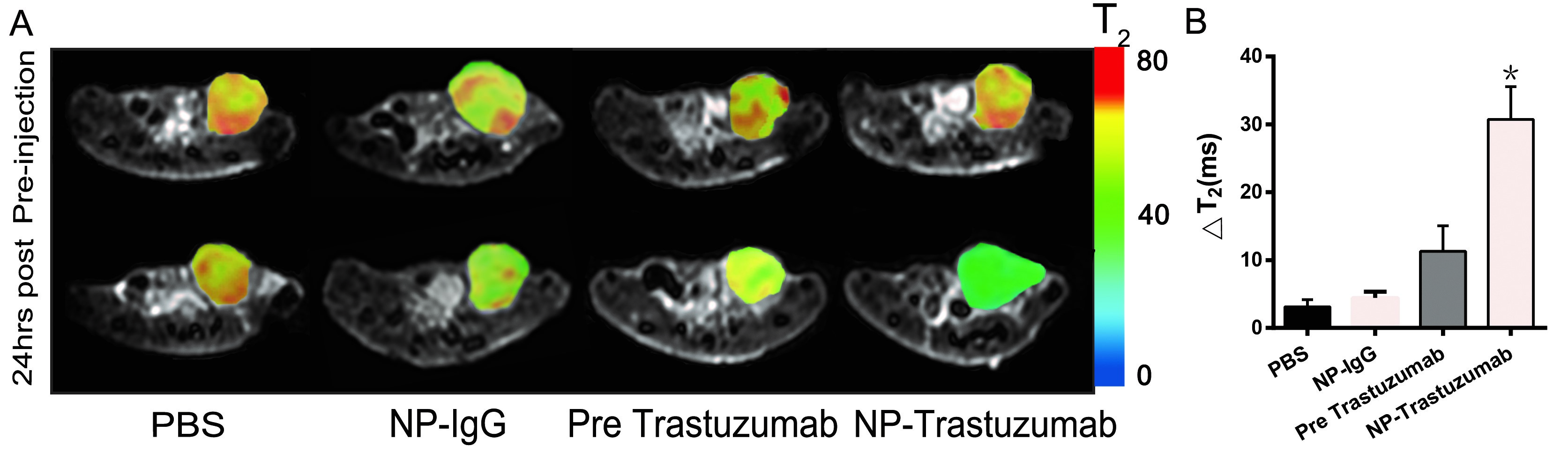
Fe3O4-trastuzumab-ICG NPs exhibited excellent physicochemical properties (Fig. 1). TEM images revealed that the NPs might specifically target HER-2 receptor and then enter the endoplasmic reticulum of SK-BR-3 cells (Fig. 2). Furthermore, MRI studies showed a marked uptake of NPs in tumor region with decreased T2 relaxation time, particularly at 24 h after injection. Moreover, significantly higher ΔT2 values (average pre-injection T2–average 24 h post-injection T2) of the tumor area were observed in mice treated with Fe3O4-trastuzumab-ICG group (30.7±4.8) ms compared to PBS group (3.1±1.1) ms, Fe3O4-IgG-ICG group (4.4±0.9) ms and trastuzumab + Fe3O4-trastuzumab-ICG group (11.3±3.8) ms (all P<0.05) (Fig. 3). Consistently, fluorescence imaging revealed a significant tumor-specific accumulation of ICG in Fe3O4-trastuzumab-ICG group 24 h after administration. Besides, the Fe3O4-trastuzumab-ICG NPs were shown to generate significant photothermal effect in tumor site; the maximum temperature reached 49.4℃ after continuous irradiation of near infrared light (Fig. 4).Discussion
HER-2 positive breast cancer is usually associated with poor prognosis. Accordingly, the correct assessment of the receptor expression is of significant importance for accurate diagnosis and treatment of HER-2 positive BC[1]. Currently, the extent of HER-2 expression in BC has been clinically evaluated either by immunohistochemistry (IHC) or by in situ hybridization (ISH) analyses. However, all of these are invasive techniques. Molecular imaging provides a real-time non-invasive way of assessing the extent of HER-2 expression before treatment, as well as a great approach for monitoring the therapeutic efficiency in patients. Moreover, the application of NPs in dual-modal molecular imaging has been reported by other studies[2-4]. In this study, Fe3O4 particles were found in periphery and endosomes of SK-BR-3 cells (Fig. 2). This suggests the effect of HER2 receptor-mediated endocytosis in the cellular uptake of Fe3O4-trastuzumab-ICG. MR T2 mapping images showed significantly higher △T2 in mice injected with Fe3O4-trastuzumab-ICG compared to PBS, non-specific Fe3O4-IgG-ICG or pre-trastuzumab group (30.7 ± 4.8 ms vs. 3.1 ± 1.1 ms, 4.4 ± 0.9 ms or 11.3 ± 3.8 ms, respectively) which suggested that Fe3O4-trastuzumab-ICG may target HER-2 positive and enter SK-BR-3 breast cancer cells in vivo (Fig. 3). Additionally, photothermal therapeutic property of ICG was integrated into the NPs for theranostics in vivo (Fig. 4). Due to technical limitations during preparation, the size distribution of NPs and the number of ligands on NPs surface may vary from batch to batch, thus leading to dramatic differences in circulation and tumor targeting[5].Conclusions
Fe3O4-trastuzumab-ICG had the potential to become an effective multifunctional imaging agent and a tool for photothermal therapy for HER-positive breast cancer.Acknowledgements
National Basic Research Program of China (973 Program, 2014CB744505) financially supported this work. We would also like to thank Xukun Li for providing technical assistance.References
[1]Mihaly Z, Gyorffy B. [HER2-positive breast cancer: available targeted agents and biomarkers for therapy response][J]. Magy Onkol,2013,57(3):147-156.
[2] Li J, You J, Dai Y, et al. Gadolinium oxide nanoparticles and aptamer-functionalized silver nanoclusters-based multimodal molecular imaging nanoprobe for optical/magnetic resonance cancer cell imaging[J]. Anal Chem,2014,86(22):11306-11311.
[3] Kosaka N, Bernardo M, Mitsunaga M, et al. MR and optical imaging of early micrometastases in lymph nodes: triple labeling with nano-sized agents yielding distinct signals[J]. Contrast Media Mol Imaging,2012,7(2):247-253.
[4] Lee J H, Jun Y W, Yeon S I, et al. Dual-mode nanoparticle probes for high-performance magnetic resonance and fluorescence imaging of neuroblastoma[J]. Angew Chem Int Ed Engl,2006,45(48):8160-8162.
[5] Mu Q, Wang H, Zhang M. Nanoparticles for imaging and treatment of metastatic breast cancer[J]. Expert Opin Drug Deliv,2017,14(1):123-136.
Figures