4258
TAT-functionalized perfluorocarbon nanoemulsion for 19F imaging of CAR T cells in vivo.1Radiology, University of California San Diego, La Jolla, CA, United States, 2Bioengineering, University of California San Diego, La Jolla, CA, United States, 3Pharmacology, University of California San Diego, La Jolla, CA, United States, 4Neurological Surgery, University of California San Francisco, San Francisco, CA, United States
Synopsis
To improve the utility of 19F MRI to detect lymphocytes in vivo, we looked into strategies to increase cell loading via the use of
Introduction
Non-invasive immune cell tracking using 19F MRI and perfluorocarbon (PFC) nanoemulsion cell labeling has made significant advances in recent years [1]. While macrophages and immature dendritic cells are highly phagocytic and have a relatively large cytoplasmic volume to accommodate labeling media, lymphocytes have low intrinsic phagocytic properties and small cytoplasmic volumes that can limit label uptake and thus imaging sensitivity. For cancer immunotherapy, chimeric antigen receptor (CAR) T cells have emerged as a viable therapy for certain liquid tumor (leukemias), and trials are underway to determine their efficacy towards solid cancers [2]. Non-invasive imaging of CAR T cells can provide crucial feedback regarding the initial cell distribution and survival in vivo and could help elucidate the modes of action. To enhance the effectiveness of CAR T cell 19F MRI, we have synthesized PFC nanoemulsion functionalized with the transactivator of transcription (TAT), cell penetrating peptide (GRKKRRQRRRPQ) to boost endocytosis of the PFC by CAR T cells in culture prior to transfer to the subject and in vivoMRI. We synthesized and characterized TAT PFC nanoemulsion for optimal and stable incorporation of TAT and evaluated uptake, viability and phenotype of in vitro labeled CAR T cells. Labeled cells were administered to a xenograft glioma mouse model to evaluate the 19F imaging sensitivity increase in vivo due to TAT-enhanced cell labeling.Methods
For synthesis, 1H-perfluoro-3,6,9-trioxadecan-1-ol is conjugated to maleimidohexanoic acid in the presence of PyBOP and base DIEA. The purified product is reacted with Cys-TAT in a H20/TFA/trifluoroethanol solution to get final product TATP with m/z=1127.4. Nanoemulsions were prepared by passing a mixture of F68 pluronic surfactant, and perfluoro-15-crown-5-ether and TATP through an LV1 microfluidiser (Fig A). Particle size of nanoemulsion was evaluated by dynamic light scattering (DLS). Cell uptake in pellets (via19F NMR) and viability (trypan blue assay) were measured in Jurkat cells and primary human CAR T cells expressing EGFRvIII receptor as described elsewhere [3]. CAR T cell viability and phenotypic expression were also evaluated pre- and post-labeling using flow cytometry, and TAT emulsion internalization was confirmed by confocal and electron microscopy. Animal experiments were approved by internal the UC San Diego IACUC committee. Female SCID mice (N=4) received bilateral sub-cutaneous injections of 5×10⁶PFC-labeled U87-EGFRvIII-Luc cells. Five days post-tumor implantation, mice received 107CAR T cell intratumoral injections. In each mouse, one tumor received CAR Ts labeled overnight with 15mg/ml TAT-PFC emulsion, and the other tumor was injected with CAR Ts labeled with control emulsion (no TAT). Two hours after intratumoral delivery, anesthetized mice were imaged at 11.7 T with a dual 1H/19F volume coil. Proton images were acquired using T2-weighted RARE (TR/TE=2000/13 ms, RARE factor 4, FOV=38×30 mm2, matrix = 256×184, 4 averages). Co-registered 19F images were acquired with a spin-density weighted RARE (TR/TE=1500/4.7 ms, RARE factor 8, matrix = 64×46, 400 averages). Absolute fluorine atoms in tumors were calculated from 19F images based on a fiducial marker in field of view using VoxelTracker software (Celsense). 1H/19F overlays were performed in ImageJ.
Results
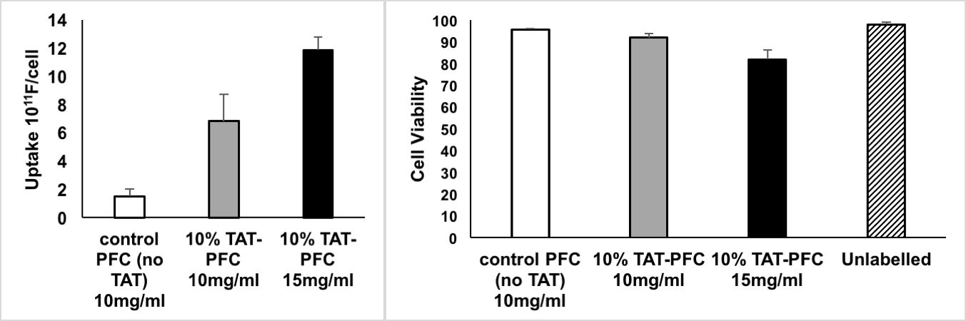
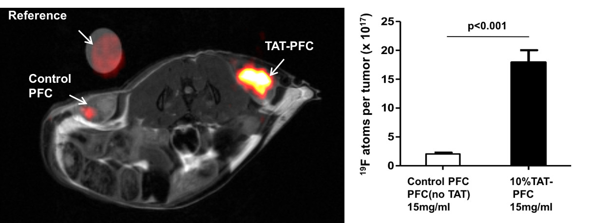
Upon formulation (Fig. A), the TAT-PFC nanoemulsion displays a mean particle size of175.6 ± 4.48 nm and PDI of 0.115±0.0026by DLS and a stable shelf life >1 month. At 10 mg/ml CAR T cell labeling for 18 hr, the 10%w/w TAT-PFC nanoemulsions have a 3-fold and 8-fold higher uptake, for 10 mg/ml and 15 mg/ml, respectively, compared to TAT-negative emulsion (Fig. B). TAT containing emulsions remained non-toxic to the cells with viability at 92% for 10 mg/ml dose compared to 97.7% for untreated cells (Fig. B). Analysis of CD3, CD4 and CD8 markers showed that CAR T cells remains unaltered after treatment with TAT-PFC (data not shown). Confocal and electron microscopy confirmed cytoplasmic localization of TAT-PFC droplets (data not shown). In vivoMRI signal (total 19F atoms measured in tumors) from TAT-labeled CAR T cells was 8-times higher than that of contralateral tumor injected with control PFC-labeled CAR T cells (Fig. C). This 8-fold sensitivity matched closely that of in vitrouptake results at 15 mg/ml, as measured by 19F NMR in cell pellets (Fig. B). Longitudinal studies are currently underway to investigate CAR T cell dynamics in vivo.
Conclusions
In this study, we show that incorporation of a TAT peptide in fluorous nanoemulsions results in multi-fold uptake improvement for non-phagocytic cells such as T cells. TAT-PFC emulsions may enable in vivo imaging of CAR T cells as they find their tumor target and provide insight into the modes of action of engineered T cell immunotherapy against cancer.
Acknowledgements
No acknowledgement found.References
[1] Chapelin, Fanny, Christian M. Capitini, and Eric T. Ahrens. "Fluorine-19 MRI for detection and quantification of immune cell therapy for cancer." Journal for immunotherapy of cancer 6.1 (2018): 105.
[2] Chang, ZeNan L., and Yvonne Y. Chen. "CARs: synthetic immunoreceptors for cancer therapy and beyond." Trends in molecular medicine 23.5 (2017): 430-450.
[3] Chapelin, Fanny, et al. "Fluorine-19 nuclear magnetic resonance of chimeric antigen receptor T cell biodistribution in murine cancer model." Scientific reports 7.1 (2017): 17748.
Figures