4257
Molecular MR imaging of Acute Inflammation using a Biochemically Responsive Iron-Based Contrast Agent1Radiology/ Martinos Center, Massachusetts General Hospital, Charlestown, MA, United States
Synopsis
The redox active MRI contrast agent Fe-PyC3A is shown to provide strong, selective contrast enhancement of acutely inflamed pancreatic tissue in a murine model. Fe-PyC3A is administered in the weakly contrasting Fe(2+) oxidation state but is converted to the 15-fold more strongly contrasting Fe(3+) state in the presence of reactive oxygen species (ROS). Fe-PyC3A does not provide contrast enhancement of normal pancreas, but provides significant contrast enhancement of inflamed pancreas. Pancreatic enhancement correlates tightly with activity levels of the pro-inflammatory biomarker myeloperoxidase. This is the first example of using metal ion redox for MR imaging of pathologic change in vivo.
Purpose
The purpose of this study was to demonstrate the capability of the redox responsive MRI contrast agent Fe-PyC3A to detect acute inflammation in vivo. Oxidative stress is a key pathologic feature of the inflammatory microenvironment, due to high levels of reactive oxygen species (ROS) and oxidase enzymes secreted by infiltrating neutrophils.1 Conventional MRI readouts of inflammation focus on structural and physiological changes, such as edema or endothelial breakdown.2 We hypothesized that Fe-PyC3A, which is converted from the weakly contrasting Fe(2+) oxidation state to the strongly contrasting Fe(3+) oxidation state in the presence ROS, could be used to map, quantify, and monitor the dynamics of inflammation at the biochemical level using contrast enhanced MRI.Methods
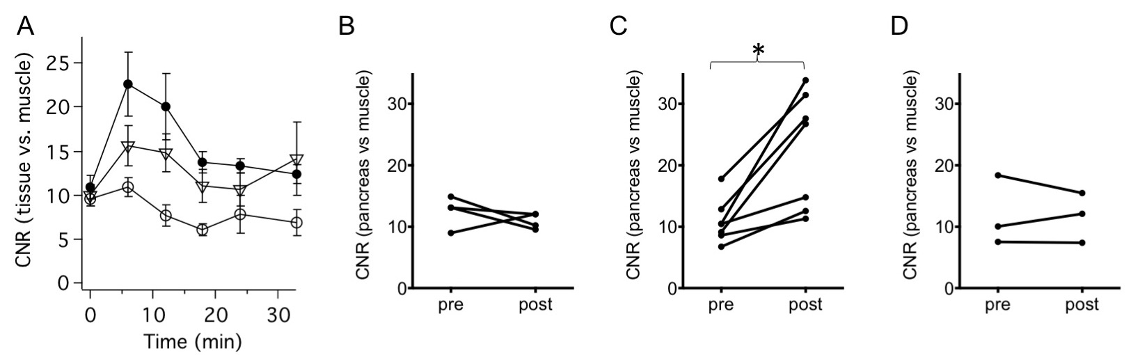
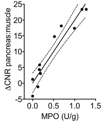
We evaluated the capability of Fe-PyC3A to detect ROS production in a murine model of acute pancreatitis (N = 7). Pancreatitis was pharmacologically induced via 6 hourly intraperitoneal injections of 0.05 mg/kg caerulean initiated 24 h prior to imaging, and the inflammatory response was amplified 3h prior to imaging via i.p. injection of 10 mg/kg lipopolysaccharide (LPS) 3 h prior to imaging.3 Control mice (N = 4) were treated with i.p. injections of saline. A second set of caerulean/ LPS treated mice were imaged with the negative control agent Mn-PyC3A, which does not undergo relaxivity change in the presence of ROS. Mice were imaged with a 2D T1-weighted FLASH sequence at 4.7T prior to and out to 30 min after intravenous injection of a 0.2 mmol/kg dose of Fe-PyC3A, or 0.02 mmol/kg Mn-PyC3A, which was administered at equal volume but at a formulation that was “T1 matched” to the Fe-PyC3A dose. Pancreas vs. muscle contrast-to-noise ratios (CNR) prior to and 6 min after contrast injection were compared by a paired t-test. After imaging the mice were euthanized and the tissues harvested for quantification of myeloperoxidase activity levels using the guaiacol spectrophotometric assay.4 Pancreas vs. muscle CNR were correlated to ex vivo readouts of myeloperoxidase activity by calculating the Pearson’s product moment correlation coefficient followed by a correlation t-test.Results
Injection of Fe-PyC3A as the Fe(2+) chelate does not provide significant contrast enhancement of the pancreas in the saline treated mice, but provides strong, selective contrast enhancement of the acutely inflamed pancreas. Fig 1 compares axial T1-weighted images at the level of the pancreas prior to and 6 min after Fe-PyC3A injection. Notice that pancreas is virtually isointense with the neighboring kidney prior to contrast injection (Figs 1A-B) and remains so 6 min after injection of Fe-PyC3A to the saline treated mice (Fig 1C). The kidney pelvis does enhance due to a high concentration of Fe complex that collects en route to urinary excretion, consistent with the large dose of contrast. On the other hand, the pancreas of the caerulean/ LPS treated mouse receives significant contrast enhancement and is rendered conspicuously hyperintense relative to the neighboring kidney 6 min after injection (Fig 1D). The time course of pancreas vs. muscle CNR following injection of Fe-PyC3A to caerulean/ LPS vs. saline treated mice, and following injection of Mn-PyC3A are shown in Fig 2A. Figs 2B-C compare CNR prior to and 6 min post contrast. Ex vivo measurements of myeloperoxidase activity correlate strongly and significantly with peak pancreas vs. muscle CNR, P <0.0001 (Fig 3).Discussion
ROS mediated conversion of weakly contrasting Fe(2+)-PyC3A to strongly contrasting Fe(3+) provides an effective means to detect acutely inflamed tissue using contrast enhanced MRI. We demonstrated that when administered as Fe(2+)-PyC3A, we can achieve strong and selective MRI contrast of acutely inflamed pancreatic tissue. No such enhancement is observed in normal pancreas, or in inflamed pancreas treated with a “T1-matched” formulation of negative control contrast agent that cannot change relaxivity in the presence of ROS. The 15-fold relaxivity change far exceeds what is possible with biochemically responsive Gd-based contrast agents. To the best of our knowledge, this is the first example of using metal ion redox for the MR imaging of pathologic change in vivo.Conclusions
Fe-PyC3A provides strong, selective contrast enhancement of acutely inflamed pancreatic tissue in a murine model of pancreatitic. Pancreas vs. muscle CNR correlates strongly and significantly with ex vivo quantitation of the pro-inflammatory biomarker myeloperoxidase.Acknowledgements
This work was supported by grants from the National Institutes of Health: HL128899, HL119145, EB022804, EB009062, RR014075, RR023385, and OD010650.References
1. Daugherty A, Dunn JL, Rateri DL and Heinecke JW. Myeloperoxidase, a Catalyst for Lipoprotein Oxidation, Is Expressed in Human Atherosclerotic Lesions. J. Clin. Invest. 1994;94(1):437-444.
2. Hammoud DA. Molecular Imaging of Inflammation: Current Status. J. Nucl. Med. 2016;57(8):1161-1165.
3. Ding SP, Ji-Chen L, and Jin C. A Mouse Model of Severe Acute Pancreatitis Induced with Caerulein and Lipopolysaccharide. World J. Gastroentrol. 2003;9(3):584-589.
4. Klebanoff SJ, Waltersdorph AM, Rosen H. Antimicrobial Activity of Myeloperoxidase. Methods in Enzymology 1984;105 (52):399-403.
Figures