4238
Detection of metabolite concentration changes in young adult volunteer brains after oral glucose administration using short-TE STEAM 1H MRS sequence at 7T1Siemens Healthineers, Tokyo, Japan, 2Siemens Healthineers, Malvern, PA, United States, 3Siemens Healthineers, Boston, MA, United States, 4Siemens Healthineers, San Francisco, CA, United States, 5Human Brain Research Center, Kyoto Univeristy, Kyoto, Japan
Synopsis
Glucose detection was examined in young adult volunteer brains (the posterior cingulate cortex) with 1H downfield MRS at 7T, and metabolite concentration changes before and c.a. 55 minutes after oral 50-g glucose administration were measured with the downfield and a conventional upfield MRS. The downfield MRS detected H1-α-glucose peak at 5.3 ppm and quantified glucose concentration as a wider range compared to that with the upfield MRS using LCModel. Detection of the H1-α-glucose peak showed a potential to quantify net glucose concentration without underestimation.
INTRODUCTION
Glucose is the energy substrate of the brain, and investigation of its metabolism is highly important for neurological research. Although proton MR spectroscopy (1H MRS) can detect glucose non-invasively, human studies have not been much reported because of low detection sensitivity of fully J-coupled peaks overlapped with other metabolites peaks1. Kaiser et al.2 developed a J-difference-edited sequence for selective detection of H2-β-glucose peaks at 3.23 ppm. However the sequence has several disadvantages of a long echo time (TE = 114 ms), sensitivity to field instability, and high specific absorption rate. Recently, an MRS scan of 1H downfield at 7.5-ppm transmitter offset frequency was introduced to detect peaks to the left of the water peak in the human brain at 7 Tesla (7T)3. As in nitrogen-containing compounds like N-acetylaspartate (NAA) and N-acetylaspartylgultamate (NAAG), H1-α-glucose peaks can be solely detected in the 1H downfield MRS, which had been performed at lower field or in small animals1. In this study, the 1H downfield MRS was introduced to quantify glucose concentration in volunteer brains at 7T in comparison to a conventional 1H upfield MRS. Moreover, metabolite concentration changes after oral glucose administration were measured using those techniques.METHODS
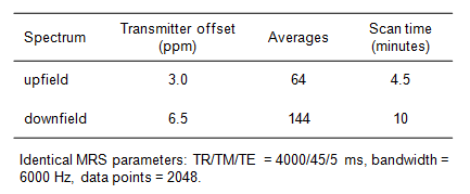
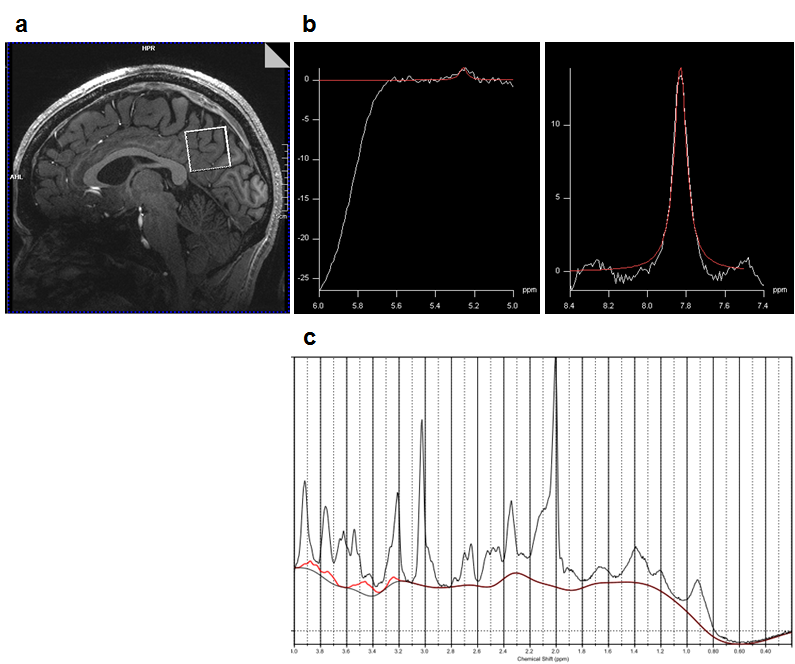
This study was approved by the IRB of our hospital, and written informed consent was obtained from all subjects. MRS scans were performed on a 7T whole-body scanner (Siemens Healthineers, Erlangen, Germany) using a single-transmit volume coil and a 32-receiver head coil (Nova Medical, MA, USA). Twenty-three healthy volunteers (16 males and 7 females, mean age 23 years, aged 20-29 years) were examined without fasting. An MRS voxel of a 3.0×3.0×3.0 (cm)3 was positioned at the posterior cingulate cortex (PCC) across the mid-sagittal plane on T1-weighted images (Figure 1a). FASTMAP shimming (prototype) and transmit amplitude adjustment were performed in the MRS voxel. 1H MR spectra were acquired using the short-TE (5 ms) STEAM pulse sequence (prototype) with water (VAPOR4) and outer volume suppressions. MRS acquisition parameters are listed in Table. 1. Seventeen volunteers (13 males and 4 females, mean age 23 years, aged 20-26 years) were scanned before and after taking 50-g oral glucose (Torelan-G50, Ajinomoto Pharmaceuticals, Tokyo, Japan) outside the magnet.
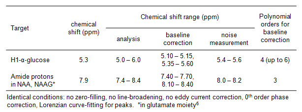
Upfield spectra were analyzed using LCModel (version 6.3-1L, LA Systems, Tokyo, Japan) with standard STEAM basis-set. Eddy current correction and water-scaling for quantification were conducted using water unsuppressed spectra5. Downfield spectra were analyzed using Syngo MR software (VB17A, Siemens Healthineers). Peak height and integral of peaks at around 5.3 and 7.9 ppm were measured with analysis conditions listed in Table. 2. Concentration of α-glucose was calculated using the integrals of those peaks as a 1:1 molar ratio of α-glucose and total NAA (NAA+NAAG) which was quantified in the upfield spectrum obtained right after the downfield scan. Statistical analysis between before and after glucose administration was conducted using Wilcoxon signed-rank test.
RESULTS
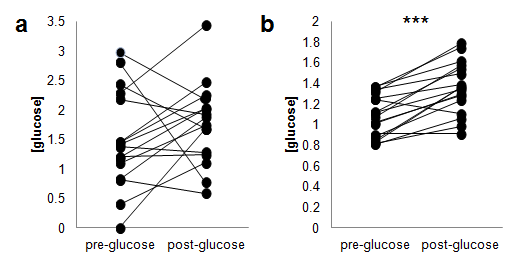
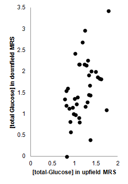
Peaks of the lowest H1-α-glucose (signal-to-noise ratio (SNR) = 2.7) among the subjects and of amide protons in NAA and NAAG in the downfield spectrum in PCC in the same volunteer are shown in Figure 1b. Mean SNR of the H1-α-glucose peak obtained before glucose administration was 6.5 (SD = 2.6, N = 21). A representative upfield PCC spectrum and a corresponding LCModel fit curve for glucose are shown in Figure 1c. Glucose concentrations in PCC are compared between before and approximately 55 minutes after oral glucose administration (Figure 2). Glucose concentrations increased significantly (P < 0.05), which were observed in the upfield MRS. When glucose concentrations from those techniques are compared, its range from the downfield spectra was wider than that from the upfield spectra (Figures 2 and 3).DISCUSSION
The H1-α-glucose peak was selectively detected at 5.3 ppm using the downfield MRS with a wider range of quantified glucose concentration compared to that from the upfield MRS in which no independent peak of glucose exists to be fit. We utilized highly sensitive amide protons at 7.9 ppm for the H1-α-glucose peak quantification. Because the H1-α-glucose peak quantification required double references of water and total NAA, accumulated errors exacerbated the glucose quantification which could not observe significant increase of glucose concentration in PCC after glucose administration. Accurate kinetic studies of cerebral energy metabolism will be available with improving glucose quantification method using the downfield H1-α-glucose peak.CONCLUSION
Detection of the H1-α-glucose peak showed a potential to quantify net glucose concentration without underestimation. Both of the downfield and the upfield 1H MRS will contribute to accurate quantification of neurochemical concentrations for cerebral glucose metabolism study.
Acknowledgements
No acknowledgement found.References
- Duarte JMN and Gruetter R. Cerebral glucose transport and homeostasis. In: Choi IY and Gruetter R. Neural Metabolism in vivo. New York: Springer; 2012. P 655-673.
- Kaiser LG, Hirokazu K, Fukunaga M et al. Detection of glucose in the human brain with 1H MRS at 7 Tesla. Magn Reson Med 2016;76:1653-1660.
- Fichtner ND, Henning A, Zoelch N et al. Elucidation of the downfield spectrum of human brain at 7 T using multiple inversion recovery delays and echo times. Magn Reson Med 2017;78:11-19.
- Oz G and Tkac I. Short-echo, single-shot, full-intensity proton magnetic resonance spectroscopy for neurochemical profiling at 4T: Validation in the cerebellum and brainstem. Magn Reson Med 2011;65:901-910.
- Gasparovic C, Song T, Devier D, et al. Use of tissue water as a concentration reference for proton spectroscopic imaging. Magn Reson Med 2006;55:1219–1226.
- Krawczyk H and Gradowska W. Characterisation of the 1H and 13C NMR spectra of N-acetylaspartylglutamate and its detection in urine from patients with Canavan disease. J Pharm Biomed Anal 2003;31:455-463.
Figures