4223
Protocol Optimization for Functional 17O-MRI of Donor Kidneys at 3T1Department of Radiology, Medical Physics, Medical Center - University of Freiburg, Faculty of Medicine, University of Freiburg, Freiburg, Germany, 2German Cancer Consortium Partner Site Freiburg, German Cancer Research Center (DKFZ), Heidelberg, Germany, 3NUKEM Isotopes GmbH, Alzenau, Germany, 4Department of Surgery – Organ Donation and Transplantation, University Medical Center Groningen, Groningen, Netherlands, 5Medical Imaging Center, University Medical Center Groningen, Groningen, Netherlands
Synopsis
Direct 17O-MRI is able to measure the dynamics of renal metabolism in a porcine kidney in an organ transplantation setup at 3T. To obtain stable SNRs above 20 over time while maintaining a spatial resolution below 8 mm, we investigated the influence of nominal spatial resolution, bandwidth and acquisition time window of a UTE-sequence with a golden-angle acquisition pattern on SNR. Signal increase of up to 25% per liter of 17O-gas was observed in a pilot experiment.
Introduction
Most organ transplants are renal transplants1–3, but even though biomarkers exist to diagnose complications after kidney transplantation4, survival rates decrease drastically with the time after transplantation5,6. One reason for post-transplantation complications is the insufficient functional characterization of the transplanted kidney. It would thus be advantageous to measure renal function in vitro to quantify the suitability of a resected kidney before implantation. Besides perfusion and renal filtration, an important renal function parameter is tissue oxygenation.
A direct method to assess the metabolic rate of oxygen consumption is dynamic 17O-MRI, which has been extensively used for metabolic measurements in the brain7–14. In this work, we propose and optimize a 17O-MR measurement protocol to spatially assess renal metabolic rates of oxygen consumption in donor organs before transplantation.
Materials and Methods
To perform robust mapping of the renal metabolic rate of oxygen consumption (RMRO2) with 17O-MRI, the acquisition parameters need to be optimized to achieve a nominal spatial resolution of $$$\Delta{x}=6\text{mm}$$$, a minimal temporal resolution of $$$\Delta{t}=2$$$ min while maintaining a $$$\text{SNR}>20$$$.
Protocol Optimization
17O-MRI protocol optimization was performed at a clinical 3T$$$~$$$MR$$$~$$$system$$$~$$$(Prisma FIT; SIEMENS, Erlangen, Germany) with a custom-built Tx/Rx$$$~$$$17O-head coil. For image acquisition a radial UTE sequence with golden-angle (GA) projection acquisition pattern16 was used. The acquired spokes were divided using a sliding window reconstruction technique such that each image covers a specified reconstruction time window $$$\Delta{t_w}$$$. Kaiser-Bessel-regridding17 of k-space data and Hanning-filtering was subsequently applied in each frame. SNR was optimized as a function of $$$\Delta{x}$$$, BW and $$$\Delta{t_w}$$$, and other imaging parameters (Tab.$$$~$$$1) were taken from previous CMRO2 experiments in human brain7.
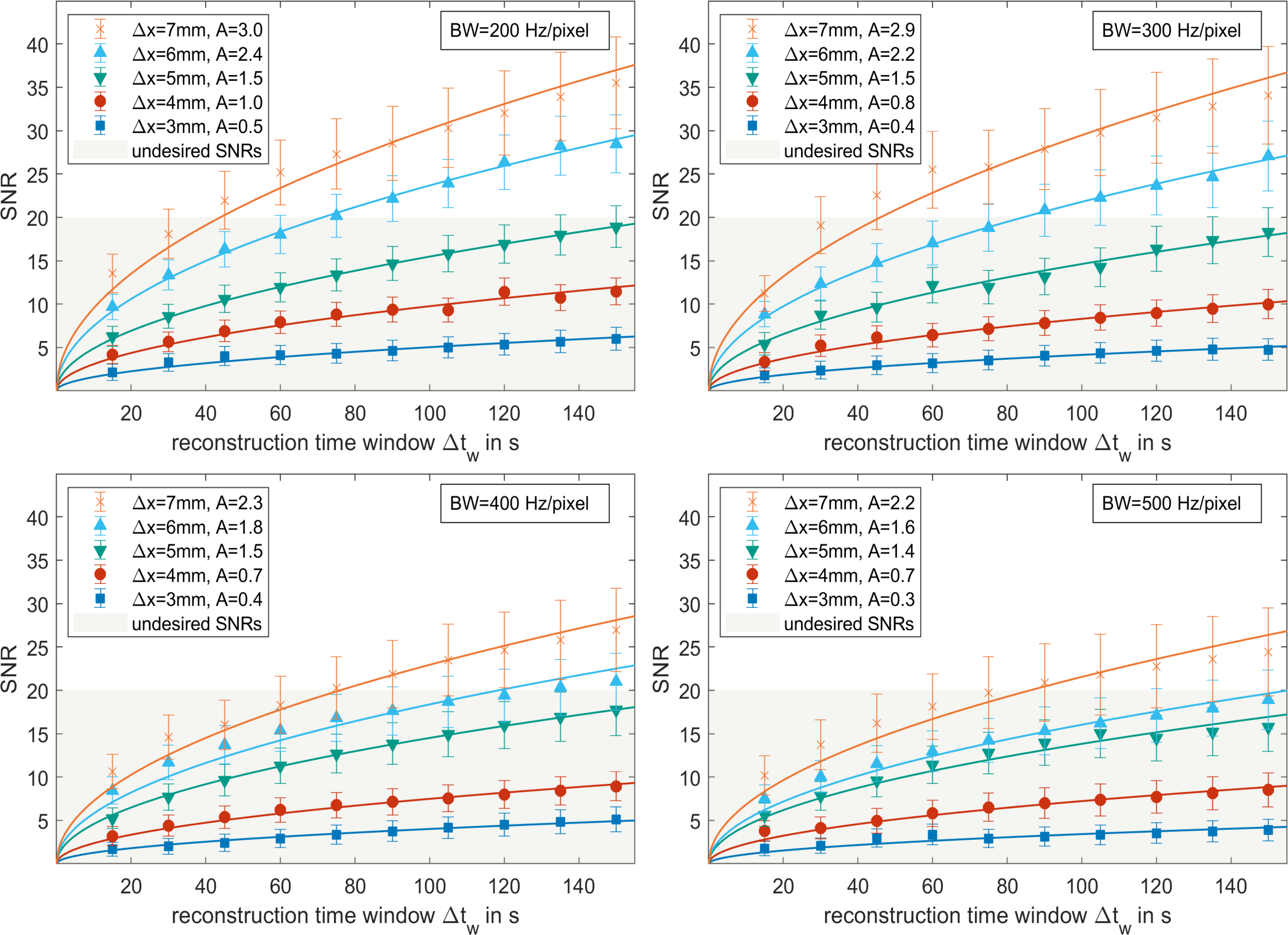
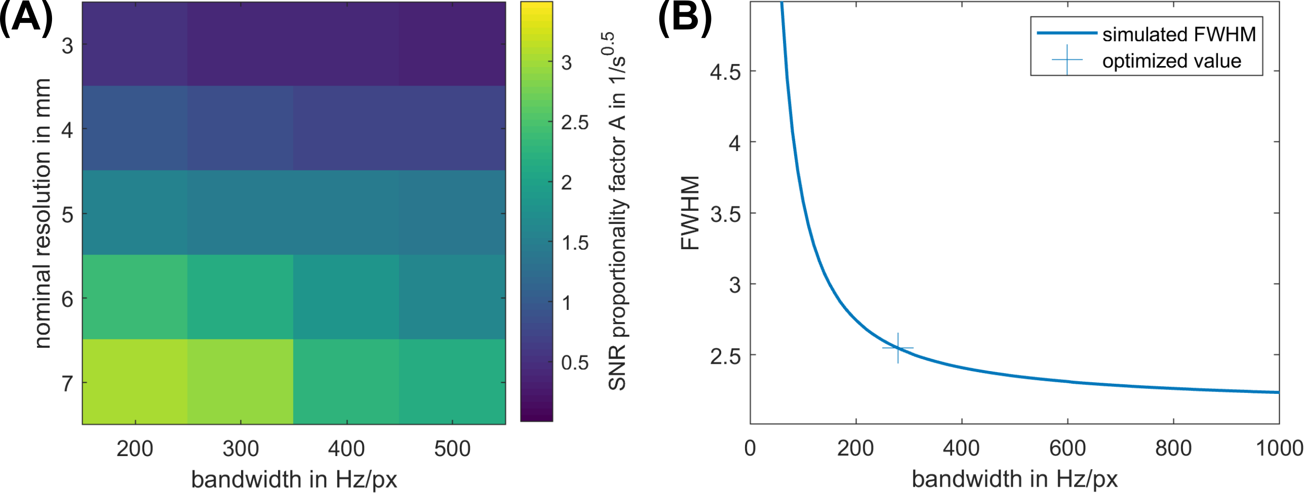
We numerically investigated the influence of the readout BW on the full-width-half-maximum (FWHM) of the point-spread-function by simulating the $$$T_2^*$$$-decay$$$~$$$($$$T_2^*=1.8\text{ms}$$$)18 during the acquisition with the given parameters and resulting gradient shapes. The influence of different combinations of BW and $$$\Delta{x}$$$ was experimentally evaluated on a homogeneous phantom with slightly larger dimensions than a kidney19 ($$$\text{vol}=450\text{mL}$$$) filled with 0.9%$$$~$$$NaCl. To scale the SNR dependency with the acquisition time, a non-linear fit $$$SNR=A\cdot\sqrt{(\Delta t_w)}$$$ was applied20.
To demonstrate that the optimized protocol is suitable for dynamic
17O-MRI, images of a oxygenated porcine kidney were acquired at a
clinical 3T MR system (Prisma; SIEMENS, Erlangen, Germany) with a custom-built
Tx/Rx$$$~$$$17O-loop$$$~$$$coil using our optimized parameters (Tab.$$$~$$$1).
Results
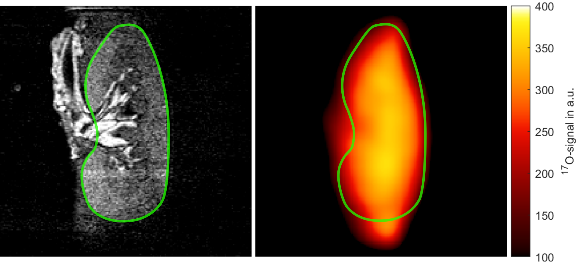
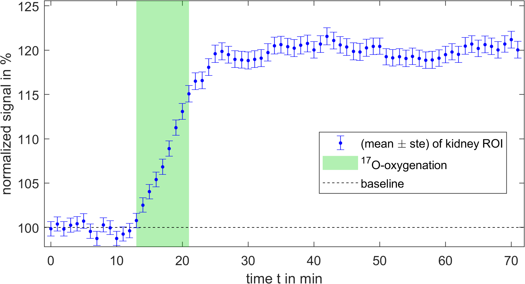
All combinations of BW and $$$\Delta{x}$$$ of the SNR dependency on $$$\Delta{t_w}$$$ showed the expected dependency on the square-root of time (Fig.$$$~$$$1, $$$R^2>0.9$$$), and $$$A$$$ decreased with BW and increased with $$$\Delta{x}$$$ (Fig.$$$~$$$2a). The simulation of the PSF (Fig. 2b) revealed a decrease of FWHM-factors with increasing bandwidth, approaching a limit of 2.2. For the lowest possible BW of 200 Hz/px in the desired parameter combinations, a FWHM-factor of 2.75 was found, while factors of below 2.5 were accomplished at BWs below 300 Hz/px. Figure$$$~$$$3 shows the anatomical 1H-MPRAGE image obtained with the body coil next to the interpolated 17O-magnitude signal averaged over the entire acquisition time. The mean kidney 17O-signal dynamically increased during 17O-oxygenation by up to 20% and remained relatively stable during the remaining time (Fig.$$$~$$$4).Discussion
In the desired parameter range ($$$\text{SNR}>20,\Delta{t}\leq120$$$), a spatial resolution of 6$$$~$$$mm could be achieved while maintaining a BW below 400$$$~$$$Hz/px. Although a BW of 200$$$~$$$Hz/px at $$$~$$$6mm resolution would result in the highest SNR values for the lowest possible nominal resolution, the associated blurring (FWHM-factor$$$~$$$2.75) would deteriorate the spatial accuracy of the measurement. Thus, $$$\Delta{x}$$$ and BW were set to 6$$$~$$$mm and 280$$$~$$$Hz/pixel (Tab.$$$~$$$1), respectively, such that images could be reconstructed with an acquisition time down to 90s while maintaining the FWHM-factor in the range of 2.5. In the pilot experiment dynamic 17O-signals could be acquired over a time course of 72$$$~$$$minutes from a porcine kidney with SNR values above 20 in the ROI. The signal increase in the area of the kidney was similar those of previous CMRO2 experiments in brain, which were in the range of 10-35%7–9,14.Conclusions
Parameter optimization of functional 17O-MRI in transplants as small as kidneys at 3T is a challenging compromise between low bandwidths and nominal resolution. Nevertheless we were able to perform a proof-of-concept experiment with the concluded parameters, which shows the possibility of dynamic 17O-MRI with SNRs above 20 and a temporal resolution down to 90$$$~$$$s. Due to direct oxygenation of the organ, the efficiency of used 17O2 of 25%/L is higher than those of previous CMRO2 experiments of 2.5-10%/L.Acknowledgements
Support from NUKEM Isotopes Imaging GmbH is gratefully acknowledged.References
1. Branger, P. & Samuel, U. Annual Report 2017. 124 (Eurotransplant International Foundation, 2017).
2. Hart, A. et al. OPTN/SRTR 2016 Annual Data Report: Kidney. Am. J. Transplant. 18, 97
3. Department of Health and Human Services, Health Resources and Services Administration, Healthcare Systems Bureau, Division of Transplantation. Annual Report of the U.S. Organ Procurement and Transplantation Network and the Scientific Registry of Transplant Recipients: UNOS (2016). Available at: https://unos.org/about/annual-report/2016-annual-report/. (Accessed: 30th October 2018)
4. Salvadori, M. & Tsalouchos, A. Biomarkers in renal transplantation: An updated review. World J. Transplant. 7, 161–178 (2017).
5. Pestana, J. M. Clinical outcomes of 11,436 kidney transplants performed in a single center - Hospital do Rim. J. Bras. Nefrol. 39, (2017).
6. Wang, J. H., Skeans, M. A. & Israni, A. K. Current Status of Kidney Transplant Outcomes: Dying to Survive. Adv. Chronic Kidney Dis. 23, 281–286 (2016).
7. Kurzhunov, D. et al. 3D CMRO2 mapping in human brain with direct 17O MRI: Comparison of conventional and proton-constrained reconstructions. NeuroImage 155, 612–624 (2017).
8. Kurzhunov, D., Borowiak, R., Reisert, M., Özen, A. C. & Bock, M. Direct estimation of 17O MR images (DIESIS) for quantification of oxygen metabolism in the human brain with partial volume correction. Magn. Reson. Med. (2018). doi:10.1002/mrm.27224
9. Niesporek, S. C. et al. Reproducibility of CMRO2 determination using dynamic 17O MRI: Direct CMRO2 Measurements: Reproducibility Study. Magn. Reson. Med. 79, 2923–2934 (2018).
10. Liu, Y. et al. High-resolution dynamic oxygen-17 MR imaging of mouse brain with golden-ratio-based radial sampling and k-space-weighted image reconstruction: Dynamic 17O-MRI of Mouse Brain. Magn. Reson. Med. 79, 256–263 (2018).
11. Zhu, X.-H. & Chen, W. In vivo 17 O MRS imaging – Quantitative assessment of regional oxygen consumption and perfusion rates in living brain. Anal. Biochem. 529, 171–178 (2017).
12. Zhu, X.-H., Zhang, Y., Wiesner, H. M., Ugurbil, K. & Chen, W. In vivo measurement of CBF using 17O NMR signal of metabolically produced H217O as a perfusion tracer: Simultaneous CBF and CMRO2 Measurement. Magn. Reson. Med. 70, 309–314 (2013).
13. Zhu, X.-H., Chen, J. M., Tu, T.-W., Chen, W. & Song, S.-K. Simultaneous and noninvasive imaging of cerebral oxygen metabolic rate, blood flow and oxygen extraction fraction in stroke mice. NeuroImage 64, 437–447 (2013).
14. Hoffmann, S. H., Radbruch, A., Bock, M., Semmler, W. & Nagel, A. M. Direct 17O MRI with partial volume correction: first experiences in a glioblastoma patient. Magn. Reson. Mater. Phys. Biol. Med. 27, 579–587 (2014).
16. Chan, R. W., Ramsay, E. A., Cunningham, C. H. & Plewes, D. B. Temporal stability of adaptive 3D radial MRI using multidimensional golden means. Magn. Reson. Med. 61, 354–363 (2009).
17. Jackson, J. I., Meyer, C. H., Nishimura, D. G. & Macovski, A. Selection of a convolution function for Fourier inversion using gridding (computerised tomography application). IEEE Trans. Med. Imaging 10, 473–478 (1991).
18. Zhu, X.-H., Merkle, H., Kwag, J.-H., Ugurbil, K. & Chen, W. 17O relaxation time and NMR sensitivity of cerebral water and their field dependence. Magn. Reson. Med. 45, 543–549 (2001).
19. Cheong, B., Muthupillai, R., Rubin, M. F. & Flamm, S. D. Normal Values for Renal Length and Volume as Measured by Magnetic Resonance Imaging. Clin. J. Am. Soc. Nephrol. 2, 38–45 (2006).
20. Brown, R. W., Cheng, Y.-C. N., Haacke, E. M., Thompson, M. R. & Venkatesan, R. Magnetic resonance imaging: physical principles and sequence design. (John Wiley & Sons, Inc, 2014).
Figures