4221
Non-invasive intracranial detection of the small molecule inhibitor BAY 1436032 in an orthotopic glioma model using 19F MR spectroscopy1Neuroradiology, University Hospital Frankfurt, Frankfurt, Germany, 2Neurooncology, University Hospital Frankfurt, Frankfurt, Germany, 3Institute for Organic Chemistry and Chemical Biology, Goethe University Frankfurt, Frankfurt, Germany, 4Research&Development Pharmaceuticals, Bayer AG Berlin, Berlin, Germany
Synopsis
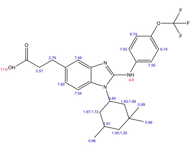
BAY 1436032 is a small-molecule inhibitor of the R132X-mutant form of IDH1 with a trifluoromethoxy-group that is currently clinically investigated in IDH1-mutant solid tumors. Non-invasive monitoring of intracranial drug delivery of BAY 1436032 in an animal model using 19F MRS is technically challenging but feasible.
Introduction
With 70–80% of astrocytomas and oligodendrogliomas harboring mutations in IDH1 genes, the mutant form of the enzyme is an attractive therapy target.1 2 Accumulating intracellular levels of (D-)2-Hydroxyglutarate (2-HG) in mutated cells presumably lead to a block in differentiation of non-transformed cells.3 BAY 1436032 is a small-molecule inhibitor of the R132X-mutant form of IDH1, inhibiting the mutation mediated generation of 2-HG. Since the inhibitor has a trifluoromethoxy-group (Fig.1) and the endogenous 19F MRI signal from the body is considered to be negligible, intracranial drug delivery might be monitored using 19F NMR/MR-Spectroscopy.Methods
BAY 1436032 was solubilized (DMSO) and administered to human serum. 19F/1H NMR spectroscopy was conducted on these samples and serum samples of a patient treated with BAY 1436032 (NCT02746081).
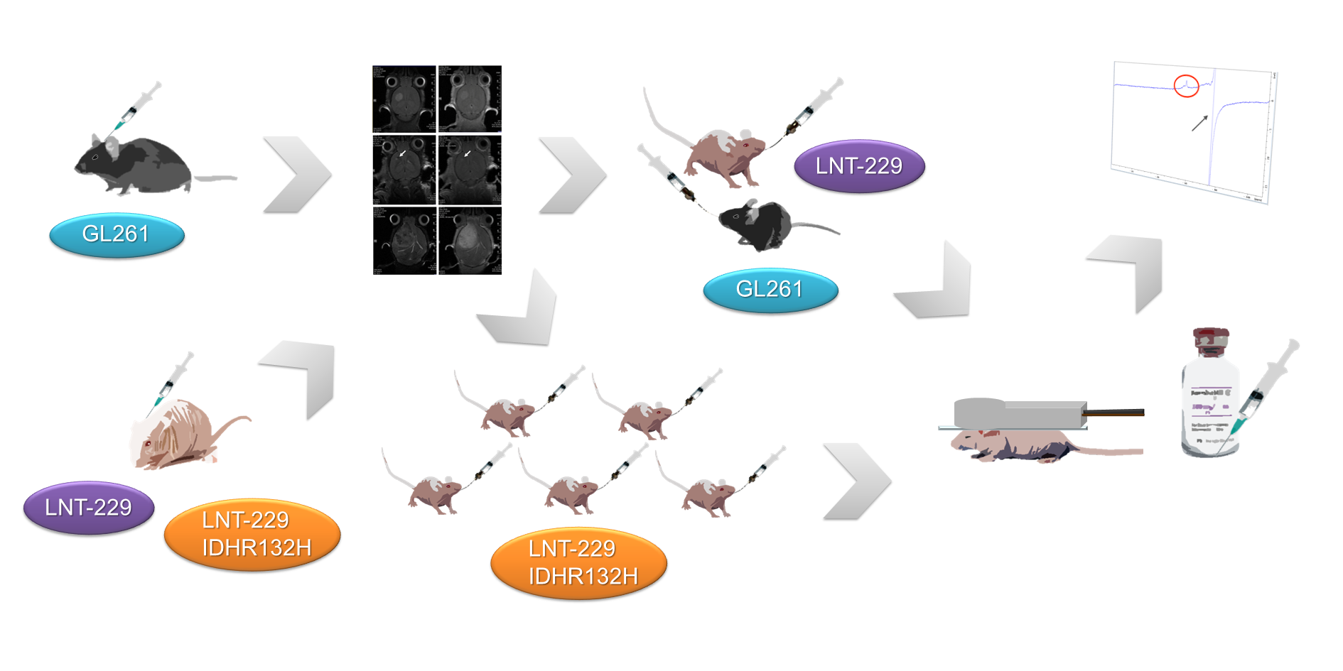
Twenty Fox/nu mice and ten C57/6N mice underwent orthotopic xenograft implantation using three glioma cell lines LNT-229, LNT-229 transfected to express mutant isocitrate dehydrogenases and GL261. MRS (1H PRESS, FID pulse, 19F single pulse) was performed on day 23 for GL261, on day 32 for LNT-229 and on days 39 and 44 for LNT-229 IDH1R132H on a small animal scanner (details Fig.2). Animals bearing an LNT-229 IDH1R132H tumor model received five days of treatment with BAY1436032 per os (75 mg/kgKG twice/day) after baseline scan and underwent follow-up scan at the end of treatment. All other animals received a single dose 3-6 hours prior to MRS (150 mg/kgKG). 19F signals (AMARES) and 1H signals (AQSES) were modeled in jMRUI. Animal brains were isolated immediately after MRS completion and separated into “tumor” and “brain” tissue. Samples were homogenized and aliquots of the supernatants analyzed by LC-MS/MS. Two patients treated with BAY1436032 in a phase I study (NCT02746081) underwent 19F MR spectroscopy on a clinical 3T scanner using a 19F transmit-receive volume coil.
Results
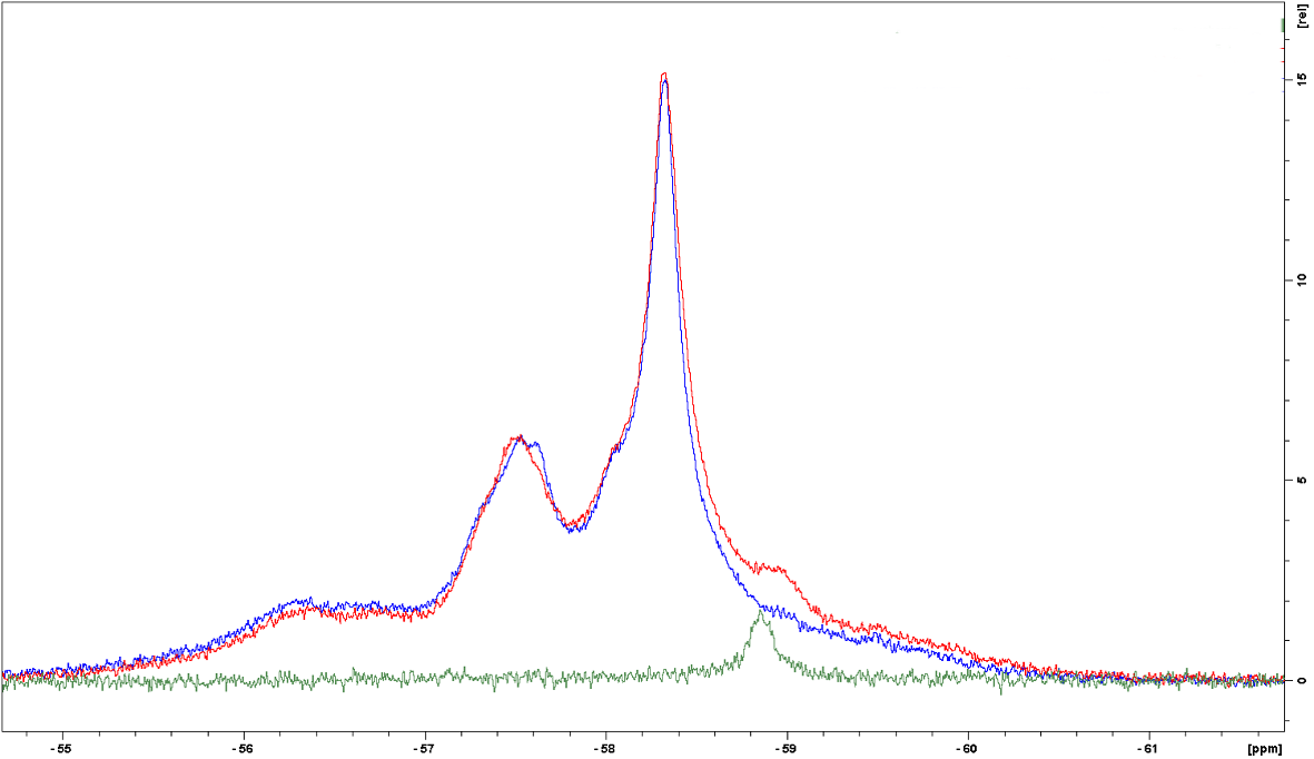
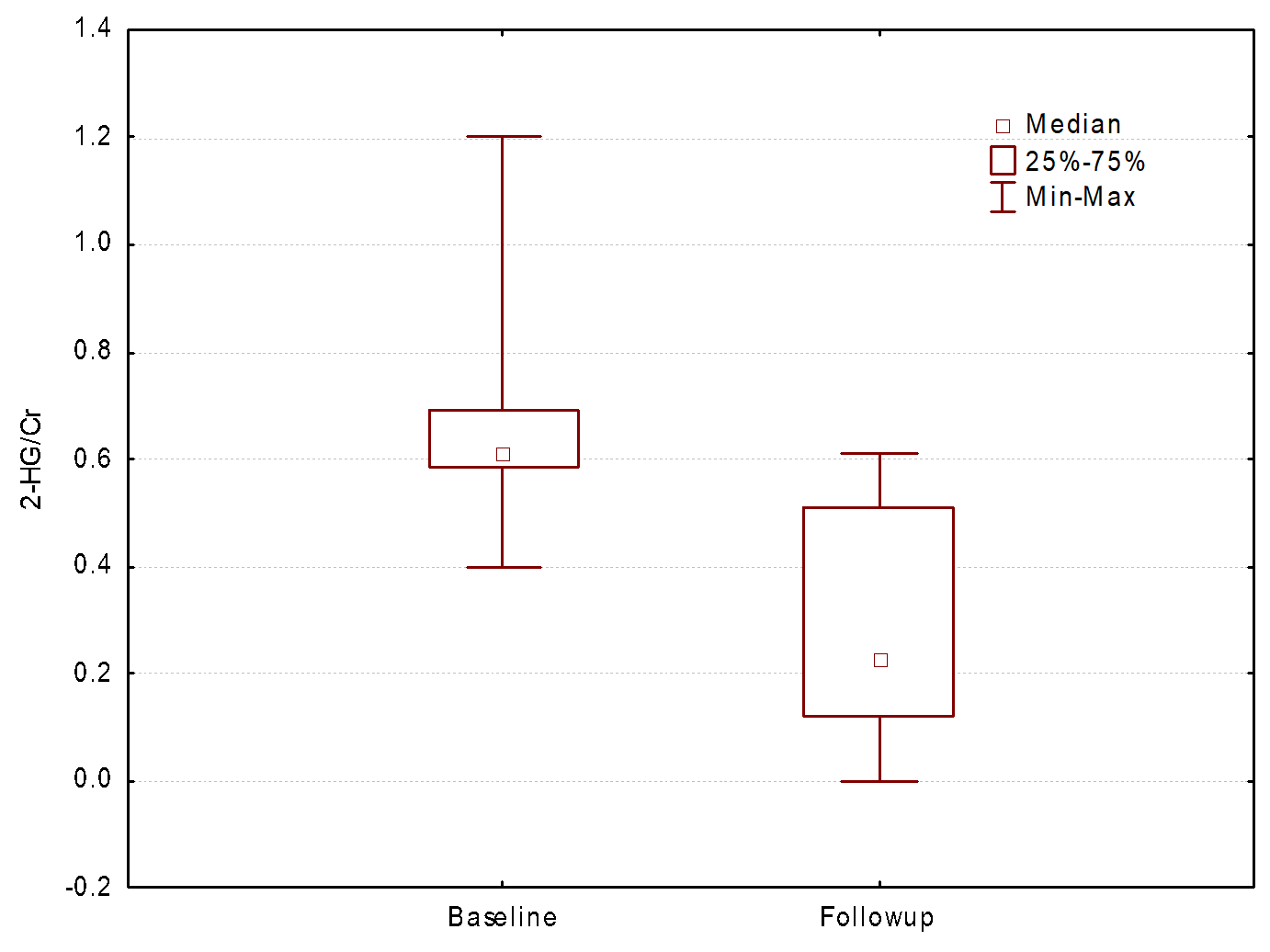
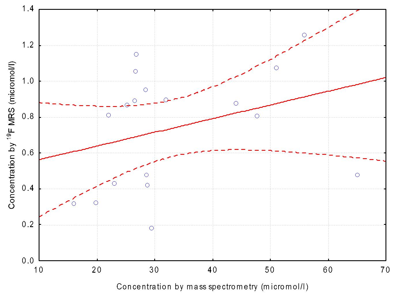
We were able to record five fluor signal peaks in patient serum samples using NMR spectroscopy, with one specifically identified as albumin after titration with HSA (Fig.3). Spectra were externally referenced to TFA at -76.55 ppm with the 19F signal of the inhibitor identified at approximately -59 ppm (free compound). 3/10 mice in the GL261 group had to be sacrificed prior to treatment due to acute and severe clinical symptoms and 2/10 in the LNT-229 IDH1R132H due to suspected meningeosis. Anatomical scans on day 26 for LNT-229 revealed tumor take in 10/10 animals, on day 33 for LNT-229 IDH1R132H in 8/10 animals and on day 23 for GL261 in 7/7 (remaining) animals. Four mice in the GL261 group and one non-tumor bearing mouse died during single pulse sequence. 19F signal could be identified in most mice at approximately -59 ppm to -60 ppm. Signal shape revealed one rather sharp w/o multiple broad peaks. Excluding animals that died during the single pulse sequence, 19F signal amplitudes for largely protein bound inhibitor showed a low positive correlation (Pearson‘s r = 0.32) with total tissue concentration determined by MS (Fig. 4). Total concentration in brain was not dependent on tumor model. Phantom experiments recording the water signal (pulse FID) were conducted to compare water signal intensity (110 [M]) to fluor signal intensity (10 [M]). Based on this calibration, approx. 1/100th of BAY 1436032 concentration in in vivo experiments can be detected by 19F MRS. Evaluation of 1H MRS showed a significant decrease in 2-HG/Cr ratios (p = 0.04; Fig. 5) from baseline to post-treatment follow-up scans in the LNT-229 IDH1R132H tumor model. SNR at 3T in patients was not sufficient for 19F signal detection.Discussion
Despite a low signal intensity of a substance with only three fluor atoms which is mainly protein bound4, 19F MRS signal of BAY 1436032 can be identified intracranial in an animal model using a 19F single pulse sequence at a 300 MHz small animal scanner. We were not able to detect differences in concentrations of mice bearing invasive tumor models with a glioblastoma phenotype and a relevant BBB integrity loss (GL261) and with a less severe breakdown in slower growing models (LNT-229). This suggests that BBB breakdown might not affect the activity of a systemic therapy with BAY 1436032. In literature there is still an ongoing controversy over the role of the BBB in uptake of targeted therapeutic agents.5 As expected, we were able to show a decrease in 2-HG concentrations from baseline to follow-up scans in the LNT-229 IDH1R132H tumor model during therapy.4 Improved SNR for human studies might be achieved at 7T.Conclusion
In vivo detection of BAY 1436032 using 19F MRS in an animal model is technically challenging, but feasible.Acknowledgements
No acknowledgement found.References
1. Yan H, Parsons DW, Jin G, et al. IDH1 and IDH2 mutations in gliomas. N Engl J Med. 2009;360(8):765-773. doi:10.1056/NEJMoa0808710.2. Bleeker FE, Lamba S, Leenstra S, et al. IDH1 mutations at residue p.R132 (IDH1(R132)) occur frequently in high-grade gliomas but not in other solid tumors. Hum Mutat. 2009;30(1):7-11. doi:10.1002/humu.20937.
3. Pusch S, Krausert S, Fischer V, et al. Pan-mutant IDH1 inhibitor BAY 1436032 for effective treatment of IDH1 mutant astrocytoma in vivo. Acta Neuropathol. 2017;133(4):629-644. doi:10.1007/s00401-017-1677-y.
4. Bayer AG. Investigator's Brochure, BAY 1436032, Version 3.0, Date 16.04.2018: Identity of Investigational Product: BAY 1436032 Indication: Cancer - mIDH1-R132X mutant malignancies; 2018.
5. Osswald M, Blaes J, Liao Y, et al. Impact of Blood-Brain Barrier Integrity on Tumor Growth and Therapy Response in Brain Metastases. Clin Cancer Res. 2016;22(24):6078-6087. doi:10.1158/1078-0432.CCR-16-1327.
Figures