4214
Longitudinal heterogeneity of post-exercise perfusion, T2*-weighted MRI and high-energy phosphate metabolism in human calf muscle quantified by interleaved multislice 1H MRI and multivoxel 31P MRS at 7T1Center for Medical Physics and Biomedical Engineering, Medical University of Vienna, Vienna, Austria, 2MR Center of Excellence, Medical University of Vienna, Vienna, Austria, 3Department of Biomedical Imaging and Image-guided Therapy, Medical University of Vienna, Vienna, Austria, 4Department of Clinical Pharmacology, Medical University of Vienna, Vienna, Austria, 5NMR Laboratory, Institute of Myology, Paris, France
Synopsis
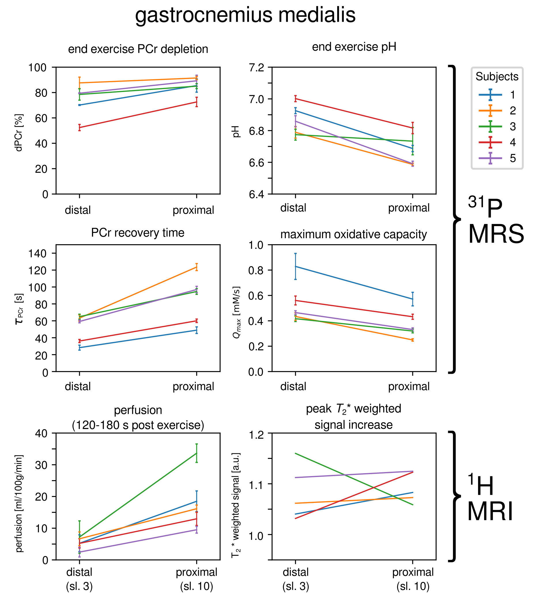
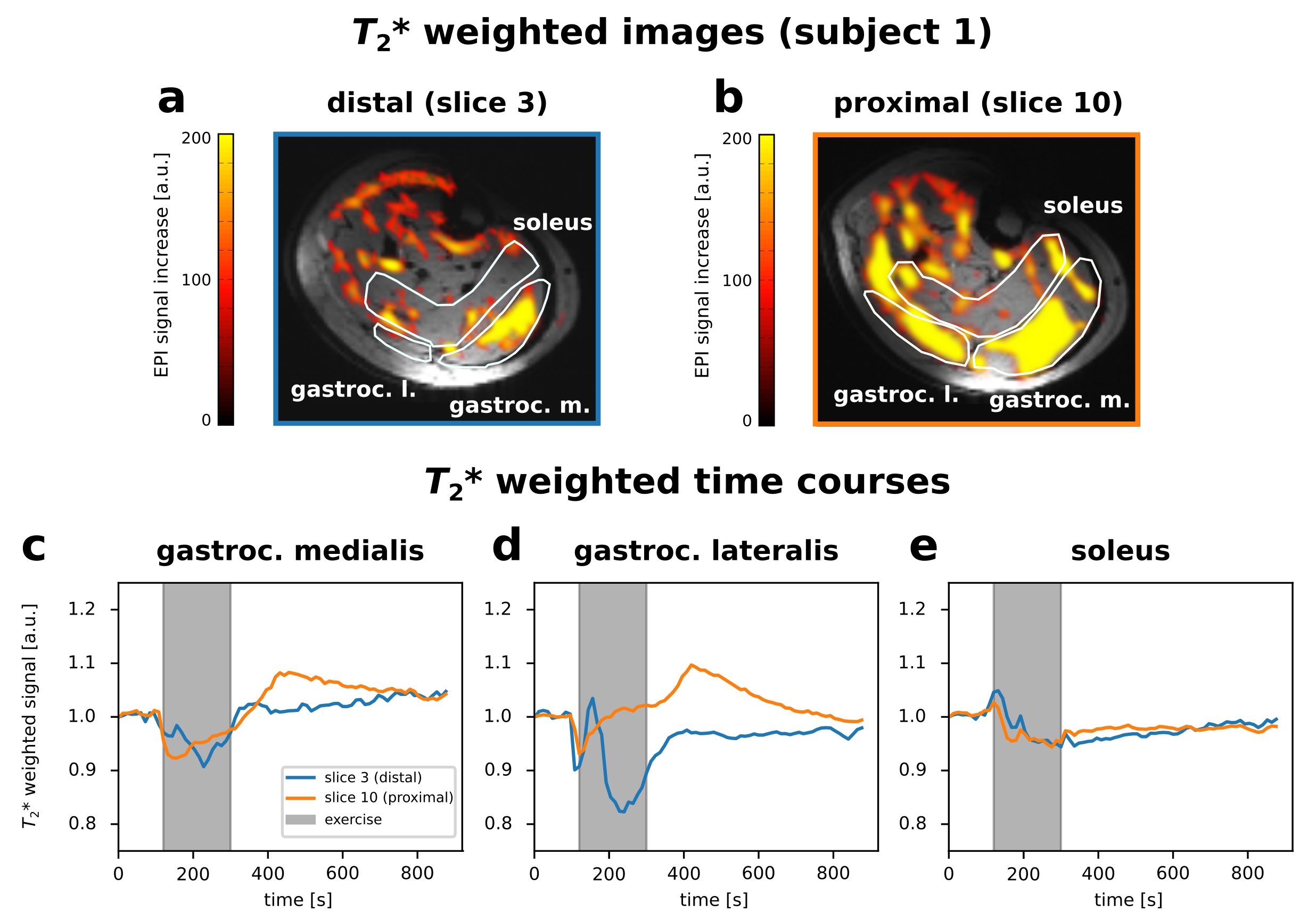
Multivoxel 31P semi-LASER was used to acquire time-resolved spectra from two positions along the gastrocnemius medialis muscle during rest, exercise and recovery. Simultaneously, quantitative perfusion- and T2*-weighted images of the calf were acquired with multislice 1H ASL MRI, in an interleaved fashion. Significantly different results were found with 31P MRS along gastrocnemius medialis: proximally, end-exercise PCr depletion was higher, PCr recovery times longer, end-exercise pH was lower and maximum oxidative capacity was also lower. Significantly higher post-exercise perfusion and slightly stronger T2*-weighted signal increase were found in proximal slices, which is consistent with the aforementioned distribution of metabolic activity.
Introduction
The distribution of workload between individual muscles leads to heterogeneous activation patterns associated with spatially distinct metabolic activity across an exercising limb. This has been taken into account in various studies employing localized 31P MR spectroscopy1-3. So far, the recruitment of tissue along the muscle has been implicitly assumed to be homogeneous, i.e. potential variations have been largely ignored. Recently, Boss, Heskamp et al.4 discovered that the oxidative capacity varies not only between muscle groups but also along the length of one specific muscle (tibialis anterior) and later confirmed a gradient of O2 supply and perfusion in sequential measurements5. We recently presented a pulse sequence combining arterial spin labelling 1H MRI and multivoxel 31P MRS, which allows for time-resolved measurements of perfusion, oxygenation and oxidative phosphorylation at multiple independent positions in an interleaved fashion6 within a single exercise-recovery experiment. Here we show first results obtained by applying this sequence to investigate human calf muscle groups spatially resolved lengthwise, during plantar flexion exercise.Methods
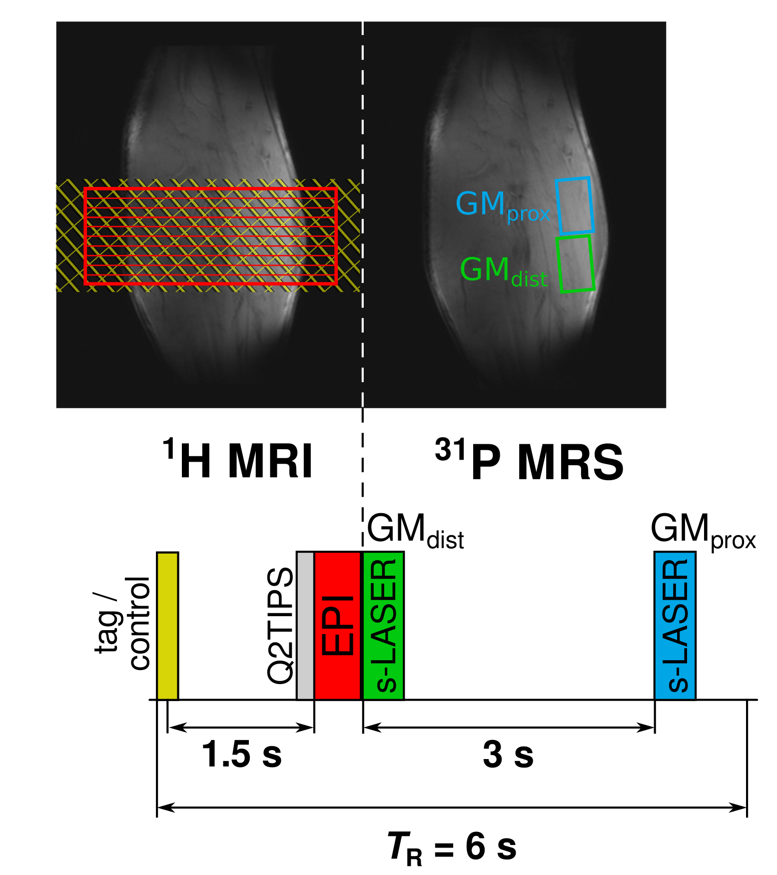
Pulsed arterial spin labeling (ASL) with FAIR tagging was applied using global/slice-selective inversion7 fully overlapping with the readout slab (Fig.1). The blood inflow time was 1.3s, followed by 200ms Q2TIPS saturation before ten EPI slices were acquired without slice gaps (TE=20ms, d=6mm) in sequential order (direction H-F). Following 1H-ASL, a multivoxel 31P semi-LASER acquisition scheme8 was applied in gastrocnemius medialis (GM) at the distal and the proximal half of the 6cm box covered by the imaging readout (Fig.1 , GMdist=GMprox=25cm³, TE=29ms) . The acquisition delay between the voxel positions was 3s. Total TR was 6s, resulting in a set of perfusion images every 12s. Five healthy volunteers were measured during 2 min rest, 3 min plantar flexion exercise at approximately 40% MVC on a non-magnetic pneumatic ergometer, and 10 min recovery. Measurements were performed at 7T (Siemens, Erlangen, Germany) using a calf shaped multichannel 1H/31P transceiver surface coil-array9. Image reconstruction and postprocessing was performed using Siemens’ ICE and Matlab for spatially resolved calculation of perfusion and T2*-weighted time courses. Processing of spectra was performed on raw data using python for phasing and channel combination. Spectral quantification was done using jMRUI with AMARES followed by monoexponential PCr recovery time fitting using gnuplot.Results
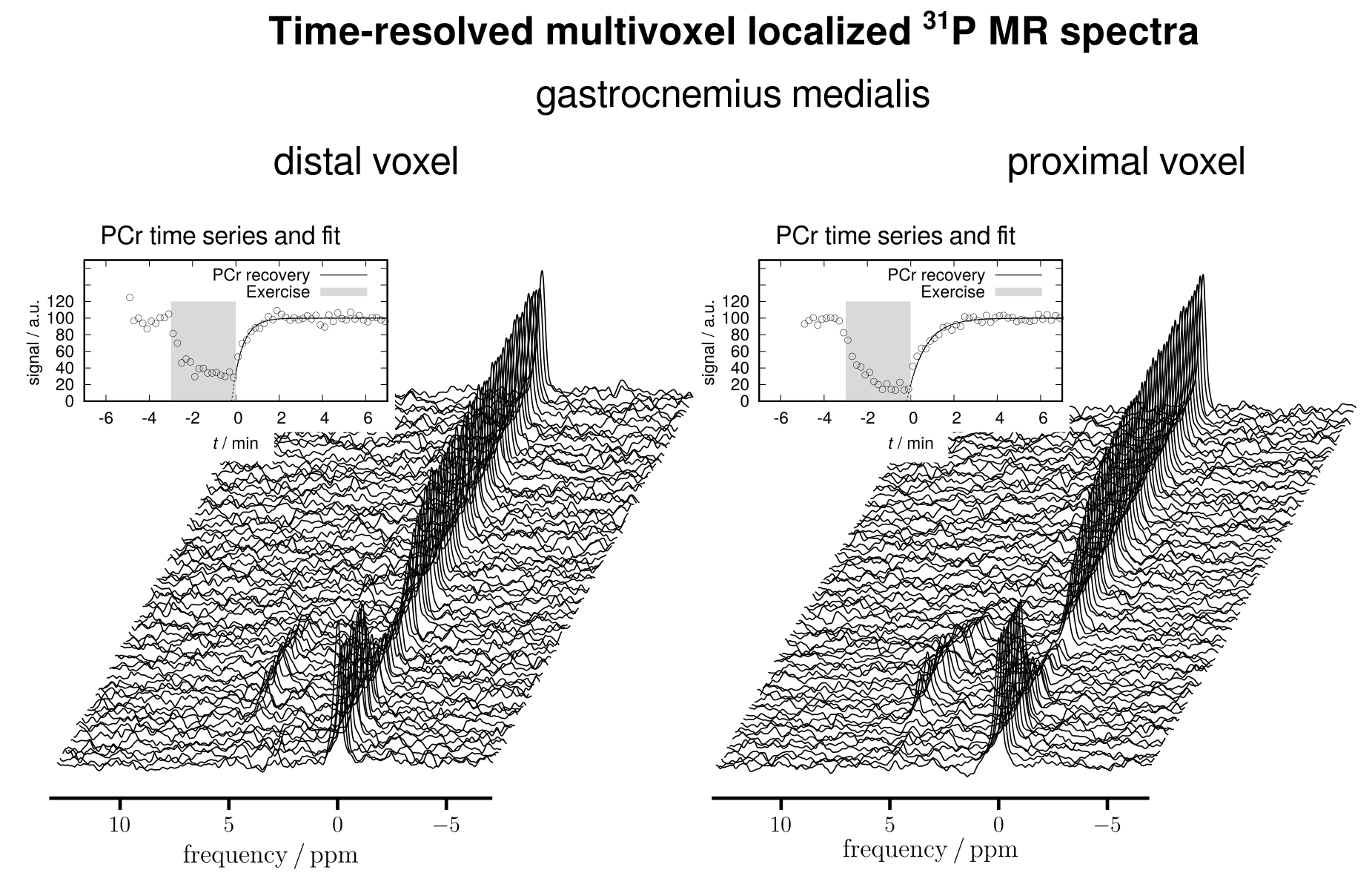
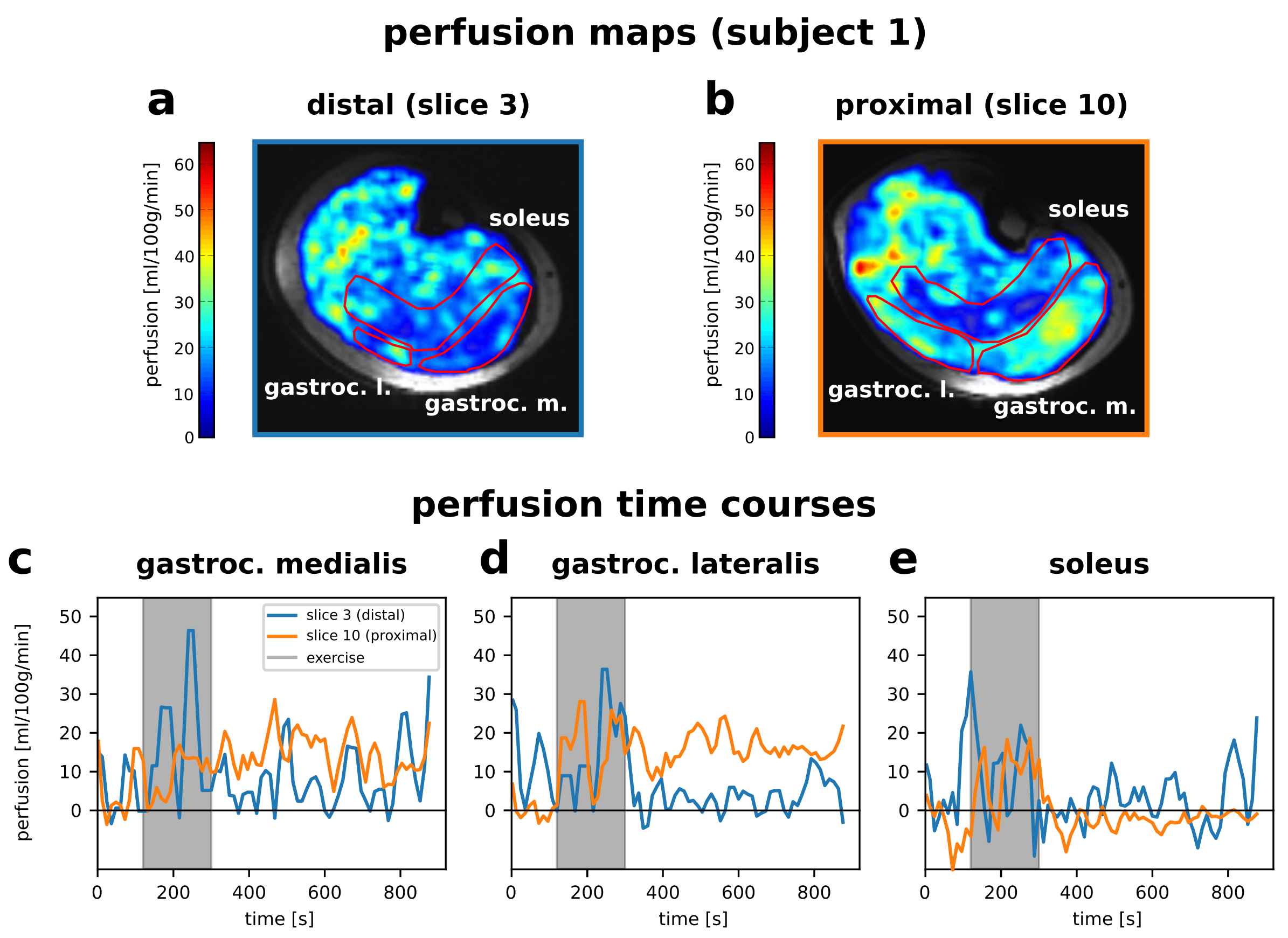
Time series 31P spectra acquired at the distal and proximal position of GM were sucessfully quantified throughout the experiment. Fig.2 shows results of one representative subject. PCr depletion, pH, PCr recovery time (τPCr) and the maximum oxidative capacity Qmax were derived from 31P spectra from both positions for each subject (Fig.3). Further, individual post-exercise perfusion (120-180s averaged during recovery) and maximum T2*-weighted signal increase in GM during recovery were calculated from 1H data acquired during the same time as 31P spectra for distal (no.3) and proximal (no.10) slices (Fig.3). Proximo-distal differences of perfusion and T2*-weighted signal increase is illustrated in Figs.4 and 5 showing post-exercise perfusion maps with perfusion time courses and T2*-weighted images with T2*-weighted time courses of GM, gastrocnemius lateralis (GL) and soleus (SOL) of one representative subject. The response to exercise in GM was significantly different between distal and proximal positions. All subjects showed proximally higher end-exercise PCr depletions and PCr recovery times in accordance with a lower end-exercise pH. Qmax was significantly lower proximally. Additionally we found a significantly higher post-exercise perfusion for all subjects in proximal compared to distal slices in GM, while differences between peak T2*-weighted data were not significant.Discussion/Conclusion
The different findings with 31P MRS at proximal and distal positions in GM (higher activation proximally) are consistent with perfusion and T2* data acquired simultaneously. This consistency inherently also supports the notion that apparent differences in perfusion at different slice positions reflect a true physiological effect and are not likely to be the result of a systematic error of the multislice protocol. Spatial differences of perfusion were stronger in GL, however, the cross-sectional area of GL strongly decreases distally, precluding placement of a distal 31P voxel. We also excluded two 1H slices at the distal end in post processing as the ROI in GL was too small. A limitation is the length of our 1H coil for tagging inflowing blood, which might affect absolute perfusion values. Our results showed higher muscle activation proximally, which is in excellent agreement with literature4-5 with the methodical surplus of simultaneous data acquisition during a single exercise-recovery experiment. In contrast to (4) we measured a more glycolytic muscle, showing significantly lower pH and hence slower PCr recovery rates proximally. This method allows for linking complementary information from a single exercise and will improve the investigation of metabolic parameters.Acknowledgements
This work has been funded by the Austrian Science Fund (FWF): I1743-B13References
1. Fiedler GB, Schmid AI, Goluch S et al. Skeletal muscle ATP synthesis and cellular H+ handling measured by localized 31 P-MRS during exercise and recovery. Sci Rep 2016;6:32037
2. Valkovic L, Chmelik M, Meyerspeer M et al. Dynamic 31P-MRSI using spiral spectroscopic imaging can map mitochondrial capacity in muscles of the human calf during plantar flexion exercise at 7T. NMR Biomed 2016;29(12):1825-1834
3. Niess F, Fiedler GB, Schmid AI et al. Dynamic multivoxel-localized 31P MRS during plantar flexion exercise with variable knee angle. NMR Biomed 2018;31(6):e3905
4. Boss A, Heskamp L, Breukels V et al. Oxidative capacity varies along the length of healthy human tibialis anterior. J Physiol 2018 596.8:1467-1483
5. Heskamp L, Lebbink F, van Uden M et al. Both oxygen supply and phosphocreatine recovery rate show proximo-distal gradients along the human tibialis anterior after exercise. Abstract #821, ISMRM 2018, Paris, France
6. Niess F, Schmid AI, Fiedler GB et al. Dynamic perfusion and T2*-weighted 1H MRI interleaved with multivoxel 31P MRS of exercising human calf at 7T, Abstract #624, ISMRM 2018, Paris, France
7. Schewzow K, Fiedler GB, Meyerspeer M et al. Dynamic ASL and T2*-Weighted MRI in Exercising Calf Muscle at 7 T- A Feasibility Study Magn Reson Med 2015;73:1190-1195
8. Niess F, Fiedler GB, Schmid AI et al. Interleaved Multivoxel 31P MR Spectroscopy Magn Reson Med 2017;77:921-927
9. Goluch S, Kuehne A, Meyerspeer M et al. A Form-Fitted Three Channel 31P, Two Channel 1H Transceiver Coil Array for Calf Muscle Studies at 7 T Magn Reson Med 2015;73(6):2379-89
10. Kemp GJ and Radda GK Quantitative Interpretation of Bioenergetic Data from 31P and 1H Magnetic Resonance Spectroscopic Studies of Skeletal Muscle: An Analytical Review. Magn Reson Q; 10:43-63
Figures