4207
Comparison of anisotropic UTE sequences for 23Na/39K imaging in the human calf at 7T1Institute of Radiology, University Hospital Erlangen, Friedrich-Alexander-Universität Erlangen-Nürnberg (FAU), Erlangen, Germany, 2Center for Medical Physics and Engineering, Friedrich-Alexander-Universität Erlangen-Nürnberg (FAU), Erlangen, Germany, 3Medical Physics in Radiology, German Cancer Research Center (DKFZ), Heidelberg, Germany
Synopsis
Anisotropically scalable UTE sequences enable a higher in-plane resolution for X-nuclei imaging. In this work, a density-adapted 3D radial cuboid acquisition scheme is compared theoretically and experimentally against a 3D acquisition-weighted density-adapted stack-of-stars (AWSOSt) design. For the use case of 23Na/39K imaging in the human calf at 7T, it was found that, when driven at similar and clinically feasible measurement times, the AWSOSt approach provides an increased SNR at the same effective resolution. Potential T2 blurring due to the additional phase encoding had no significant effect in this study. For both sequences a good suitability for anisotropic X-nuclei MR imaging in muscular tissue was confirmed.
Introduction
Through the striving towards ultrahigh field strengths in MR tomography, X-nuclei applications are becoming increasingly feasible. Nevertheless, low concentration and low sensitivity, paired with short T2* relaxation times, require ultrashort echo time imaging (UTE) techniques with large voxel volumes to achieve a reasonable signal-to-noise ratio (SNR) within clinical measurement times. In suitable tissue types, higher in-plane resolution can be achieved by extending the voxel length in slice direction.1 Different anisotropically scalable radial UTE sequence schemata are available and inhibit distinct characteristics.2 For the application of 23Na/39K imaging in the human calf at 7T with non-selective excitation, we compare a density-adapted 3D radial cuboid acquisition scheme (DA-3D-RAD-C)3 to a 3D acquisition-weighted density-adapted stack-of-stars scheme (AWSOSt)4,5, to assess their applicabilities and differences.Methods
DA-3D-RAD-C samples k-space with every readout from the center. Its projectional acquisitions can start directly after the RF pulse and are scaled to the surface of a cuboid. AWSOSt samples a cylindrical k-space and consists of several stacked, density-adapted 2D stars with varying TE, also allowing anisotropic scaling (Fig.1). In order to quantify the effective resolution of the considered sequences within the mentioned scope of application, the point spread functions (PSF) of the two schemes are simulated and compared (Fig.2). With this, the full width at half maximum (FWHM) can be applied to compare the effective resolutions of the different approaches. Building upon this, 23Na/39K MRI measurements were performed at a 7T whole-body system (MAGNETOM 7T Terra, Siemens Healthineers, Erlangen, Germany), using a dual-tuned 23Na/39K birdcage coil with inner diameter 20cm (RAPID Biomedical, Rimpar, Germany). Clinically feasible protocols were set up for both sequences and nuclei, each limited to 7:30min. Parameters: TRO=10ms, TE/TR(23Na)=0.30/40ms, TE/TR(39K)=0.55/20ms(phantom)&40ms(in vivo). The effective resolutions were chosen to Δx(23Na)=(3.0x3.0x15.1)mm3, Δx(39K)=(9.7x9.7x36.3)mm3 and scaled to each sequence’s nominal resolution by the respective FWHM of the PSF (Cubic: (1.21x1.21x1.21)px3, Cylindric: (1.41x1.41x1.21)px3). In a phantom study, the SNR was evaluated by the signal statistics of two separate ROIs of a single image (Fig.3).6 The signal was determined in a phantom containing 4% agarose gel, with 50mM NaCl and 120mM KCl. The determined SNRs were normalized via the simulated PSFs, incorporating decay and delay parameters (c.f. Fig.2). Actual spatial resolution and sensitivity to off-resonances were classified through acquisitions of a resolution phantom, filled with a 50mM NaCl, 150mM KCl solution, pierced by acrylic glass bars ranging from 2-10.5mm (Fig.4). Finally, to evaluate the performance of both sequences for 23Na/39K MRI of calf muscle tissue, two male healthy subjects were examined (Fig.5).Results
Simulations of the PSFs (Fig.2) show a larger FWHM of the AWSOSt in X/Y direction and a narrower PSF in Z direction, compared to the DA-3D-RAD-C under the given measurement application. Considering these results into the computation of the SNR (Fig.3), AWSOSt shows higher normalized ratios for both nuclei, also when including the additionally induced T2 blurring of the phase encoding in the calculation. Profile sections through a resolution phantom (Fig.4) confirm the agreement of the calculated effective resolutions between both sequences and the determined scaling factors. In addition, an increased susceptibility of the AWSOSt method to B0 inhomogeneities in the resolution phantom can be observed. The applicability of anisotropic imaging sequences in general and DA-3D-RAD-C versus AWSOSt in particular for 23Na/39K MRI of the human calf muscles are shown in Fig.5 for two healthy volunteers.Discussion
Evaluating the FWHMs of the simulated PSFs (Fig.2), it can be seen that in the present measurement setup, despite short relaxation times, only negligible T2 blurring occurs despite the additional phase encoding of AWSOSt. The fact, that the AWSOSt PSF remains comparatively narrow in slice direction and a generally more homogeneous distribution of the sampling points in k-space at high anisotropies lead to a higher SNR at the AWSOSt acquisition, which was confirmed in the phantom study. Furthermore, this approach reaches a complete Nyquist coverage at a lower number of projections, avoiding noise-like undersampling artifacts at shorter measurement times. However, limitations arise from the possibility of aliasing in the Cartesian stacking direction of AWSOSt.Conclusion
In this work, we assessed the characteristics, similarities and distinctions of the anisotropically scalable, non-selective UTE sequences DA-3D-RAD-C versus AWSOSt for 23Na/39K MRI of human calf muscle at 7T. A generally valid, good applicability for both sequence types was confirmed. When employing AWSOSt, the discussed characteristics such as delay time and coil FOV must be considered. If these comply with the planned measurement setup, this approach is able to provide an increased SNR for an unchanged effective resolution.Acknowledgements
No acknowledgement found.References
1. Larson, Peder EZ, Paul T. Gurney, and Dwight G. Nishimura. "Anisotropic field-of-views in radial imaging." IEEE transactions on medical imaging 27.1 (2008): 47-57.
2. Konstandin, Simon, and Armin M. Nagel. "Measurement techniques for magnetic resonance imaging of fast relaxing nuclei." Magnetic Resonance Materials in Physics, Biology and Medicine 27.1 (2014): 5-19.
3. Nagel, Armin M., et al. "3D density-adapted projection reconstruction 23Na-MRI with anisotropic resolution and field-of-view." Proceedings of the International Society for Magnetic Resonance in Medicine. 2012.
4. Qian, Yongxian, and Fernando E. Boada. "Acquisition‐weighted stack of spirals for fast high‐resolution three‐dimensional ultra‐short echo time MR imaging." Magnetic resonance in medicine 60.1 (2008): 135-145.
5. Konstandin, Simon, et al. "Two-dimensional radial acquisition technique with density adaption in sodium MRI." Magnetic resonance in medicine 65.4 (2011): 1090-1096.
6. Dietrich, Olaf, et al. "Measurement of signal‐to‐noise ratios in MR images: influence of multichannel coils, parallel imaging, and reconstruction filters." Journal of Magnetic Resonance Imaging: An Official Journal of the International Society for Magnetic Resonance in Medicine 26.2 (2007): 375-385.
7. Nagel, Armin M., et al. "39K and 23Na relaxation times and MRI of rat head at 21.1 T." NMR in Biomedicine 29.6 (2016): 759-766.
Figures
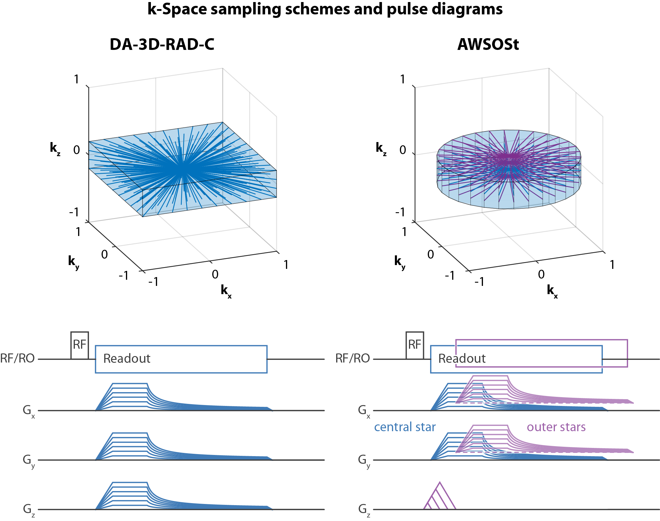
Figure 1:
DA-3D-RAD-C: Through its cuboid surface, resolution and FOV can be adjusted anisotropically. Compared to the spherical variant, the Cartesian k-space is thus sampled more completely (i.e. higher effective resolution), which conversely leads to a more inhomogeneous distribution of the sampling points (i.e. reduced SNR).
AWSOSt: The central star can start readout at minimum TE, while the outer stars are only delayed as long as required by their preceding phase encoding. The respective delay times are dependent on imaged nucleus, voxel length, echo time, gradient system and stimulation limits. For our setup, maximal delays of 250µs and 130µs were required for 23Na/39K MRI, respectively.
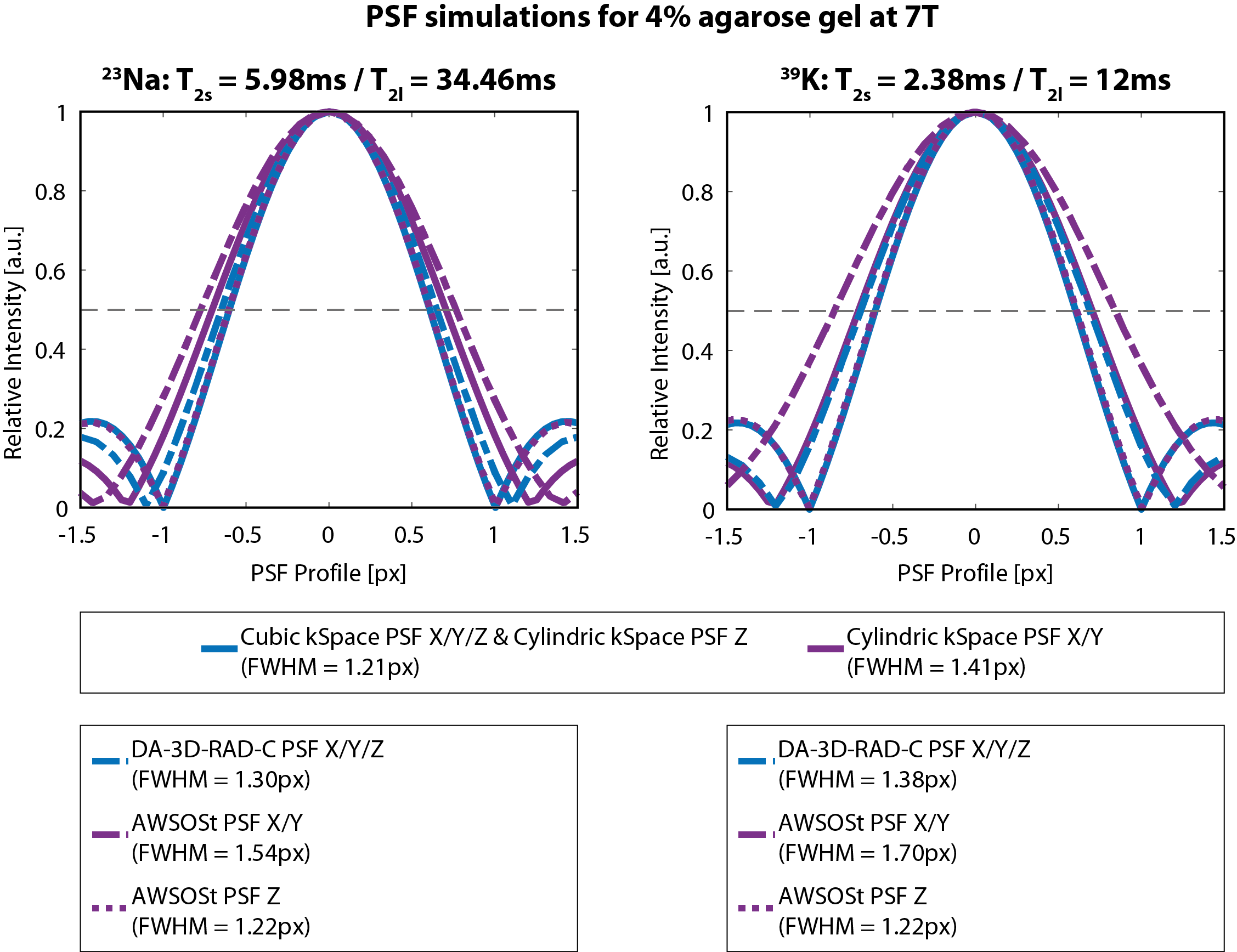
Figure 2:
Simulation of point spread functions: In a first step, the influence of the basic sampling forms (cubic/cylindrical) onto the FWHMs of the PSFs was investigated. Subsequently, T2 decay and delay of the required phase-encoding gradients were included in the computation. As decay parameters, values for a 4% agarose gel were applied, with short biexponential relaxation constants within the order of magnitude of human calf muscle tissue.7 Further simulation parameters were aligned to the applied measurement protocols as well.
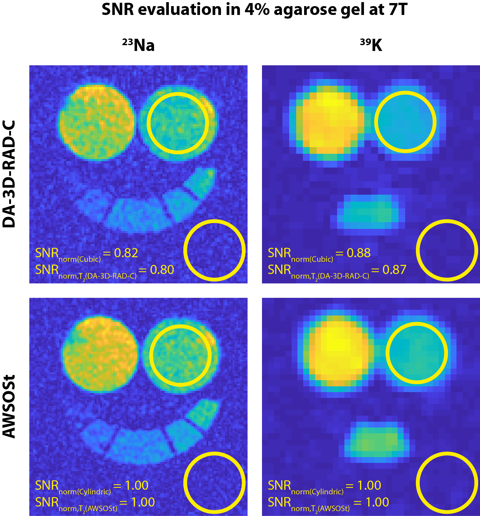
Figure 3:
Phantom measurements for SNR evaluation: In a 4% agarose gel phantom, with known relaxation times, the respective SNR was determined. This was then normalized to the effective voxel volumes from the PSF simulations (without and with decay/delay) and compared between the two acquisition schemes. While the 39K acquisitions were all acquired above their Nyquist limits, the 23Na DA-3D-RAD-C acquisition only reached 42% of its Nyquist projections in the given time frame. The reference phantoms contain 10, 20, 25, 30 and 40mM NaCl with additional 150mM KCl in the central phantom. The left bottle phantom contained 50mM NaCl and 150mM KCl solution.
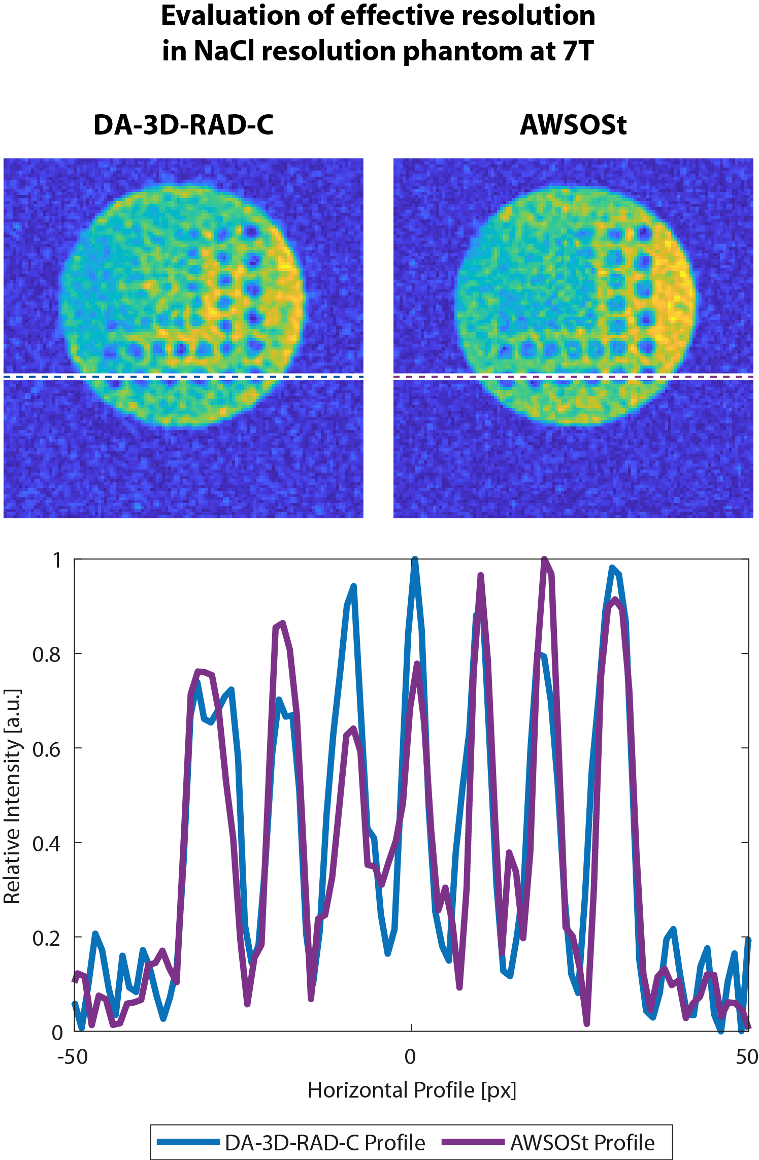
Figure 4:
23Na images of a NaCl resolution phantom with rods ranging from 2.5 to 10mm. A comparison of the two images confirms, that both acquisition schemes yield similar effective spatial resolution. The profile section confirms this finding. Artifacts within the plastic rods in the AWSOSt acquisition indicate a higher sensitivity of this acquisition scheme to B0 inhomogeneities.
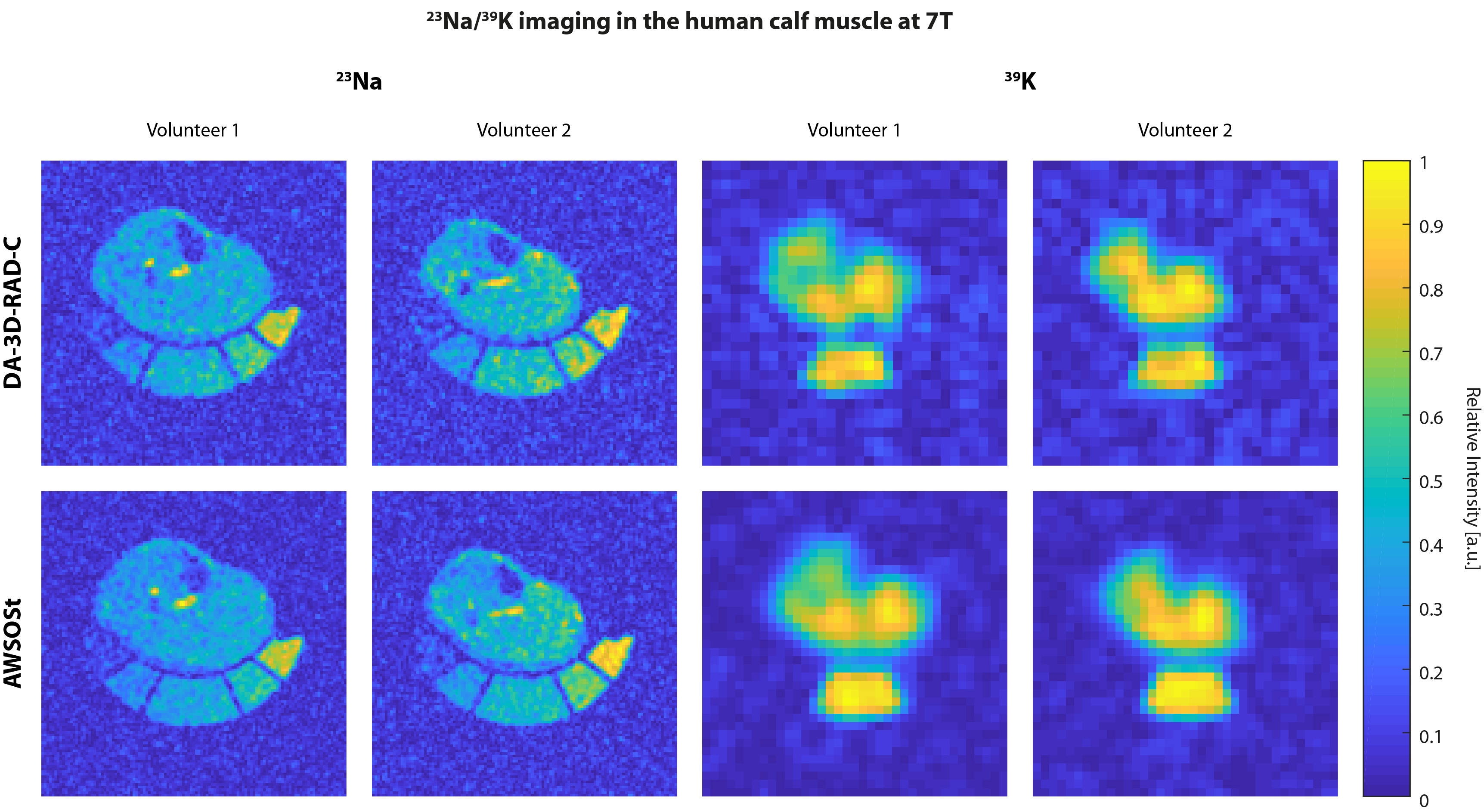
Figure 5:
23Na/39K images of the human calf muscle: Two healthy volunteers were imaged with both sequences under investigation, in order to demonstrate their feasibility at in vivo measurements. Especially in anisotropically structured tissue, such as muscular fiber, increasing the in-plane resolution at the expense of a higher slice resolution can considerably improve image quality. The increased SNR of the AWSOSt sequence could, for instance, be traded for an even better in-plane resolution. By combining 23Na with 39K MRI at high in-plane resolutions, new valuable insights might be gained.