4205
B0 shimming for X-nuclei imaging at 7 T using 23Na B0 maps1Institute of Radiology, University Hospital Erlangen, Friedrich-Alexander-Universität Erlangen-Nürnberg (FAU), Erlangen, Germany, 2Max Planck Institute for Biological Cybernetics, Tübingen, Germany, 3Center for Medical Physics and Engineering, Friedrich-Alexander-Universität Erlangen-Nürnberg (FAU), Erlangen, Germany, 4Division of Medical Physics in Radiology, German Cancer Research Center (DKFZ), Heidelberg, Germany
Synopsis
B0 inhomogeneities are a major challenge at ultra-high magnetic field strengths (B0 ≥ 7 T), as they can lead to strong image artifacts. In this work, a B0 shimming routine that is based on 23Na B0 maps was implemented for applications in X-nuclei imaging where no 1H MRI data can be acquired and therefore, no vendor-provided shimming routines can be used. The proposed 23Na ConsTru shimming routine showed an improvement in B0 homogeneity comparable to the vendor-provided GRE Brain shimming routine both in phantom and in vivo measurements using 23Na B0 map acquisition times less than 1 minute.
Introduction
At ultra-high magnetic field strengths (B0≥ 7 T), B0 inhomogeneities are a major challenge as they can lead to strong image artifacts. Especially for quantitative measurements as well as advanced acquisition techniques such as multiple quantum filtration,1,2 a homogeneous B0 field is indispensable. In X-nuclei imaging, B0 shimming is usually performed using 1H MRI based shimming routines provided by the manufacturers of the MRI system. However, if an X-nuclei RF coil without 1H channel is used, B0 shimming usually cannot be performed using vendor-provided shimming routines. The aim of this work was to implement a B0 shimming routine that is based on 23Na B0 maps and to evaluate its performance.Methods
The implemented B0 shimming routine consists of four steps:
- 23Na image acquisition using a double-echo 3D density-adapted radial readout (DA-3D-RAD),3 resulting in two phase images $$$\Phi_1$$$ and $$$\Phi_2$$$ corresponding to echo times TE1 and TE2.
- Phase unwrapping 4 of the images ($$$\Phi_{1,unwrapped}$$$, $$$\Phi_{2,unwrapped}$$$).
- Calculation of the B0 deviation map according to $$\Delta B_0 = \frac{\Phi_{2,unwrapped}- \Phi_{1,unwrapped}}{\gamma_{Na}\left(TE_2-TE_1\right)}\qquad\text{(Eq. 1)}$$ with the gyromagnetic ratio of sodium ($$$\gamma_{Na} = $$$11.27 MHz/T).
- Solution of the shim problem 5 $$\left(A\cdot C\right)\cdot b = B_0\qquad\text{(Eq. 2)}$$ Here, $$$A$$$ is the matrix of the ideal shim fields described by spherical Harmonics, $$$C$$$ is the decomposition coefficient matrix modeling the real shim fields,5 $$$b$$$ is the vector of the shim currents to be determined and $$$B_0$$$ is the map calculated in Step 3. To solve Eq. 2, the ConsTru algorithm as proposed by Nassirpour et al. 6 was chosen due to its robustness with respect to low SNR data as expected for in vivo 23Na B0 maps.
Measurements were performed at a 7 T Magnetom Terra system (Siemens Healthineers, Erlangen, Germany) equipped with third order shim coils. For the validation of the implemented shimming routine, a double-resonant 32Na/1H head coil (Rapid Biomedical, Rimpar, Germany) was used. B0 maps of a spherical phantom containing 137 mM NaCl in 5% Agarose were acquired both with the 23Na DA-3D-RAD sequence and a 1H GRE-B0-mapping sequence. Parameters: 23Na: TR = 50 ms, TE1/2 = 0.3/5.8 ms, FA = 49°, nominal spatial resolution Δx= (5 mm)3, Gaussian Filter; 1H: TR = 304 ms, TE1/2 = 2.99/4.60 ms, FA = 17°, Δx = (4 mm)3, 50 slices, FOV = 240x240x200 mm3, TAcq = 38 s. The dependency of the 23Na shim on the B0 map acquisition duration was examined by varying the number of radial projections between 500 and 12,000, corresponding to acquisition times of 25 s to 10 min. To assess the 23Na shimming results, the vendor-provided GRE Brain shimming routine was used as reference (TR = 4.3 ms, TE1/2 = 1.02/3.06 ms, FA = 10°, Δx = (4.4 mm)3, 52 slices, FOV = 282x282x274 mm3, TAcq = 10 s). Additionally, shimming with B0 maps acquired using 23Na MRI (TAcq = 50 s and 5 min) and shimming using the vendor-provided shim were performed on a healthy volunteer. Reconstruction and post-processing of the sodium data sets was performed using MATLAB (TheMathworks, Natick, USA).
Results
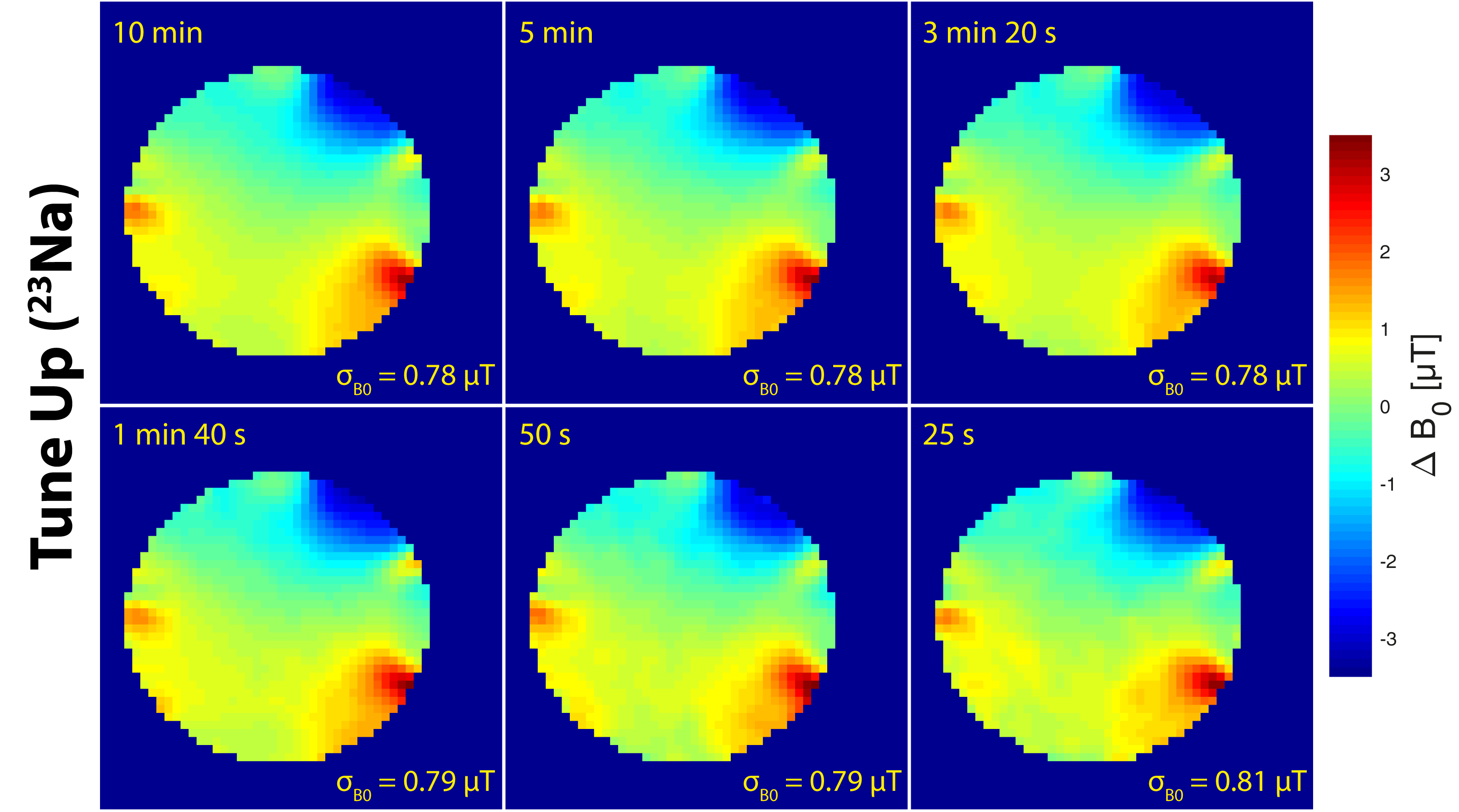
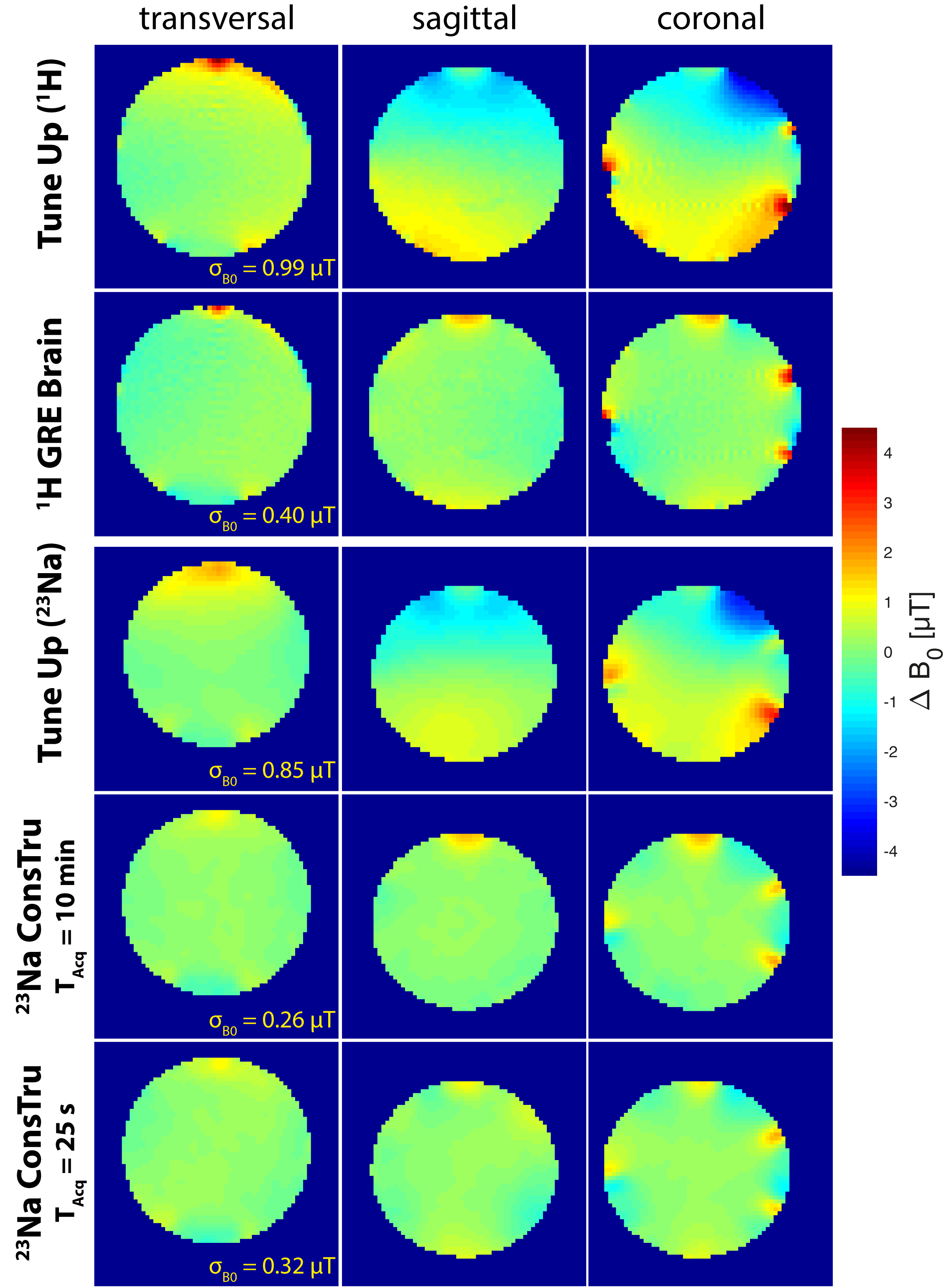
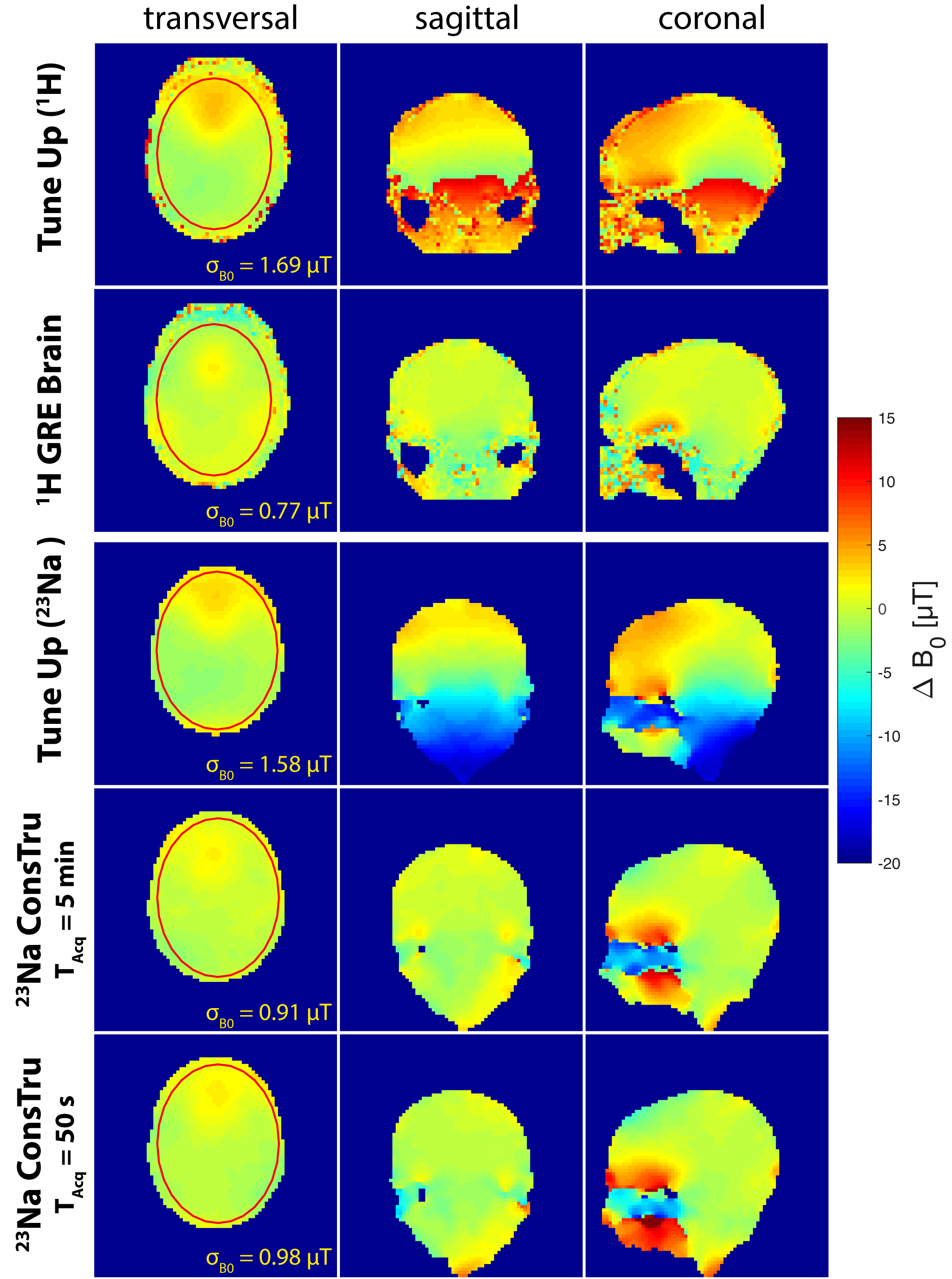
B0 maps of the spherical phantom acquired using 23Na MRI show similar quality for all acquisition times (10 min to 25 s; Figure 1). Even when acquiring B0 maps with strong radial undersampling, reasonable B0 maps can be reconstructed. 1H and 23Na B0 maps acquired using the Tune Up shim values, as well as shim values calculated using the 1H GRE Brain shim and the 23Na ConsTru shim algorithm can be found in Figure 2. For the 23Na shim, B0 maps with acquisition durations of 10 min and 25 s were used. B0 variations over the phantom were reduced from 0.99 µT to 0.40 µT using the vendor-provided 1H GRE brain shim. The 23Na MRI based ConsTru shim showed a similar performance (0.85 µT to 0.26 µT (TAcq = 10 min)/0.32 µT (TAcq = 25 s)). Results for the in vivo measurement are shown in Figure 3. Again, the 23Na ConsTru algorithm results in comparable homogeneity as the GRE Brain shim, even for the short acquisition time (50 s).Discussion and Conclusion
B0 maps acquired with 23Na imaging show a B0 variation similar to 1H B0 maps and can therefore be used for the calculation of shim values. With the proposed 23Na ConsTru shimming routine, an improvement of the B0 homogeneity comparable to the vendor-provided GRE Brain shim routine could be achieved. Even when reducing the acquisition time of the 23Na B0 maps to less than 1 minute, similar ΔB0 distributions and resulting shim values were found. These results are promising for future applications, where no 1H MRI data can be acquired (e.g. single-tuned X-nuclei RF coils).Acknowledgements
No acknowledgement found.References
- Gast LV, Gerhalter T, Hensel B, Uder M, Nagel AM. Double quantum filtered 23Na MRI with magic angle excitation of human skeletal muscle in the presence of B0 and B1 inhomogeneities. NMR in Biomed 2018;e4010.
- Tanase
C, Boada FE. Triple‐quantum‐filtered imaging of sodium in presence of B0
inhomogeneities. J Magn Reson 2005; 174(2):270‐278.
- Nagel AM, Laun FB, Weber MA, et al. Sodium MRI using a density-adapted 3D radial acquisition technique. Magn Reson Med 2009; 62:1565–1573.
- Maier F, Fuentes D, Weinberg JS, Hazle JD, Stafford RS. Robust Phase Unwrapping for MR Temperature Imaging Using a Magnitude-Sorted List, Multi-Clustering Algorithm. Magn Reson Med 2015; 73(4):1662–1668.
- Chang P, Nassirpour S, Henning A. Modeling Real Shim Fields for Very High Degree (and Order) B0 Shimming of the Human Brain at 9.4 T. Magn Reson Med 2018; 79:529-540.
- Nassirpour S, Chang P, Fillmer A, Henning A. A Comparison of Optimization Algorithms for Localized In Vivo B0 Shimming. Magn Reson Med 2018; 79:1145-1156.
Figures