4204
Ultrahigh Field (14.1 Tesla) 1H/23Na MRI of the Rat Tail In Vivo: A Comparison Study1Biomedical and Mechanical Engineering, The Pennsylvania State University, State College, PA, United States, 2State College High School, State College, PA, United States, 3Biomedical Engineering, The Pennsylvania State University, State College, PA, United States
Synopsis
This study verified the ability to acquire high resolution sodium and proton MR images of rat tail intervertebral discs in vivo. Utilizing the hardware developed in a previous study, female Fischer 344 rats were imaged in vivo. Resolutions of 78um isotropic and 500um isotropic were achieved for proton and sodium images respectively prior to zerofilling. Comparisons between in vivo and ex vivo images showed similar anatomical and biochemical results. However, slight differences in the sodium images were observed. Sodium concentration was estimated using blood in the ventral tail artery as a reference.
Introduction
Degenerative Disc Disease (DDD) is the gradual change of compositional, structural, and mechanical properties of the intervertebral disc that affects its function. One of the initial signs of DDD is a decrease in proteoglycan (PG) content. These large proteins are highly negatively charged and attract free cations such as sodium.1,2 Proteoglycan content also has a significant effect on T1, T1⍴, and T2. Therefore, quantitative MRI may be used to detect early stage disc degeneration.1-3 There are several limitations to quantitative proton MRI techniques such as T1⍴, T2, and gagCEST mapping, despite correlating well with proteoglycan content.1,3
A strong, linear correlation exists between sodium and PG concentration, meaning sodium MRI can potentially detect changes in composition before structural changes occur [1]. Ultrahigh field sodium MRI, paired with high resolution proton MRI, may provide a better alternative for evaluating structural and compositional disc changes in vivo for longitudinal studies [2]. Previous work demonstrated the feasibility of obtaining high resolution MRI images of intervertebral discs in ex vivo rat tails at 14.1T. This study utilizes the hardware and techniques developed previously to acquire in vivo rat tail images for comparison of structure and composition.
Methods
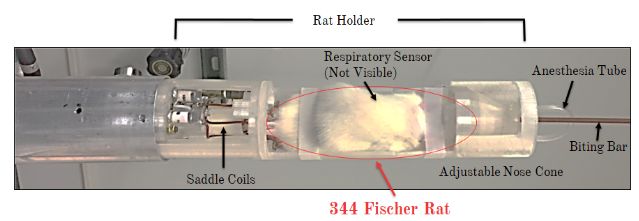
All imaging was conducted on a 14.1T micro imaging system utilizing the dual saddle coil MRI probe discussed in previous work. Due to the small bore size (5.5cm inner diameter of gradients), F344 female Fischer rats were chosen for in vivo imaging. An adjustable animal holder was designed in Solidworks (Dassault Systèmes, Waltham, MA) and 3D printed. This holder was compatible with the current hardware. An adjustable nose cone, biting bar, and respiratory monitor were integrated into the system.
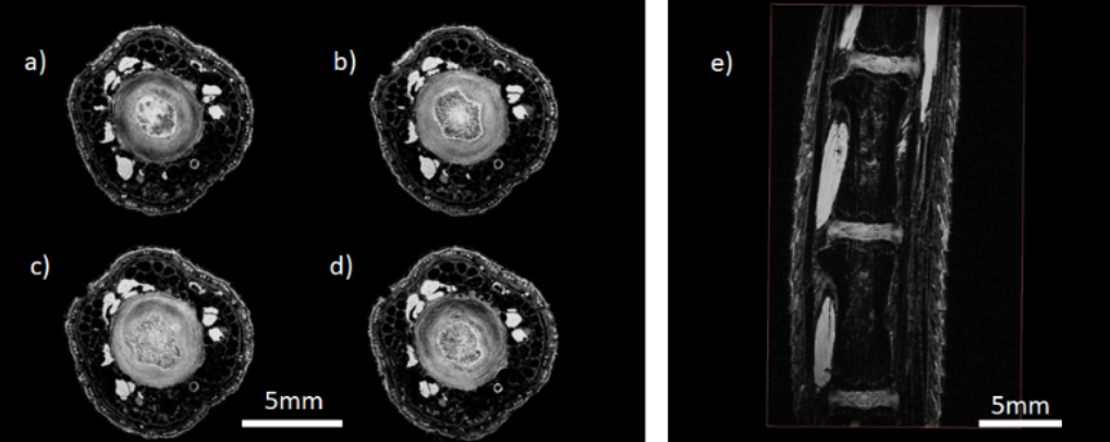
Rats were anesthetized using 1 - 3% isoflurane as prescribed in the approved IACUC protocol. T2-weighted proton images were first acquired for sodium localization and anatomy using a standard 3D gradient echo sequence (TE = 4.54ms, TR = 40.00ms, FOV: 20mm x 10mm x 10mm, matrix: 256 x 128 x 128, resolution = 78μm3, averages = 6, scan time = 1 hour 5 min). The resolution and number of averages were reduced in vivo to lower the scan time. Sodium images were then acquired using a modified 3D gradient echo sequence. A 70μs hard pulse with no slice select gradient was used to minimize the echo time (TE = 0.98ms, TR = 30.00ms, FOV: 32mm x 20mm x 20mm, matrix: 64 x 40 x 40, resolution = 500μm3, averages = 200 , scan time = 2 hours 40 min). A higher resolution was possible in vivo by optimizing the sequence parameters. Images were reconstructed in Matlab (The Mathworks Inc., Natick, MA).
Results
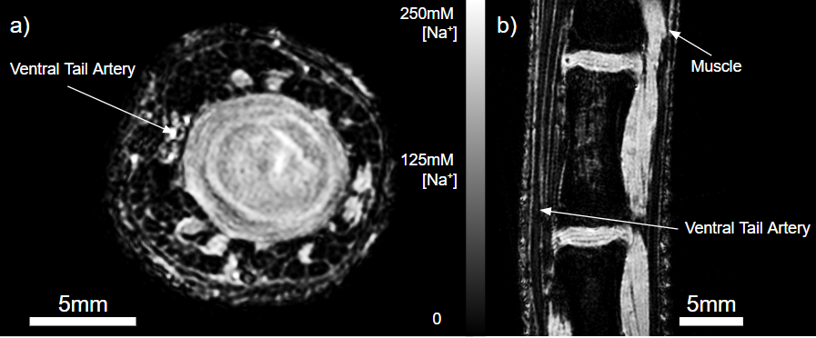
The in vivo proton images showed similar anatomy to the ex vivo images. Muscle bundles and the IVDs had high signal intensity. Skin layers, blood vessels, and bone were also visible in both cases with less signal. However, the lower resolution of the in vivo images resulted in loss of structure within the IVD. Laminae of the annulus fibrosus (AF) were resolved in both images but the collagen fibers of the nucleus pulposus (NP) not resolved in vivo.
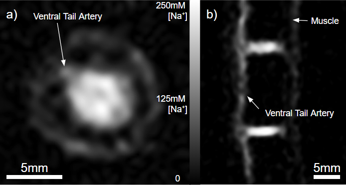
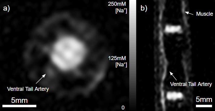
Several differences were observed between the in vivo and ex vivo sodium images. A sharper distinction between the AF and the NP was present in vivo with the NP having the highest signal intensity. Muscle had much lower signal in vivo than ex vivo. Furthermore, the ventral tail artery was visible in the in vivo images but not in the ex vivo images due to perfusion. Using the sodium concentration of blood in the tail artery as a reference, the relative sodium concentration in the disc could be determined. The sodium concentration was estimated to be about 200 mmol which is consistent with the expected concentration in vivo.
Discussion and Conclusion
Based on these results, we determined that in vivo sodium imaging of rat tail IVDs is feasible and could be an effective method for measuring the progression of disc degeneration. The in vivo sodium and proton images were very similar to the ex vivo images. However there were differences in signal intensity found in muscle which may be due to the lack of concentration gradient ex vivo. A decrease in signal was also observed at the center of the disc which may be due to a high concentration of notochordal cells. Before direct concentration can be extracted from the sodium images, B1 inhomogeneities must be accounted for. Future research will involve inducing disc degeneration in rats for a longitudinal study using the imaging techniques developed in this study.
Acknowledgements
Department of Mechanical and Nuclear Engineering, Penn State
The Huck Institutes of the Life Sciences, Penn State
Dr. Daniel H Cortes, PI, The Biomechanics and Imaging Lab, Penn State
References
[1] Borthakur, A., Melon, E., Niyogi, S., Witschey, W., Kneeland, J. B., & Reddy, R. (2006). Sodium and T1r MRI for molecular and diagnostic imaging of articular cartilage. NMR in Biomedicine, 19, 781–821. https://doi.org/10.1002/nbm
[2] Burstein, D., Bashir, A., & Gray, M. L. (2000). MRI techniques in early stages of cartilage disease. Investigative Radiology, 35(10), 622–638. https://doi.org/10.1097/00004424-200010000-00008
[3] Wang, C., McArdle, E., Witschey, W., Elliott, M., Reddy, R., & Borthakur, A. (2009). Validation of Sodium MRI of Intervertebral Disc Degeneration. Proceedings 17th Scientific Meeting, International Society for Magnetic Resonance in Medicine, 35(5), 294. https://doi.org/10.1097/BRS.0b013e3181b32d3b
Figures