4199
Neurostimulation Safety Investigations using Reference, Posable Anatomical Models1Foundation for Information Technology in Society, Zurich, Switzerland, 2Max Plank Institute, Tuebingen, Germany, 3Swiss Federal Institute of Technology (ETHZ), Zurich, Switzerland
Synopsis
Investigations about the risk of Peripheral Nerve Stimulation (PNS) in magnetic resonance imaging (MRI) are fundamental to mitigate potential safety issues related to the new generation of MRI scanners that use larger Larmor fields and faster/higher gradients. In this study we evaluate the potential of in silico studies for determining the thresholds of PNS when taking into account the anatomical detail and electrophysiology of the peripheral nervous system. We used the latest hybrid electromagnetic (EM) and neuronal simulators combined with our recently released neuro-functionalized Virtual Population model Yoon-sun V4.0. The vision is to utilize such systems for pulse sequence optimization to reduce PNS, the revision of low frequency exposure guidelines, etc..
Introduction
Current
advances in MRI-based techniques to reduce imaging time and increase contrast,
require higher Larmor field intensities (7 ‒ 10T) and faster gradient
switching and higher gradient intensities. Important projects, such as the
Human Brain Connectome1,
critically depend on such developments. These novel imaging techniques may pose safety issues related to the enhanced risk of radio frequency (RF) tissue
heating and unwanted peripheral (or cardiac) stimulation. In 2016, our group pioneered
research2 on the use of hybrid computational neuro-electrophysiology and
electromagnetics to quantify gradient-induced contexts stimulation performing coupled
EM and neuronal simulations centered on different computational human body
models inclusive of realistic nerve trajectories. While that study provided results
in qualitative and quantitative agreements with experiments about specificity
of thresholds to pulse sequences, gradient units, human BMI and position as
well as sites of spike initiation, shortcomings of the models (nerves were
modeled according to anatomy textbooks, not from patient/anatomy-specific
segmentations) limited the significance of our predictions. Here we present results
obtained with the first neuro-functionalized Virtual Population (ViP) model3,
“Yoon-sun V4.0”. The current work extends the work initiated in 2016 and provides
additional insights into both the effects of typical body postures in MRI (e.g.,
position of hands and arms) and the identification of fiber type (i.e., sensory
or motor fibers) specific stimulation thresholds.
Methods
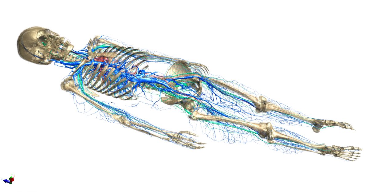
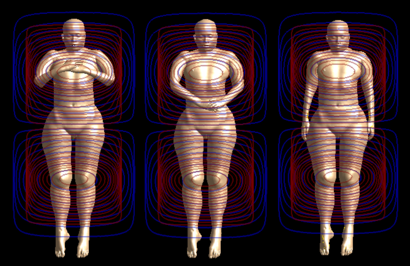
The neuro-functionalized phantom Yoon-sun V4.0 was created on the basis of high-resolution cryosection images from the Visible Korean Human project4 (Figure 1). All major peripheral nerves were segmented and spline nerve trajectories were automatically created by computing the centerlines from the surface models of the nerves. Three typical body postures were considered (Figure 2) with respect to arm positions as well as three different landmark positions of the whole body within the MRI scanner (head-, heart-, and pelvis-centered). The modelling pipeline was implemented in Sim4Life (ZMT Zurich MedTech AG, Switzerland). Nerve trajectories were assigned to electrophysiological models of myelinated sensory and motor axons (e.g., MRG5). EM simulations were executed using the Magneto-Quasistatic solver. Neurostimulation was investigated considering 3D EPI/ 3D Spiral waveforms typically used for brain imaging. Furthermore, the effect of gradient waveforms by individual and combined gradient units were included. Stimulation thresholds and site of spike initiation were identified for each nerve trajectory. Results are compared with experimental values and recently published computational work6.Results and Discussion
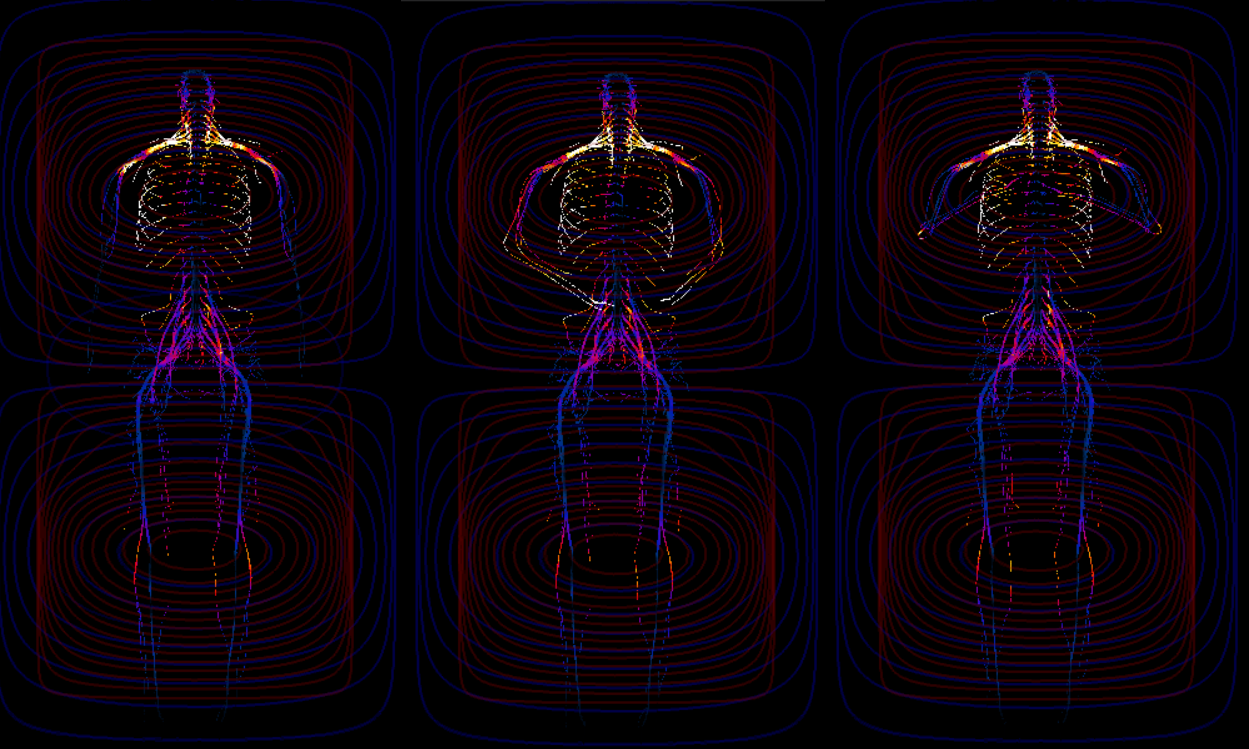
The developed workflow allows to analyses all the different permutations of coil units, postures and landmark positions with respect to the MRI scanner. Figure 3 illustrates an example of E-field exposure extracted at the nerve entities for the x-gradient coil unit. Positions of the hands and arms affect the local E-field exposure on nerves, especially in the region of the arms and hands and around the brachial plexus, where typically neurostimulation is experienced in experiments. Regions of large E-field variations are also observed in the different postures that can be related to potential sites of neurostimulation. Precise identification of sites of spike initiation and quantification of stimulation thresholds will be provided by the hybrid EM-neuronal simulations currently in execution.
Conclusions
The newly developed Yoon-sun V4.0 model permits quantitative predictions of the risk of PNS in MRI environments and arbitrary postures. After confirming the predictive power of hybrid EM-electrophysiological simulations with more experimental data, we foresee applications in 1) scanning guidelines (posture, position) to reduce unwanted stimulation, 2) pulse sequence optimization that can also include stimulation reduction as goal or constraint (in addition to image quality, scan time, etc.), and 3) should further results confirm mechanistic observations akin to those from [7], the low frequency exposure guidelines (e.g. ICNIRP, etc.) will require revision. The proposed modeling could be a valuable tool for that purpose.Acknowledgements
This project supported by the Swiss-Korean project NEUROMAN (CTI 25290.1 PFLS-LS).References
[1] Nowogrodzki A. The world's strongest MRI machines are pushing human imaging tonew limits. Nature. 2018 Nov;563(7729):24-26
[2] Cassara, A.M., Neufeld, E., Hagberg, G., Guidon, M., Scheffler, K. & Kuster, N. Peripheral Nerve Stimulation in MRI: Insights from a three level analysis and coupled EM-electrophysiological simulations in neuro-functionalized human models. In Proceedings of the 25th International Society for Magnetic Resonance in Medicine (ISMRM), Honolulu, USA, April 22-27, 2017
[3] Gosselin MC, Neufeld E, Moser H, Huber E, Farcito S, Gerber L, Jedensjö M, Hilber I, Di Gennaro F, Lloyd B, Cherubini E, Szczerba D, Kainz W, Kuster N. Development of a new generation of high-resolution anatomical models for medical device evaluation: the Virtual Population 3.0. Phys Med Biol. 2014 Sep 21;59(18):5287-303
[4] Park JS, Chung MS, Hwang SB et al. Visible Korean human: improved serially sectioned images of the entire body. IEEE Trans Med Imaging 2005, 24(3):352-60
[5] Gaines JL, Finn KE, Slopsema JP, Heyboer LA, Polasek KH. A model of motor and sensory axon activation in the median nerve using surface electrical stimulation. J Comput Neurosci. 2018 Aug;45(1):29-43
[6] Davids M, Guérin B, Malzacher M, Schad LR, Wald LL. PredictingMagnetostimulation Thresholds in the Peripheral Nervous System using Realistic Body Models. Sci Rep. 2017 Jul 13;7(1):5316
[7] Neufeld E, Cassará AM, Montanaro H, Kuster N, Kainz W. Functionalized anatomical models for EM-neuron Interaction modeling. Phys Med Biol. 2016 Jun 21;61(12):4390-401
Figures