4179
Planning Current Nulling for Worst Case SAR reduction in a Cardiac Guidewire Scenario at 1.5T1School of Biomedical Engineering & Imaging Sciences, King's College London, London, United Kingdom
Synopsis
Interventional MRI using long conductive guidewires, risks high Local SAR and reduced image quality. This work expands on previous work relating current ‘nulling’ techniques to reducing worst case SAR, by investigating the importance of wire placement and current sensing planning. Results showed that two very distinct wires displayed common features but differences in behaviour do not allow for a generic ‘nulling’ plan. Also, there is a trade-off between removing current-generating modes, and reducing B1+ efficiency that subsequently increases RF power to achieve necessary image quality. Overall, a careful study of the system can help find the optimal ‘nulling’ plan.
Introduction
Interventional MRI using conductive guidewires (hereafter called ‘wires’) risks high local SAR and poor image quality due to B1 enhancement. These originate from uncontrolled wire current. It has been demonstrated that currents can be minimised by ‘decoupling’ the wire from the transmit system using parallel transmit architecture.1,2 Such methods rely on independent current measurements, with physical ‘sensors’1 to make measurements independently of the MRI system. Sensor positioning affects how well this can work, as demonstrated previously.2
In this work EM simulations are used to further investigate the importance of sensor position and number while looking for common features between two distinct exemplar wire configurations.
Methods
Current ‘nulling’ starts by performing a singular value decomposition (SVD) on matrices built from current measurements; the singular vectors identify modes of the transmit coil that couple to the wire, and the singular values reflect their strength. Meanwhile, the maximum eigenvalue of a Q-matrix3,4 can be used to reflect worst case SAR (wcSAR) at its location. We used EM simulations (Sim4Life) to explore these two methods.
Two 8-channel Tx/Rx cardiac RF coils were simulated: one short; one long. Both on a model (Duke - ITIS ViP3.1), at 64MHz, with a simplistic or a realistic wire inside, resulting in four scenarios (Fig.1). The wires (core:∅ = 1mm; insulating layer: 0.1mm thickness) had their distal 5mm stripped of insulation. The Huygens Box method, implemented in Sim4Life, was applied, to reduce computation time. Analysis was performed in MATLAB.
Eigenvalue decomposition of a Q-matrix near the wiretip was used to determine wcSAR before ‘nulling’. J was sampled along the wire to simulate current sensor readings. The true coupling modes were identified using SVD of the entire current distribution. The SVD was repeated using one, two and three ‘sensor’ measurements of J, ranging from 100mm outside the body to the wiretip. For each sensing configuration, the Q-matrix was projected onto the sub-space defined by excluding the strongest detected coupling modes. Eigenvalue decomposition of this projected Q-matrix, near the wiretip, quantifies the remaining wcSAR after ‘nulling’. Coil efficiency must also be taken into account to determine wcSAR under useful imaging conditions. For that, a magnitude least squares B1+ shimming algorithm5 was applied to the heart, and all wcSAR values normalised to the square of the average shimmed B1 + in that region.
Results & Discussion
Fig.2 illustrates the currents associated with each mode: all scenarios have a minimum of 3 modes generating significant wire-currents. The vertical bar on each figure quantifies the wcSAR when modes are removed: the top value is wcSAR of the entire system, the next one is wcSAR without the highest current mode, etc, clearly showing that coupling modes contribute unequally. If true modes are identified, removing the degrees of freedom associated with the highest or, in some cases, two highest modes would reliably reduce wcSAR.
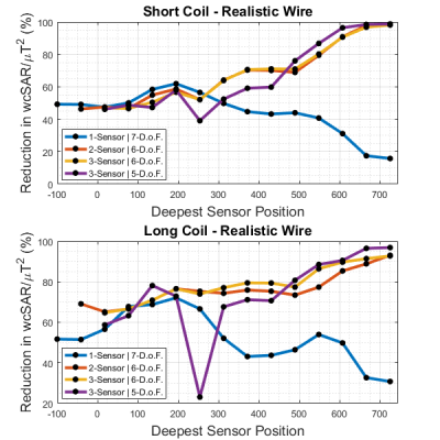
In reality, these modes are estimated from a few point-wise measurements, meaning the identified modes won’t be the true modes that lead to optimal ‘nulling’. Fig.3 and 4 show reduction in wcSAR as a function of number of ‘sensors’, their locations and the number of degrees of freedom remaining. Despite the dominance of a single mode seen in Fig.2, removal of one degree of freedom cannot reliably reduce wcSAR except when the ‘sensors’ are close to the wiretip. Removal of two degrees of freedom, reduces wcSAR while providing good B1+ efficiency, except for the short coil+simplistic wire scenario that only yields significant results when at least one ‘sensor’ is close to the wiretip. Finally, removing three degrees of freedom results in a reliable wcSAR reduction but it hinders B1+ efficiency, resulting in a comparable, sometimes worse outcome under imaging conditions. Again, the short coil+simplistic wire is an exception: in the regions where removing two degrees of freedom wasn’t enough, removing three works best.
Conclusion
We’ve expanded on previous quantification of wcSAR using a current measurement ‘nulling’ approach. It had been shown1 that at least two sensors were required to ‘null’ currents for a simplistic wire geometry. To proceed, a realistic wire was simulated to search for common features. This showed that increasing the number of ‘sensors’ helps in identifying modes of the system but removing degrees of freedom to mitigate wcSAR reduces B1+ efficiency. For all scenarios, but one, removing the two most problematic degrees of freedom yielded the best results. Taken together, the similarities and differences in the simulations studied show there might be reasonable latitude in planning current sensor placement for suppression of currents on guidewires, but the degree of geometry approximation that allows for such latitude would need to be assessed before a reliable nulling approach can be defined.Acknowledgements
JNT is funded by MRC CASE studentship [MR/P01674X/1]. This work was supported by the Wellcome EPSRC Centre for Medical Engineering at Kings College London [WT 203148/Z/16/Z], MRC strategic grant [MR/K006355/1], MRC development pathways funding scheme [MR/N027949/1], and by the National Institute for Health Research (NIHR) Biomedical Research Centre based at Guy’s and St Thomas’ NHS Foundation Trust and King’s College London. The views expressed are those of the authors and not necessarily those of the NHS, the NIHR or the Department of Health.References
1. Etezadi-Amoli M, Stang P, Kerr A, Pauly J, Scott G. Controlling radiofrequency-induced currents in guidewires using parallel transmit. Magn Reson Med. 2015;74(6):1790-1802. doi:10.1002/mrm.25543.
2. Teixeira J. N., et al. Exploring the impact of nulling currents on a cardiac guidewire in reducing worst case SAR at 1.5T. In Proceedings of the 26th Annual Meeting of ISMRM, Paris,France, 2018. Abstract 0299
3. Graesslin, I., Homann, H., Biederer, S., Börnert, P., Nehrke, K., Vernickel, P., Mens, G., Harvey, P. and Katscher, U. A specific absorption rate prediction concept for parallel transmission MR. Magn Reson in Med. 2012; 68(5):1664-1674. doi: 10.1002/mrm.24138
4. Eichfelder, G. and Gebhardt, M. Local specific absorption rate control for parallel transmission by virtual observation points. Magn Reson in Med. 2011; 66(5): 1468-1476. doi: 10.1002/mrm.22927
5. Setsompop K, Wald LL, Alagappan V, Gagoski BA, Adalsteinsson E. Magnitude least squares optimization for parallel radio frequency excitation design demonstrated at 7 Tesla with eight channels. Magn. Reson. Med. 2008; 59: 908–915.
Figures


