4166
Ultimate Intrinsic Specific Absorption Rate Efficiency1Electrical and Electronics Engineering, Bilkent University, Ankara, Turkey, 2National Magnetic Resonance Research Center (UMRAM), Ankara, Turkey, 3Center for Magnetic Resonance Research, University of Minnesota, Minneapolis, MN, United States, 4Industrial Engineering Department, Bilkent University, Ankara, Turkey
Synopsis
Peak local specific absorption rate (SAR) is a limiting factor for many high and ultrahigh field MRI applications. Therefore, improving the SAR performance of the transmitters is the scope of many studies. In this study, we used the SAR efficiency (B1+/sqrt(peak local SAR)) as a metric to evaluate the SAR performance of transmitters and investigated its upper bound, defined as Ultimate Intrinsic SAR Efficiency (
Introduction
Demand for high-field (HF, B0≥3T) MRI is continuously increasing due to its numerous benefits such as significant increase in the SNR1,2. However, peak local SAR becomes a limiting factor in many applications.3,4 Using transmit array (TxArray) coils with properly designed elements may provide a good solution to this issue.5,6
Various TxArray coil designs such as an 8-channel loop array at 3T,7 a 16-channel loop-dipole array at 7T,8 and a 10-channel fractionated dipole array at 10.5T9 were proposed to overcome the SAR issue. However, researchers are still seeking for new designs to improve the SAR performance of the transmitters and introduced the SAR-efficiency10 (B1+/sqrt(peak local SAR)) as a metric for this performance.
In this study, we utilize EM analytic relations to determine an upper bound for the SAR-efficiency inside a cylindrical homogeneous sample. We introduce this upper bound as the ultimate intrinsic SAR-efficiency (UISARE). For proof of calculations, we compared the results at 1.5T and 10.5T with the results achieved using a commercial EM-simulator.
Theory and Method
The SAR-efficiency at the point of interest (POI) ri can be defined as follows:
$$\xi =\frac{{{s^T}({r_i})\alpha}}{{\sqrt{{\alpha^H}R({r_p})\alpha}}}$$
where $$${\alpha}$$$ is a complex vector contains coefficients of the cylindrical harmonics. $$$s({r_i})$$$ contains H1+’s corresponding to each cylindrical harmonic with unit coefficient at the POI inside the cylindrical sample. $$$R({r_p})$$$ is the local SAR matrix11 at the position rp where the peak local SAR occurs.
To maximize the SAR-efficiency at the POI, the following optimization problem can be defined12:
$$\begin{array}{*{20}{c}} {{\rm{min}}}&{{\alpha^H}R({r_p})\alpha}&{}&{\alpha\in {C^{2MN\times1}}}\\ {}&{}&{}&{R\in{C^{2MN\times2MN}}}\\ {{\rm{s.t.}}}&{{s^T}({r_i})\alpha=1}&{}&{s\in{C^{2MN \times1}}} \end{array}$$
M and N are the numbers of the cylindrical harmonics in ϕ- and z-directions, respectively. The peak local SAR is defined as P0. Since R’s are positive semi-definite matrices, the minimization problem above is convex and can be solved for a global optimum. We applied the KKT conditions13 to obtain the optimal coefficients and peak local SAR as follows:
$$\begin{array}{l}\alpha=\frac{{{R^{-1}}({r_p}){s^*}({r_i})}}{{{s^T}({r_i}){R^{ -1}}({r_p}){s^*}({r_i})}}\\{P_0}=\frac{{{s^T}({r_i}){R^{-1}}({r_p}){s^*}({r_i})}}{{{{\left|{{s^T}({r_i}){R^{-1}}({r_p}){s^*}({r_i})} \right|}^2}}}\end{array}$$
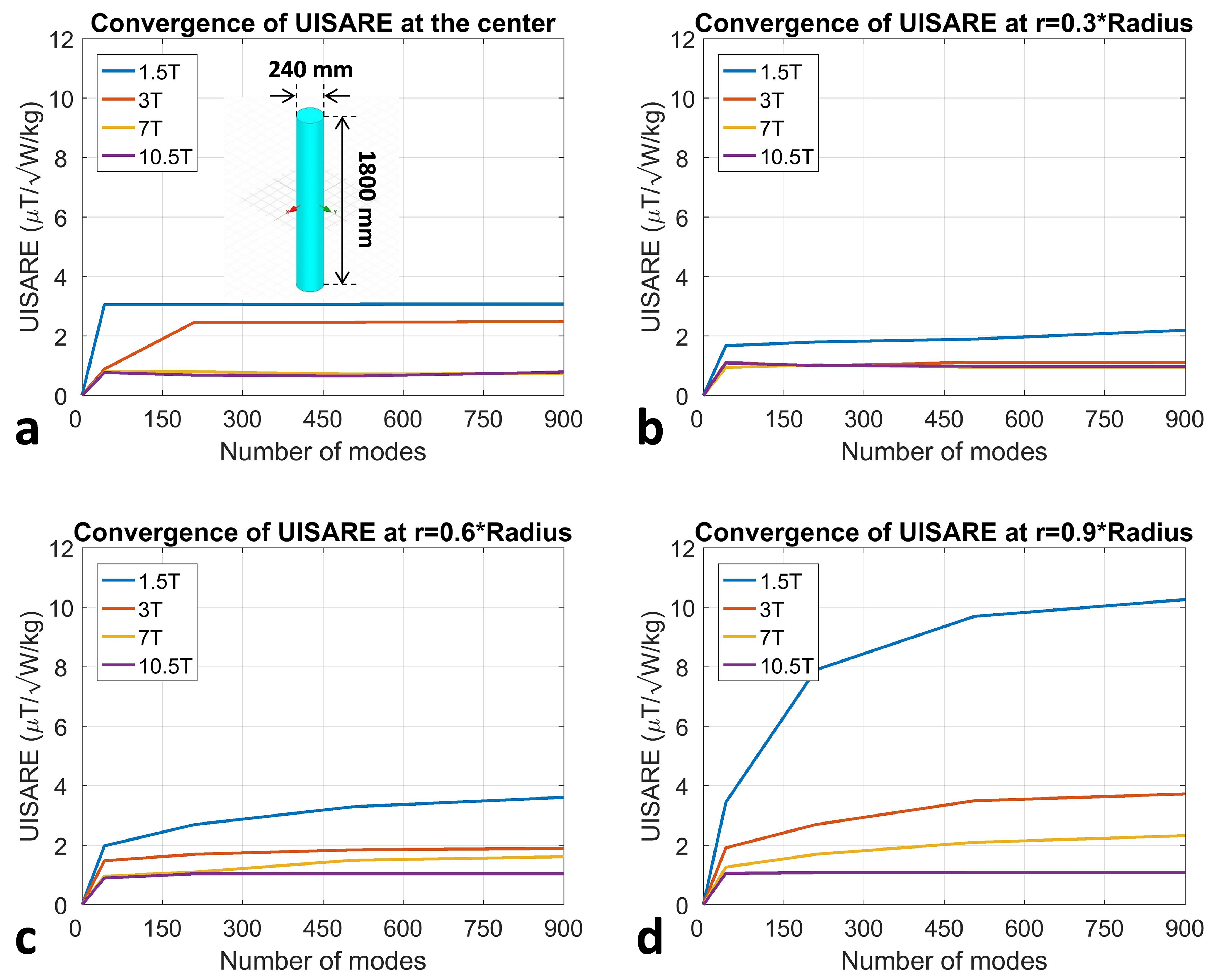
In this work, a uniform cylindrical sample electrical properties of εr=80 and σ=0.6S/m was used for calculations. We investigated how many cylindrical modes would be needed to reach the UISARE for different B0 field-strengths ranging from 1.5T to 10.5T. Further, at each field strength, UISARE was calculated for different POIs inside the sample.
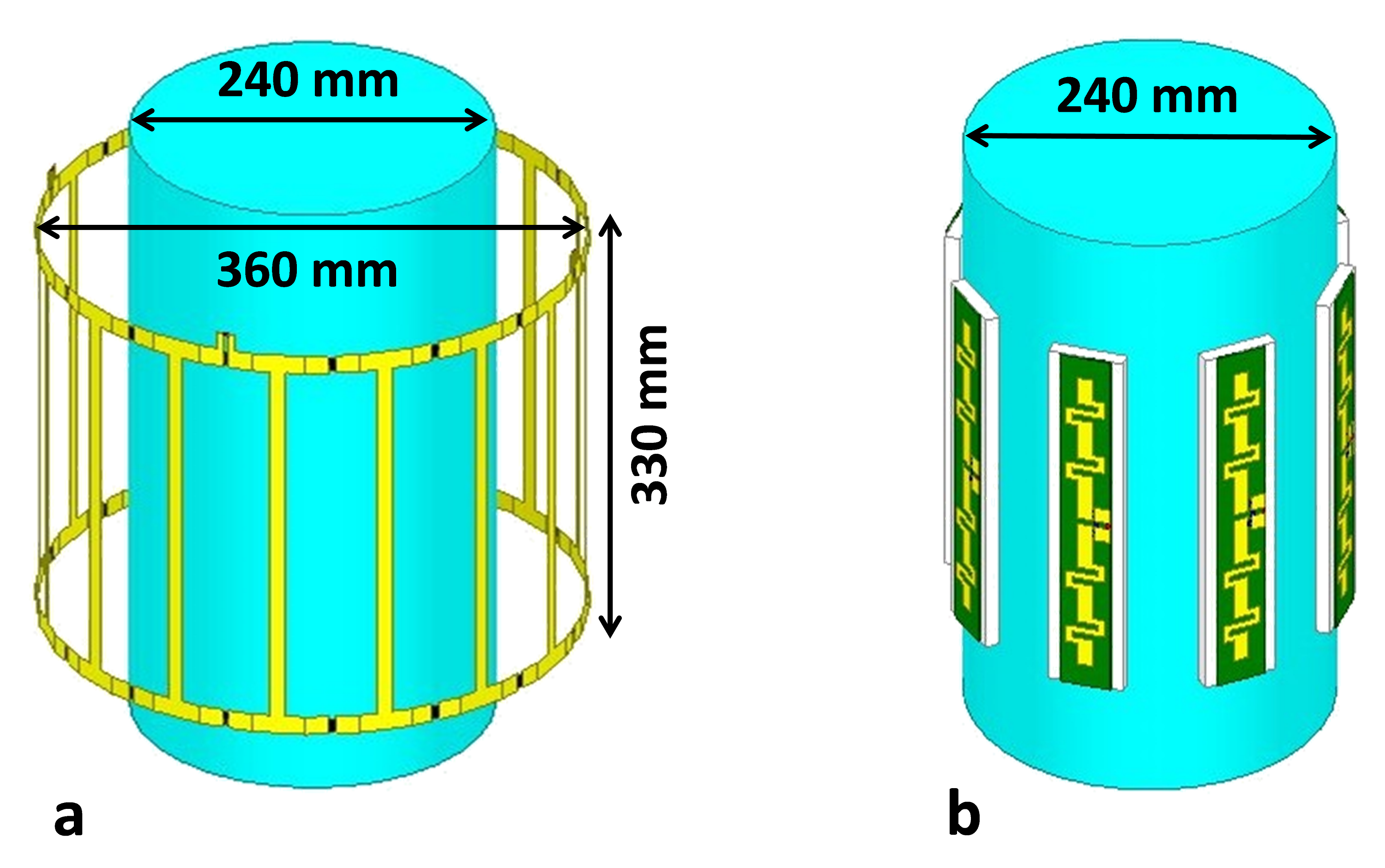
For proof of the calculations, the-EM simulation of a quadrature-excited birdcage coil (Fig.1a) was performed using an EM-simulator (HFSS) at 1.5T and the result was compared to the UISARE estimated for the center of the sample.
Eventually, the UISARE at the center of the sample at 10.5T was compared to the SAR-efficiency achieved using an 8-channel TxArray coil of fractionated dipoles (Fig.1b). Note that, this array was driven by an RF shimming solution calculated with virtual observation points14 to maximize SAR-efficiency at the origin.
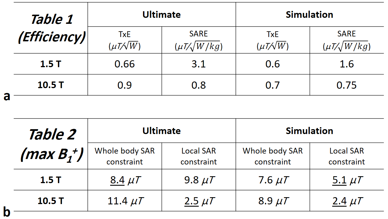
For both 1.5T and 10.5T cases, to determine the limiting factor of the excitation, UISARE at the center was compared to the ultimate intrinsic transmit efficiency15-17 (UITXE) as well. Furthermore, the 1g-averaging was chosen as the local-averaging to reduce the calculation time.
Results
Fig.2 shows the number of cylindrical modes that is necessary for convergence of the UISARE in different field strengths for different POIs on the central transverse plane.
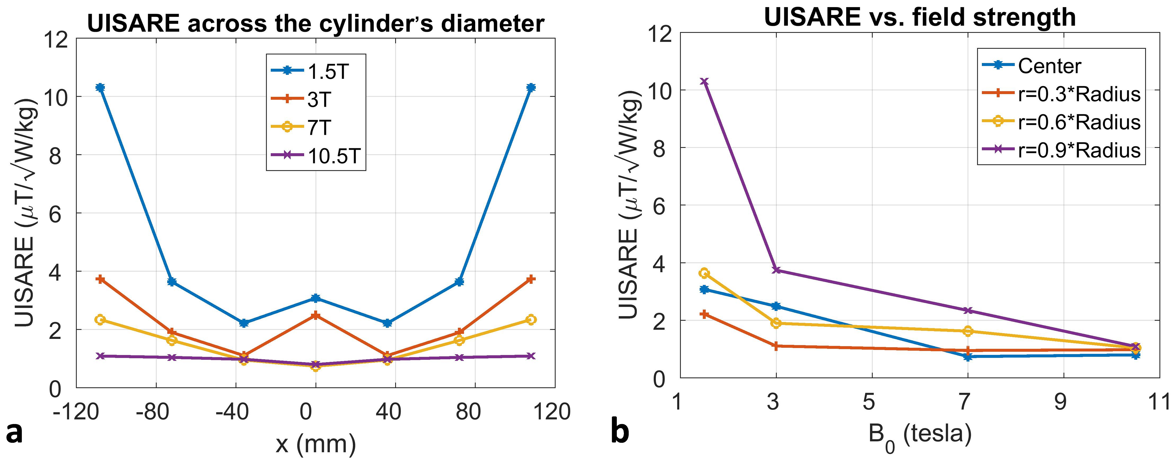
Utilizing adequately large number of modes, Fig.3a shows the UISARE at different POIs on the x-axis for each of B0 values, separately. Correspondingly, Fig.3b shows the UISARE versus B0 values at different POIs.
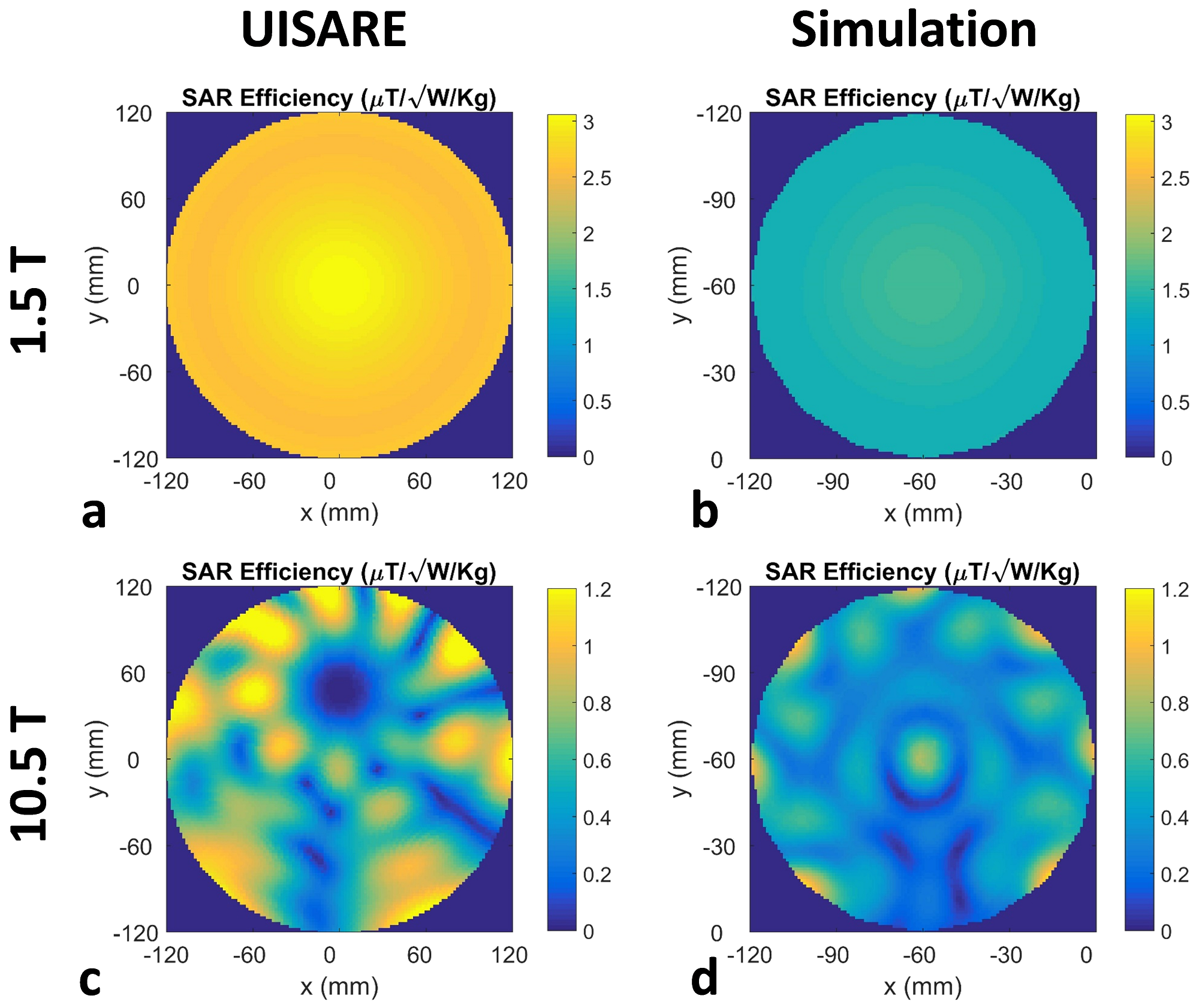
Fig.4 shows the SAR efficiency maps in 1.5T and 10.5T on a central transverse plane obtained by using the UISARE at the origin and performing EM simulations on the structures of Fig.1.
Considering 2 W/kg and 10 W/kg limits18 for the whole body and local SAR, respectively, Fig.5a shows that the ultimate whole body SAR16,17 and local SAR restrict the maximum B1+ at the center at 1.5T and 10.5T, respectively. However, for the simulated birdcage coil, local SAR is the limiting factor. Fig.5b shows the upper bounds of B1+ that can be excited at the center of the 80kg cylindrical uniform sample with consideration of corresponding SAR limits in each case.
Discussion and Conclusion
In this study, we considered the SAR-efficiency as a metric for SAR performance of the transmitters, and calculated the UISARE as an upper bound for this metric. Results show that UISARE can almost be achieved in UHF (B0≥7T) using the recently used TxArray coils with proper shimming strategy. However, at a lower field strength (B0≤3T) the upper bound is significantly higher than the SAR efficiencies achievable with the commonly used birdcage coils. On the other hand, the results show that the limiting cases at 1.5T and 10.5T are whole body and local SAR constraint, respectively.
Our future work will be to characterize the ideal current patterns17 for UISARE and study the UISARE in realistic body models19.
Acknowledgements
X.W. was supported by NIH grants U01 EB025144 and P41 EB015894 (PI K. Ugurbil).References
1. Edelstein WA, Glover GH, Hardy CJ, Redington RW. The intrinsic signal‐to‐noise ratio in NMR imaging. Magnetic resonance in medicine. 1986 Aug 1;3(4):604-18.
2. Ocali O, Atalar E. Ultimate intrinsic signal‐to‐noise ratio in MRI. Magnetic resonance in medicine. 1998 Mar;39(3):462-73.
3. Hoult DI. Sensitivity and power deposition in a high‐field imaging experiment. Journal of Magnetic Resonance Imaging. 2000 Jul;12(1):46-67.
4. Collins CM, Smith MB. Calculations of B1 distribution, SNR, and SAR for a surface coil adjacent to an anatomically‐accurate human body model. Magnetic Resonance in Medicine: An Official Journal of the International Society for Magnetic Resonance in Medicine. 2001 Apr;45(4):692-9.
5. Adriany G, Van de Moortele PF, Wiesinger F, Moeller S, Strupp JP, Andersen P, Snyder C, Zhang X, Chen W, Pruessmann KP, Boesiger P. Transmit and receive transmission line arrays for 7 Tesla parallel imaging. Magnetic Resonance in Medicine: An Official Journal of the International Society for Magnetic Resonance in Medicine. 2005 Feb;53(2):434-45. 6. Metzger GJ, Snyder C, Akgun C, Vaughan T, Ugurbil K, Van de Moortele PF. Local B1+ shimming for prostate imaging with transceiver arrays at 7T based on subject‐dependent transmit phase measurements. Magnetic Resonance in Medicine: An Official Journal of the International Society for Magnetic Resonance in Medicine. 2008 Feb;59(2):396-409.
7. Alagappan V, Wiggins GC, Potthast A, Setsompop K, Adalsteinsson E, Wald LL. An 8 channel transmit coil for transmit SENSE at 3T. InProceedings of the 14th Annual Meeting of ISMRM, Seattle, WA, USA 2006.
8. Ertürk MA, Raaijmakers AJ, Adriany G, Uğurbil K, Metzger GJ. A 16‐channel combined loop‐dipole transceiver array for 7 T esla body MRI. Magnetic resonance in medicine. 2017 Feb;77(2):884-94.
9. Ertürk MA, Wu X, Eryaman Y, Van de Moortele PF, Auerbach EJ, Lagore RL, DelaBarre L, Vaughan JT, Uğurbil K, Adriany G, Metzger GJ. Toward imaging the body at 10.5 tesla. Magnetic resonance in medicine. 2017 Jan;77(1):434-43.
10. Raaijmakers AJ, Italiaander M, Voogt IJ, Luijten PR, Hoogduin JM, Klomp DW, van Den Berg CA. The fractionated dipole antenna: A new antenna for body imaging at 7 T esla. Magnetic resonance in medicine. 2016 Mar;75(3):1366-74.
11. Graesslin I, Homann H, Biederer S, Börnert P, Nehrke K, Vernickel P, Mens G, Harvey P, Katscher U. A specific absorption rate prediction concept for parallel transmission MR. Magnetic resonance in medicine. 2012 Nov;68(5):1664-74.
12. Eryaman Y, Guerin B, Adalsteinsson A, Wald LL. The ultimate local SAR in MRI. In Proceeding of the 21st Annual Meeting ISMRM, Salt Lake City, Utah, USA. 2013 April.
13. Boyd S, Vandenberghe L. Convex optimization. Cambridge university press; 2004 Mar 25.
14. Eichfelder G, Gebhardt M. Local specific absorption rate control for parallel transmission by virtual observation points. Magnetic Resonance in Medicine. 2011 Nov;66(5):1468-76.
15. Georgakis I, Polimeridis AG, Lattanzi R. Ultimate intrinsic transmit efficiency for RF shimming. In Proceeding of the 26th Joint Annual Meeting ISMRM-ESMRMB, Paris, France. 2018 June.
16. Kopanoglu E, Erturk VB, Atalar E. Analytic expressions for the ultimate intrinsic signal‐to‐noise ratio and ultimate intrinsic specific absorption rate in MRI. Magnetic resonance in medicine. 2011 Sep;66(3):846-58.
17. Lattanzi R, Sodickson DK. Ideal current patterns yielding optimal signal‐to‐noise ratio and specific absorption rate in magnetic resonance imaging: computational methods and physical insights. Magnetic resonance in medicine. 2012 Jul;68(1):286-304.
18. International Electrotechnical Commission. International standard, medical equipment—IEC 60601-2-33: particular requirements for the safety of magnetic resonance equipment for medical diagnosis.
19. Guérin, B., Villena, J.F., Polimeridis, A.G., Adalsteinsson, E., Daniel, L., White, J.K., Wald, L.L., 2017. The ultimate signal-to-noise ratio in realistic body models. Magnetic Resonance in Medicine 78, 1969-1980.
Figures