4146
A Technique for Measuring Long-Term Alterations in Pulmonary Gas Uptake under Free-Breathing in a Murine Model of Radiation-Induced Lung Injury1Radiology, University of Pennsylvania, Philadelphia, PA, United States
Synopsis
Computed tomography (CT), pulmonary function tests (PFT) and positron-emission tomography (PET) are all currently used to assess lung structure and function clinically. However, the sensitivity and safety of these modalities limit their use in cases of progressive lung disease, where longitudinal measurements are of value for both therapeutic planning and monitoring response to treatment. Hyperpolarized 129Xe (HXe) MRI provides robust, specific information about the lungs and is suitable for longitudinal measurements. In this study, we demonstrate an HXe MRI technique for detecting and assessing alterations in pulmonary gas uptake in a mouse model of RILI under spontaneous respiration.
Introduction
Pulmonary function tests (PFTs) have been the standard for longitudinal assessments of lung function in patients with progressive lung diseases such as chronic obstructive pulmonary disease (COPD), after lung transplantation surgery, or after radiation therapy (RT) in cases of thoracic malignancies. However, while safe, PFTs cannot localize or specify the underlying cause of functional decline1. Hyperpolarized 129Xe (HXe) MRI provides both structural and functional information about the lung with a higher sensitivity and specificity than PFTs and without imparting ionizing radiation, making it an ideal modality for longitudinal assessments of lung function. Preclinical and clinical studies have already demonstrated HXe MRI’s utility for detecting and assessing progressive lung diseases such as radiation-induced lung injury (RILI)2,3 and COPD4, but small animal studies are typically cross-sectional rather than longitudinal because of the difficulty of maintaining proper ventilation under anesthesia and the consequent necessity of a terminal tracheostomy or intubation procedure. Here, we demonstrate a technique for measuring alterations in pulmonary gas uptake longitudinally in a free-breathing mouse model of lung cancer and RILI. Alveolar septal wall thickening was evaluated using the HXe MRI dissolved-to-gas ratio5, which increased dramatically during the course of the study.Methods
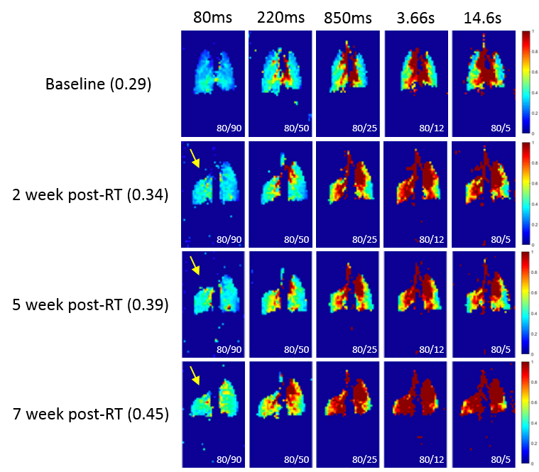
Genetically modified C57BL/6 mice were induced with lung cancer (n = 3) as previously described6, and cancer onset was monitored via weekly imaging. Mice were full-thorax irradiated at 15 Gy using a Small Animal Radiation Research Platform (SARRP) once cancer onset was detected, and were incubated until imaging commenced. Mice were anesthetized using 1-2% isoflurane, secured in an animal cradle equipped with a nose cone possessing two gas delivery and one gas exhaust port, and placed within a dual-tuned 1H/129Xe coil. Imaging was performed using a 9.4T vertical-bore micro-imaging MRI system (Bruker Inc.). Enriched 129Xe was polarized using a prototype commercial optical pumping system (XeBox-E10 Xemed, LLC, Durham, NH), stored in a tedlar bag, and held within a sealed chamber. A normoxic mixture of O2/air/isoflurane/129Xe was supplied through the nose cone at an overall rate of 120 ml/min (20ml/min for 129Xe) during imaging, which lasted approximately 35 min. Gas- and dissolved-phase image sets were acquired using a custom phase-encode/flip-angle table in conjunction with a chemical shift imaging (CSI) pulse sequence (TR/TE = 80ms/0.5ms, FA = 6°, 12°, 25°, 50°, 90°, FOV = 35x20mm2, matrix size = 64x36, projection). Images were divided into eight groups of k-space points and acquired in an interleaved manner, with oversampling near k = 0 to mitigate motion artifacts and to ensure that animals’ different breathing states were equally represented in each image. Each TR/FA combination was converted to an ‘effective T1RF’ for respective images to approximate the destruction of HXe gas signal solely due to RF-induced relaxation7. A mask was applied to gas-phase images prior to quantifying the dissolved-to-gas ratio, and regions outside the lung were excluded.Results
Figure 1 shows representative dissolved-to-gas ratio maps for images acquired at five distinct TR/FA combinations that correspond to distinct T1RF values at 2, 5 and 7 weeks post-irradiation. Mean dissolved-to-gas ratios (left, parenthesis) were calculated from images acquired with a T1RF of 80ms in order to highlight septal tissue signal contributions and avoid signal contamination by vascular blood.Discussion
The observed increase in dissolved-to-gas ratio over the period studied suggests additional dissolved 129Xe in the septal tissue compartment, likely due to increased septal wall thickness resulting from the onset of radiation-induced pneumonitis and/or fibrosis. An increase in the dissolved-to-gas ratio for longer T1RF values is also seen across timepoints, suggesting either a decline in diffusion rate, significantly longer uptake time of xenon into septal and parenchymal tissues, and/or increased volume in the irradiated lung. However, given that mice present a single dissolved-phase peak corresponding to both HXe in parenchymal tissue and red blood cells, further work is needed to elucidate the compartments HXe dissolves into.Conclusion
We successfully demonstrated a technique to detect long-term alterations in lung microstructure post-irradiation using HXe MRI by measuring the dissolved-to-gas ratio in a free-breathing mouse model of lung cancer and RILI. The trend toward increased dissolved gas signal after irradiation is consistent with previous demonstrations in a rat model.Acknowledgements
The transgenic mouse model and cell lines used were kindly provided by Dr. Diane Lim from the Department of Sleep Medicine at the University of Pennsylvania located in Philadelphia, PA, United States.References
[1] Smith, L., et al. American Journal of Respiratory and Critical Care Medicine, 2018:197(3):397
[2] Fox, M. S., et al. Medical Physics, 2014:41(7):072302
[3] Zanette, B., et al. Advances in Radiation Oncology. 2017:2(3):475
[4] Qing, K., et al. NMR in Biomedicine. 2014:27(12):1490
[5] Driehuys, B., et al. PNAS, 103(48):18278
[6] Sheen,M. R., et al. Open Life Sciences 10:854
[7] Ruppert, K., et al. Magnetic Resonance in Medicine, 10.1002/mrm.27538
Figures