4135
Accelerated Spiral-IDEAL for Gas and Dissolved Phase Hyperpolarized 129Xe in the Lungs of Healthy Human Volunteers: Effect of k-space Calibration Data on Image Quality and Gas Exchange Analysis1Translational Medicine, The Hospital for Sick Children, Toronto, ON, Canada, 2Medical Biophysics, University of Toronto, Toronto, ON, Canada
Synopsis
Parallel imaging with multi-channel receivers provides flexible acceleration of scan duration and is an important tool in both clinical and research settings. The application of parallel imaging to hyperpolarized 129Xe MRI may be useful in improving the clinical utility of parametric gas exchange mapping by reducing breath-hold durations or allowing for the acquisition of additional temporal/spatial information. In this work, we investigate the effect of k-space calibration data on parallel imaging reconstruction of time-resolved measurements of hyperpolarized 129Xe uptake using spiral-IDEAL and quantify resultant changes in the estimation of lung physiology.
Introduction
The solubility of xenon in biological tissues such as the lung parenchyma/blood plasma (T/P) and red blood cells (RBCs) and compartmentally-dependent changes in chemical shift allow for the investigation of gas exchange dysfunction using hyperpolarized (HP) 129Xe MRI1. Time-resolved measurements of 129Xe uptake may be used to model important alveolar structure/physiology, however this has typically been limited to global spectroscopic analysis due to the inherent challenges associated with the low signal of dissolved 129Xe2–5. Recently, methods employing time-resolved imaging of dissolved 129Xe uptake have been explored6–9, including approaches such as spiral-IDEAL10,11. Unfortunately, repeated imaging weighted to the uptake of 129Xe typically requires lengthy acquisitions which are of particular concern due to clinical breath-hold constraints (<16s). Acceleration with parallel imaging may be useful in addressing these concerns for clinical application9. In this study we explore the effect of k-space calibration data on accelerated image quality and associated quantitative metrics extracted from these images using compartmental modeling of gas exchange.Methods
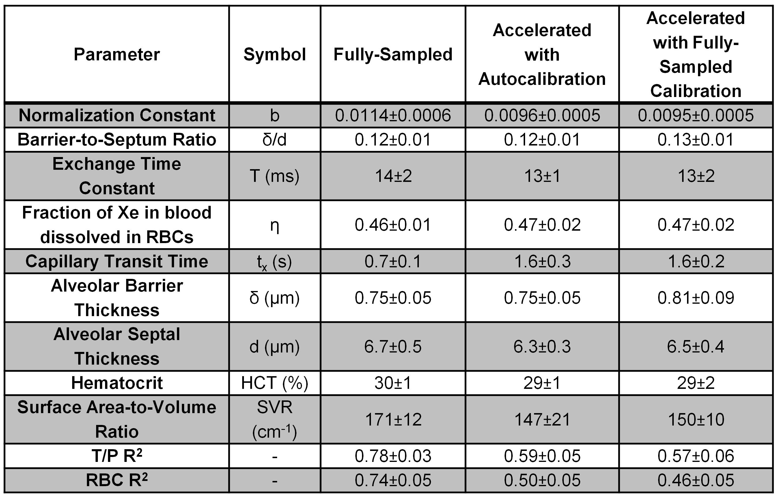
For this proof-of-concept study, a healthy adult volunteer was imaged with approval of the Research Ethics Board at The Hospital for Sick Children. Experiments were performed on a clinical 3T scanner (Skyra, Siemens GmbH, Erlangen, Germany) using an elliptical birdcage transmit coil with a flexible 8-channel receive array (Rapid Biomedical, Rimpar, Germany). Subjects were imaged with a custom spiral-IDEAL sequence for gas exchange-weighting with five gas exchange delay times (15ms, 25ms, 50ms, 100ms, 200ms) as previously described9 (FOV=48×48cm2, matrix=32×32, BW=100kHz, Nshot=6, ΔTE=230μs, Tacq=11s). Enriched (~86%) 129Xe was polarized to ~10% (Model 9800, Polarean, Durham, NC). For acceleration, the sequence was modified to prospectively remove every second interleaf shortening the acquisition to 6s and imaged with a separate dose of 129Xe. Analysis was performed in MATLAB (MathWorks, Natick, MA). Spectrally-resolved images were processed and normalized as previously described9. The Model of Xenon Exchange (MOXE)2 was used to model xenon uptake on parametric basis yielding gas exchange parameter maps as described previously9,12. Accelerated data were reconstructed using the iterative self-consistent parallel imaging reconstruction for arbitrary k-space (SPIRiT)13. A 30×30 region of gas-phase k-space was used for calibration. Calibration data from both the fully-sampled and undersampled gas images were used to compare the effect of calibration on SPIRiT reconstruction and subsequent MOXE parameters. The SPIRiT kernel size was 7×7 for image-space reconstruction.Results
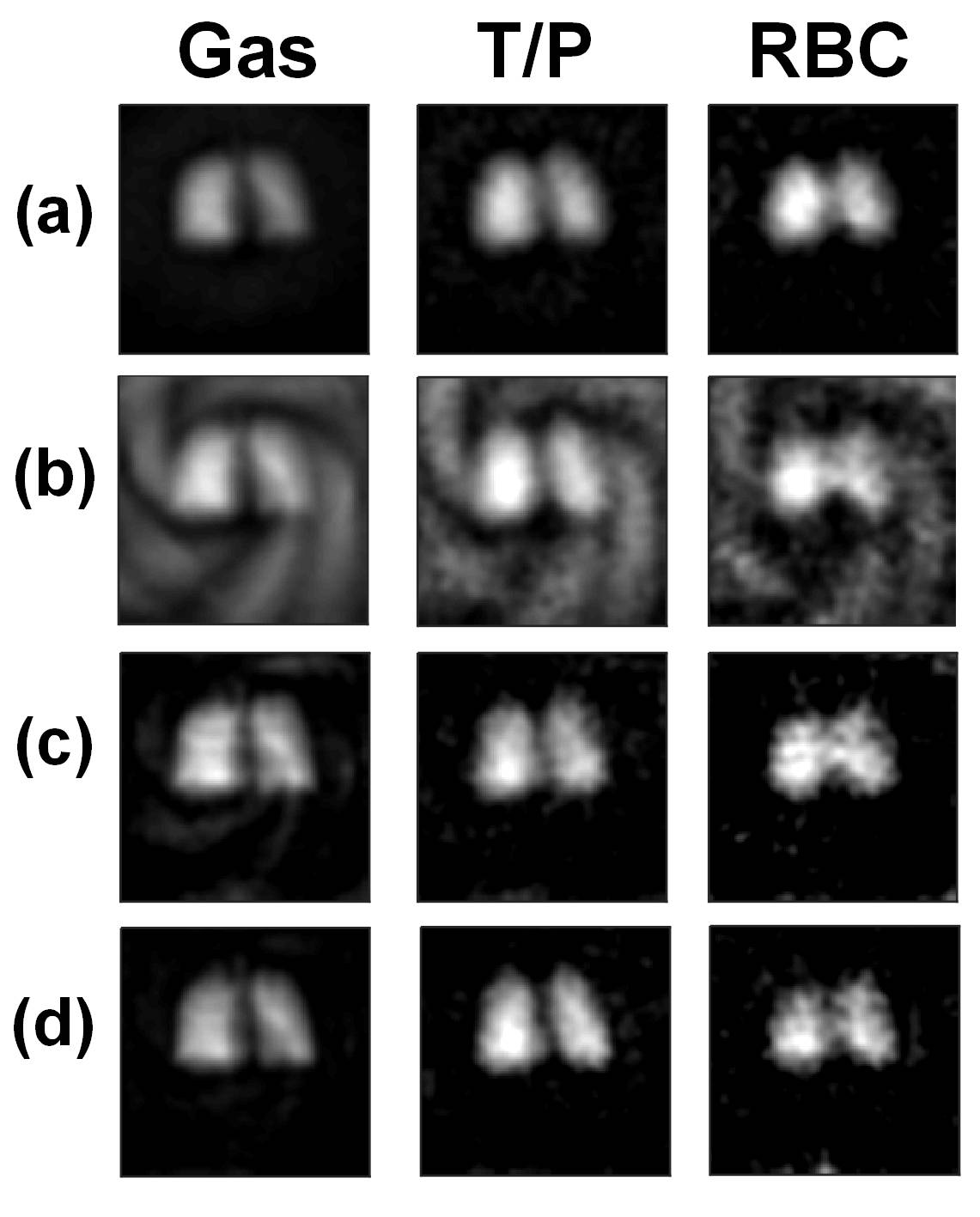
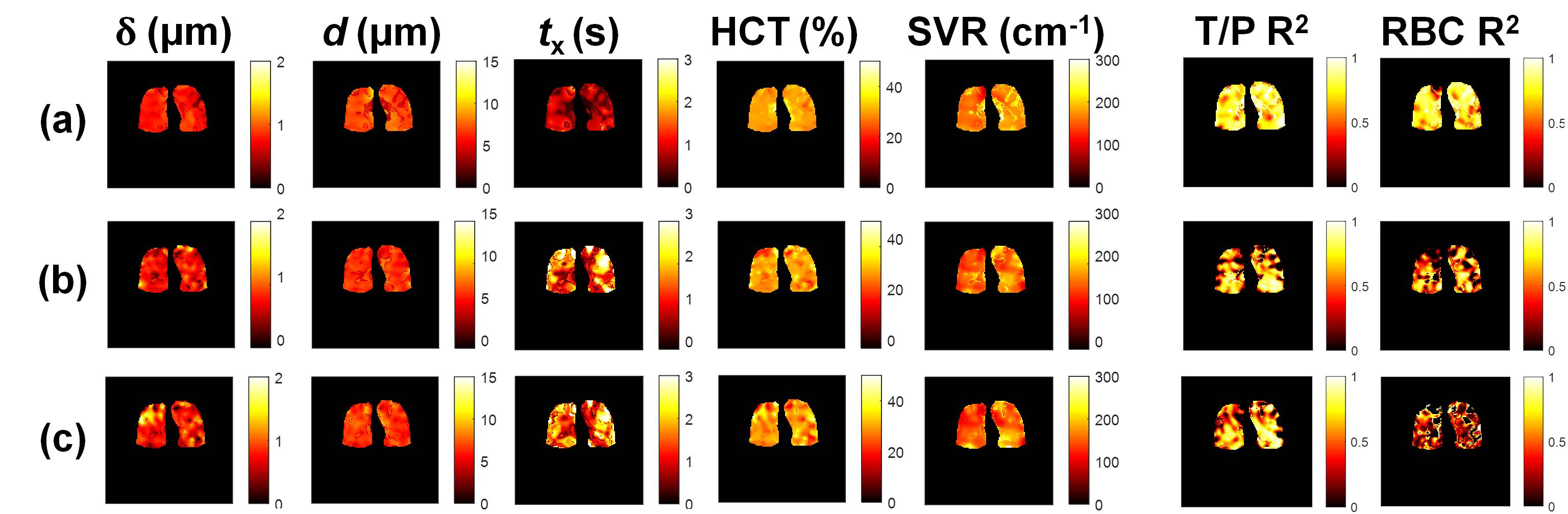
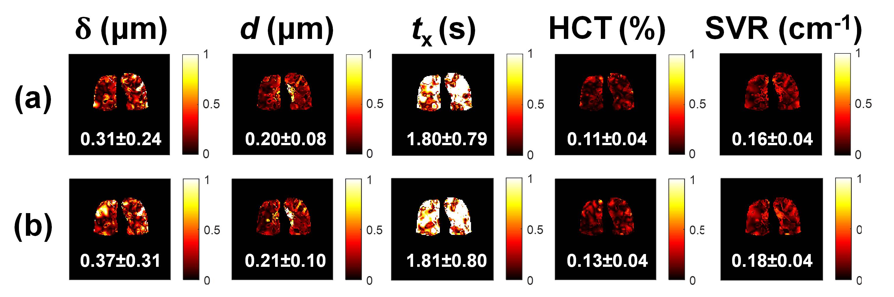
Fig.1 shows reconstructed spiral-IDEAL images. Image-wide physiological estimates extracted using MOXE are shown in Table 1. Parametric maps of MOXE parameters are shown in Fig. 2. T/P and RBC R2 are observed to be reduced after acceleration using both sets of calibration data, leading to large relative differences compared to full-sampled MOXE maps (Fig. 3).Discussion
This study demonstrates the feasibility of accelerating time-resolved spiral-IDEAL analysis of gas exchange for parametric mapping of important lung physiology. However, accelerated MOXE maps with autocalibration exhibit reduced R2 and somewhat large relative differences compared to fully-sampled data. This is presumably caused by the reduced SNR due to undersampling. In this work we explored the effect of using more robust calibration data from the centre of the fully-sampled data set to aid in the parallel imaging reconstruction. At this time, we observed a modest improvement in spiral-IDEAL image quality (Fig. 1d), although the inclusion of calibration data from the fully-sampled data did not appreciably change the MOXE parameter estimation compared to undersampled autocalibration data. It is possible that with the current modest achievable signal intensities and limited data acquired per image, the SPIRiT reconstruction only yields minimal improvements with more robust calibration data. Therefore perhaps 2× undersampling for this particular sequence design is pushing SPIRiT to the limit of recoverable image quality. With further optimizations, such as increased polarization or a decreased undersampling factor, the relative differences between fully-sampled and accelerated data should be reduced. In future, reduced scan durations provided by acceleration may be useful for patients having difficulty complying with breath-hold constraints. Alternatively, the reduced scan time (and HP magnetization depletion) may be traded to acquire more data (i.e. additional gas exchange timepoints or volumetric information). Nevertheless, more testing in an increased number of participants is required to fully understand the effect of acceleration on gas exchange modelling.Conclusion
Application of fully-calibrated parallel imaging reconstruction to accelerate time-resolved MRI of dissolved HP 129Xe uptake is feasible and yielding modest improvements in image SNR. Fully calibration does not appear to change quantitative gas exchange metrics extracted from spiral-IDEAL data, compared to autocalibration. This approach has potential moving forward to reduce the acquisition times of gas exchange mapping, thereby improving clinical utility.Acknowledgements
Work supported by NSERC (RGPIN 217015-2013) and CIHR (MOP 123431). Special thanks to Nikhil Kanhere, Elaine Stirrat, Marcus Couch, Andras Lindenmaier, Yonni Friedlander, Rosie Lye, and Manoj Singh for assistance with imaging experiments. B.Z supported by a Research Training Competition (RESTRACOMP) award from The Hospital for Sick Children.References
1. K. Ruppert, J.R. Brookeman, K.D. Hagspiel, B. Driehuys, and J.P. Mugler, “NMR of hyperpolarized 129Xe in the canine chest: Spectral dynamics during a breath-hold,” NMR Biomed. 13(4), 220–228 (2000).
2. Y. V Chang, “MOXE: A model of gas exchange for hyperpolarized 129Xe magnetic resonance of the lung,” Magn. Reson. Med. 69(3), 884–890 (2013).
3. Y. V Chang, J.D. Quirk, I.C. Ruset, J.J. Atkinson, F.W. Hersman, and J.C. Woods, “Quantification of human lung structure and physiology using hyperpolarized 129Xe,” Magn. Reson. Med. 71(1), 339–344 (2014).
4. N.J. Stewart, G. Leung, G. Norquay, et al., “Experimental validation of the hyperpolarized 129Xe chemical shift saturation recovery technique in healthy volunteers and subjects with interstitial lung disease,” Magn. Reson. Med. 74(1), 196–207 (2015).
5. N.J. Stewart, F.C. Horn, G. Norquay, et al., “Reproducibility of quantitative indices of lung function and microstructure from 129Xe chemical shift saturation recovery (CSSR) MR spectroscopy,” Magn. Reson. Med. 77(6), 2107–2113 (2017).
6. A.L. Kern, M. Gutberlet, K. Qing, et al., “Regional investigation of lung function and microstructure parameters by localized129Xe chemical shift saturation recovery and dissolved-phase imaging: A reproducibility study,” Magn. Reson. Med. E-Pub ahead of print (2018) [Published online ahead of print] 10.1002/mrm.27407.
7. A.L. Kern, M. Gutberlet, A. Voskrebenzev, et al., “Voxel-based mapping of lung microstructural parameters using hyperpolarized 129Xe dissolved-phase imging in healthy volunteers and chronic obstructive pulmonary disease patients,” Magn. Reson. Med. E-Pub ahead of print (2018) [Published online ahead of print] 10.1002/mrm.27559.
8. O. Doganay, M. Chen, T. Matin, et al., “Hyperpolarized 129Xe Gas Exchange Imaging of the Human Lung using IDEAL with Spiral k-space Sampling,” Proc. Intl. Mag. Reson. Med. 26, 4471 (2018).
9. B. Zanette and G. Santyr, “An Interleaved Spiral-IDEAL Approach for Physiological Gas Exchange Imaging using a Single Breath-Hold of Hyperpolarized 129Xe,” Proc. Intl. Mag. Reson. Med. 26, 4468 (2018).
10. F. Wiesinger, E. Weidl, M.I. Menzel, et al., “IDEAL spiral CSI for dynamic metabolic MR imaging of hyperpolarized [1-13C] pyruvate,” Magn. Reson. Med. 68(1), 8–16 (2012).
11. O. Doganay, T. Wade, E. Hegarty, C. McKenzie, R.F. Schulte, and G.E. Santyr, “Hyperpolarized 129 Xe imaging of the rat lung using spiral IDEAL,” Magn. Reson. Med. 76(2), 566–576 (2016).
12. B. Zanette, E. Stirrat, S. Jelveh, A. Hope, and G. Santyr, “Physiological gas exchange mapping of hyperpolarized 129Xe using spiral-IDEAL and MOXE in a model of regional radiation-induced lung injury,” Med. Phys. 45(2), 803–816 (2018).
13. M. Lustig and J.M. Pauly, “SPIRiT: Iterative self-consistent parallel imaging reconstruction from arbitrary k-space,” Magn. Reson. Med. 64(2), 457–471 (2010).
Figures