4127
R2* decay rates of hyperpolarized dissolved-phase 129Xe as a novel biomarker of fibrotic lung injury1Medical Physics, University of Wisconsin, Madison, Madison, WI, United States
Synopsis
Spectroscopic imaging of the hyperpolarized xenon-129 dissolved in the pulmonary tissues and blood can probe regional gas exchange within the lung. However, estimates of the frequency shifts and decay rates of the dissolved-phase signal from whole-lung spectroscopy are confounded by the large magnetic field inhomogeneities of the lung. In this work, we develop a regularized, model-based reconstruction that estimates the spectral properties of pulmonary HP 129Xe to improve image quality. Importantly, we show that the estimated r2* decay rates of dissolved-phase compartments differ substantially from those estimated in whole lung spectroscopy and are a significant biomarker of gas-exchange.
Introduction
Hyperpolarized (HP) xenon-129 has been used to probe gas exchange in the lung through imaging and spectroscopy of 129Xe dissolved into the lung tissues and bloodstream [1]. Due to its large electron cloud, the chemical shift of 129Xe is sensitive to its molecular environment. This has been exploited previously to separate spectral components of the dissolved 129Xe into red blood cell (RBC) and barrier tissue compartments [2,3]. Estimates of the spectral properties of the dissolved-phase compartments (chemical shifts and r2* relaxation rates) necessary for this separation have thus far only been derived from whole lung spectroscopy, leading to values that are confounded by the large magnetic field inhomogeneities of the lung.
In this work, we employ a model-based, regularized reconstruction of dissolved-phase HP 129Xe to estimate both the spin density maps and the spectral properties of the RBC, barrier, and gas compartments. The multi-frequency nature of the reconstruction also resolves gaseous HP 129Xe excited off-resonance during dissolved-phase acquisition. We show a reduction in image artifacts by estimating the spectral properties, as well as a significant correlation between the estimated decay rates and pulmonary function tests reflecting global surface area to volume available for gas exchange.
Methods
8 healthy subjects (aged 45-69 years, mean 60, standard deviation 8) and 15 patients with idiopathic pulmonary fibrosis (IPF) (aged 56-77 years, mean 67, standard deviation 6) underwent HP 129Xe MRI and the diffusing capacity of carbon monoxide (DLCO) pulmonary function test.
Image acquisition
Spectroscopy was performed prior to imaging with TR=20ms for 200 repetitions using 200mL of HP 129Xe diluted with 800mL N2. Gas and dissolved-phase MRI were acquired at 1.5T within a single 15s breathhold after inhalation of 1L of HP 129Xe using a 3D radial sequence with 4 echoes (0.9ms, 2.0ms, 3.1ms, 4.2ms). Transmit and receive frequencies were alternated between the gas and RBC resonances, acquiring the same projection angles in k-space for both [2].
Image reconstruction
Gas and dissolved-phase images were reconstructed and spectral properties were estimated directly from the acquired data by solving the following optimization problem.
$$\rho_i(\vec{x}),f_i,r_{2i}^*,\psi(\vec{x}),w = argmin \sum_{t} ||s_t(\rho_i(\vec{x}),f_i,r_{2i}^*,\psi,w) - y_t||_2^2 + R(\rho_i(\vec{x}),\psi(\vec{x}))$$
Regularization ($$$R$$$) is included with the L2-norm of the spatial finite differences of the spin densities of each species ($$$\rho_i$$$) and B0 field map ($$$\psi$$$) to reduce noise and enforce smoothness, respectively. $$$s_t$$$ is the forward operator, modelling the k-space data as follows:
$$s_t(\rho_i,f_i,r_{2i}^*,\psi,w) = w_{TR} \sum_{species} e^{(2\pi i f_i - r_{2i}^*)t} FT[e^{2\pi i\psi(\vec{x})t}\rho_i(\vec{x})]$$,
where $$$w_{TR}$$$ is a correction factor accounting for the decay of the hyperpolarized longitudinal magnetization with each TR; $$$FT$$$ is the non-uniform Fourier transform; and $$$f_i$$$ and $$$r_{2i}^*$$$ are the off-resonance frequencies and transverse decay rates of each species, initialized from spectroscopy.
Optimization performed iteratively, alternating between minimizing with respect to the spatial variable ($$$\rho_i$$$,$$$\psi$$$) using the CG_Descent algorithm [4], and the species properties ($$$f_i$$$,$$$r_{2i}^*$$$) using a trust-region-reflective algorithm. Reconstruction is first performed for the on resonance gas-phase data (a single species; n=1), generating an initial estimate of the B0 map. The dissolved-phase and off-resonance gas reconstruction problem is initialized using this estimate.
Analysis
The Pearson correlation was calculated between the dissolved-phase measurements and DLCO, and the Wilcoxon-Ranksum test was used to determine significant differences between measurements in healthy normals and IPF patients.
Results
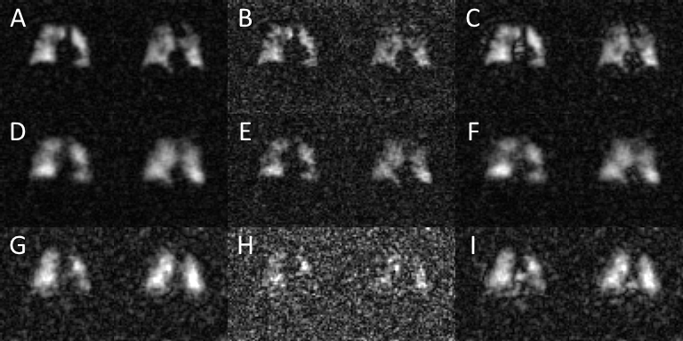
Reconstructed images of off-resonant gas excitation, barrier, and RBC are shown in Figure 1. The use of regularization results in a significant reduction in noise with minimal blurring of the image. Furthermore, by estimating the spectral properties from the acquired imaging data, image artifacts are significantly reduced.
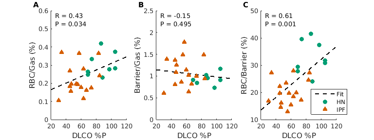
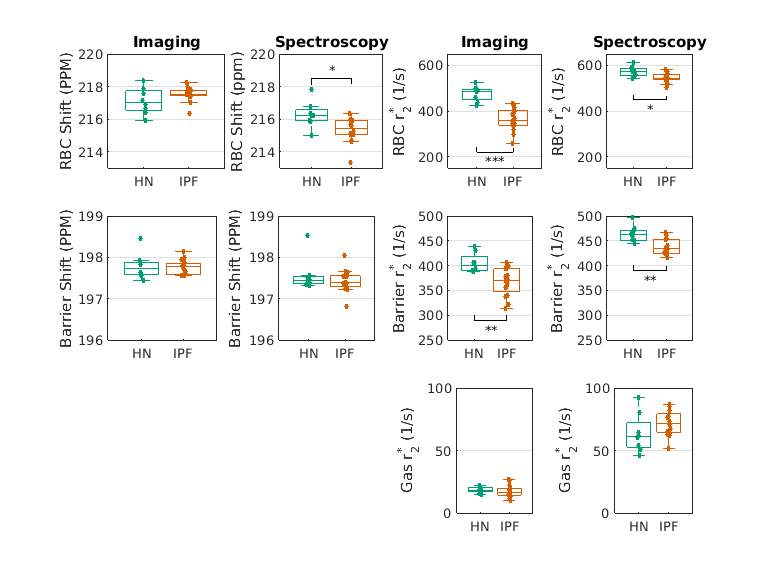
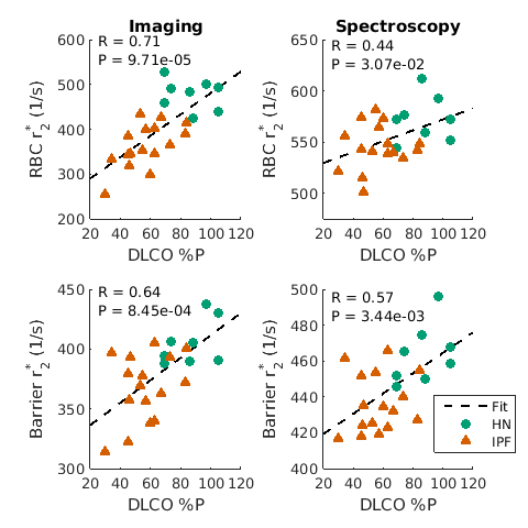
Measures of the RBC-to-Gas and RBC-to-Barrier ratios show significant correlation with DLCO (Figure 2), as has been reported previously. Spectral properties estimated from imaging data show substantial differences from those of whole lung spectroscopy (Figure 3) in both the chemical shifts and the r2* values. r2* values were substantially lower than previous estimates, with significant differences between healthy subjects and IPF patients. Furthermore, estimated dissolved-phase r2* values were significantly correlated with DLCO (Figure 4), showing higher correlations than those seen in the dissolved-phase ratios or in properties derived from whole lung spectroscopy.
Discussion and Conclusion
Increased exchange among compartments with different chemical shifts has been shown to lead to more rapid dephasing of the measured signal [5], providing a mechanism to explain the strong correlation between r2* and DLCO. The results suggest the relaxometry of dissolved-phase HP 129Xe can provide significant insight into gas exchange of the lung. Additionally, we have shown that by estimating spectral properties as part of the image reconstruction process, we can improve image quality in dissolved-phase HP 129Xe MRI.
Acknowledgements
The authors would like to acknowledge funding and support from the Pulmonary and Metabolic Imaging Center at the University of Wisconsin, Madison, and NIH/NCRR S10 OD016394.References
1. Driehuys B, Cofer GP, Pollaro J, et al (2006) Imaging alveolar-capillary gas transfer using hyperpolarized 129Xe MRI. Proc Natl Acad Sci U S A 103:18278–18283. doi: 10.1073/pnas.0608458103
2. Kaushik SS, Robertson SH, Freeman MS, et al (2016) Single-breath clinical imaging of hyperpolarized (129)Xe in the airspaces, barrier, and red blood cells using an interleaved 3D radial 1-point Dixon acquisition. Magn Reson Med 75:1434–1443. doi: 10.1002/mrm.25675
3. Qing K, Ruppert K, Jiang Y, et al. Regional Mapping of Gas Uptake by Blood and Tissue in the Human Lung using Hyperpolarized Xenon-129 MRI. Journal of magnetic resonance imaging : JMRI. 2014;39(2):346-359
4. William W. Hager and Hongchao Zhang. 2006. Algorithm 851: CG_DESCENT, a conjugate gradient method with guaranteed descent. ACM Trans. Math. Softw. 32, 1 (March 2006), 113-137.
5. McConnell HM. Reaction Rates by Nuclear Magnetic Resonance. J. Chem. Phys. 1958;28:430–431 doi: 10.1063/1.1744152.
Figures