4111
Simultaneous multi-slice single-shot DWI compared to routine read-out-segmented DWI for evaluation of breast lesions1Radiology and Nuclear Medicine, Radboudumc, Nijmegen, Netherlands
Synopsis
The aim of this study was to compare a prototype simultaneous multi-slice single-shot echo planar imaging (SMS-ss-DWI-EPI) sequence with conventional readout-segmented echo-planar imaging (rs-DWI-EPI) for diffusion-weighted imaging of the breast at 3T magnetic resonance imaging (MRI). A reader study was conducted to evaluate image quality, lesion conspicuity and BI-RADS® score. Our results show that although the image quality with the conventional rs-DWI-EPI is superior, malignant lesions have improved visibility with the SMS-ss-DWI-EPI sequence.
Introduction
The addition of diffusion-weighted imaging (DWI) to contrast-enhanced breast MRI improves the classification of breast lesions, which leads in turn to an increased positive predictive value of biopsies. Consequently, DWI with evaluation of the corresponding apparent diffusion coefficient (ADC) is included in most state-of-the-art breast MRI protocols1. The echo train of the readout-segmented echo-planar imaging-based DWI sequence (rs-DWI-EPI) was shortened to reduce distortion and improve the resulting image quality. However, this sequence results in a lower signal-to-noise ratio (SNR) than single-shot echo planar imaging (ss-EPI)2. In practice, detection of lesions on DWI is often problematic due to a relatively low lesion conspicuity. To improve the detectability of lesions and the speed of acquisition, a prototype DWI sequence, the simultaneous multi-slice single-shot DWI-EPI (SMS-ss-DWI-EPI), was developed. In this study we compare this prototype sequence with rs-DWI-EPI at 3T, in terms of image quality (IQ), lesion conspicuity, and breast imaging reporting and data system (BI-RADS®) score.Methods
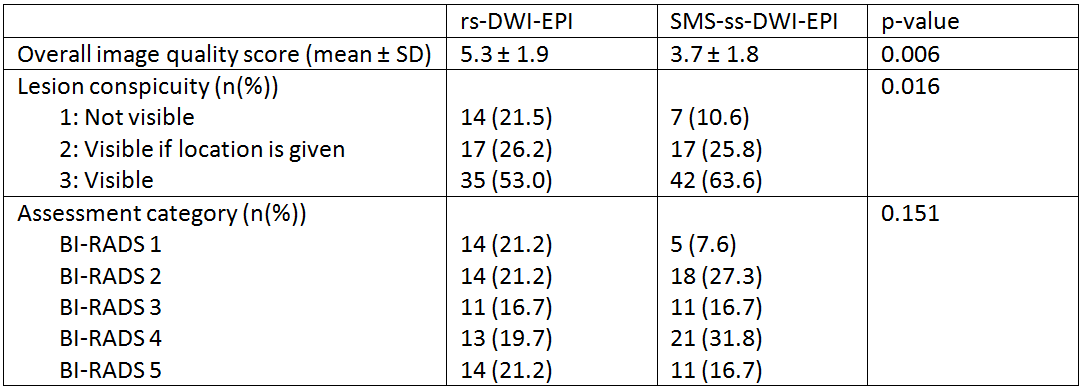
From September 2017 to August 2018, 15 women with known breast cancer or suspicious breast lesions were included, after providing signed informed consent. Women were scanned with the conventional rs-DWI-EPI and the SMS-ss-DWI-EPI during the same clinical examination on a 3T MAGNETOM Skyra system (Siemens Healthcare, Erlangen, Germany) using a 16-channel bilateral breast coil. Parameters of the rs-DWI-EPI sequence were: TR: 5450 ms, TE: 57 ms, FoV: 340 mm, voxel size: 1.2x1.2x5 mm3, acquisition time: 4:23 min, b-values: 50, 850 s/mm2, SPAIR fat suppression. Parameters of the SMS-ss-DWI-EPI sequence were: TR: 4000 ms, TE: 70 ms, FoV: 360 mm, voxel size: 0.9(i)x0.9(i)x4 mm3, acquisition time: 2:45 min, b-values: 50, 400, 800 s/mm2, SPAIR fat suppression. In addition, the clinical protocol included one pre- and five post-contrast administration regular T1-weighted Dixon acquisitions, ultrafast T1-weighted TWIST acquisitions during the inflow of contrast, and a T2 weighted Dixon acquisition. In total, 33 lesions (27 malignant, 5 benign and 1 unknown) were detected on the contrast-enhanced series and described in the clinical MRI reports. Two dedicated breast radiologists (4 and 10 years of experience with breast MRI) independently scored both sequences for overall IQ (1: extremely poor to 9: excellent). All lesions were also independently evaluated for conspicuity (1: not visible, 2: visible if location is given, 3: visible), and a BI-RADS score® (1 to 5) was given for each lesion. Statistical analysis was performed in SPSS using the Wilcoxon signed-rank test.Results
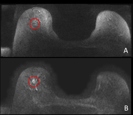
Results are presented in Table 1. Overall IQ was significantly higher for the conventional rs-DWI-EPI than for the SMS-ss-DWI-EPI (p=0.006). Lesion conspicuity scores were significantly higher for SMS-ss-DWI-EPI (p=0.016). Benign lesions had similar conspicuity with both sequences while malignant lesions had significantly higher conspicuity with SMS-ss-DWI-EPI (p=0.027) (for example, see Figure 1). There was no significant difference in BI-RADS® scores (p=0.151) between the two sequences.Discussion
Although the conventional rs-DWI-EPI sequence results in better IQ, in general ss-EPI results in a higher SNR, which may lead to better visibility of malignant lesions with SMS-ss-DWI-EPI. This might eventually improve the clinical value of DWI in addition to contrast enhanced breast MRI. Simultaneous Multi-Slice (SMS) ensures that slices are excited simultaneously with a multiband pulse, which leads to a reduced acquisition time. In our protocol, the combination of ss-EPI and SMS results in a higher spatial resolution while still having a shorter acquisition time than the conventional sequence. The higher achievable spatial resolution may be an important factor for the improved lesion visibility, and conspicuity of malignant lesions. This may make the SMS approach suitable for fast screening and diagnosis of breast cancer. Still, further development of the SMS-ss-DWI-EPI sequence is needed for improved IQ and even better lesion conspicuity. Extension of the data pool and evaluation by additional readers is pending.Conclusion
Despite the perceived poorer image quality of the SMS-ss-DWI-EPI sequence, malignant lesions are better visualized using this sequence. When image quality and conspicuity are further improved, this technique might enable improved lesion detection on unenhanced diffusion weighted breast MRI.Acknowledgements
We thank Elisabeth Weiland (Siemens Healthcare, Erlangen, Germany) for providing the prototype SMS sequence and the optimized protocol.References
1. Partridge SC, DeMartini, WB, Kurland BF, et al. Quantitative Diffusion-Weighted Imaging as an Adjunct to Conventional Breast MRI for Improved Positive Predictive Value. American Journal of Roentgenology. 2009;193(6):1716-1722.
2. Wisner DJW, Rogers N, Deshpande VS, et al. High-Resolution Diffusion-Weighted Imaging for the separation of Benign From Malignant BI-RADS 4/5 Lesions Found on Breast MRI at 3T. Journal of Magnetic Resonance Imaging. 2014;40:674-681.
Figures