4110
Clinical Feasibility of Reduced Field-of-View Diffusion-weighted Magnetic Resonance Imaging with Computed Diffusion-weighted Imaging Technique in Breast Cancer Patients1Radiology, Dong-A University Hospital, Busan, Korea, Republic of
Synopsis
We evaluated the clinical feasibility of the reduced field-of-view diffusion weighted imaging (rFOV DWI) with computed DWI technique in patients with breast cancer by performing a comparison and analysis of the inter-method agreement among the acquired rFOV DWI (rFOVA), rFOV DWI with computed DWI technique (synthetic rFOV DWI; rFOVS), and dynamic contrast-enhanced (DCE) magnetic resonance imaging (MRI) in patients with breast cancer. We found that the rFOV DWI and rFOV DWI with computed DWI technique showed nearly equivalent level of image quality required for analysis of the tumors and lesion conspicuity compared with DCE MRI.
Synopsis
We evaluated the clinical feasibility of the reduced field-of-view diffusion weighted imaging (rFOV DWI) with computed DWI technique in patients with breast cancer by performing a comparison and analysis of the inter-method agreement among the acquired rFOV DWI (rFOVA), rFOV DWI with computed DWI technique (synthetic rFOV DWI; rFOVS), and dynamic contrast-enhanced (DCE) magnetic resonance imaging (MRI) in patients with breast cancer. We found that the rFOV DWI and rFOV DWI with computed DWI technique showed nearly equivalent level of image quality required for analysis of the tumors and lesion conspicuity compared with DCE MRI.Introduction
Diffusion-weighted imaging (DWI) is one of the most frequently used magnetic resonance imaging (MRI) sequences in breast cancer patients. However, bilateral DWI is limited by magnetic susceptibility and chemical shift artifacts, low signal-to-noise ratio (SNR), and low resolution.1-3 Therefore, various techniques have been suggested for minimizing these drawbacks. Reduced field-of-view (rFOV) DWI can improve the image quality with decreased artifacts and relatively high SNR and resolution compared to conventional DWI.4-8 And computed DWI technique has recently emerged, which can improve the SNR and reduce artifacts.9 We focused on the merits of combining the advantages of the two DWI MRI techniques in the breast cancer patient. We hypothesized that a high resolution of various b-value images could be obtained by applying the above two techniques while reducing imaging acquisition time. Therefore, the purpose of this study was to evaluate the clinical feasibility of rFOV DWI with computed DWI technique in patients with breast cancer by performing a comparison and analysis of the inter-method agreement among the acquired rFOV DWI (rFOVA), rFOV DWI with computed DWI technique (synthetic rFOV DWI; rFOVS), and DCE MRI.Methods
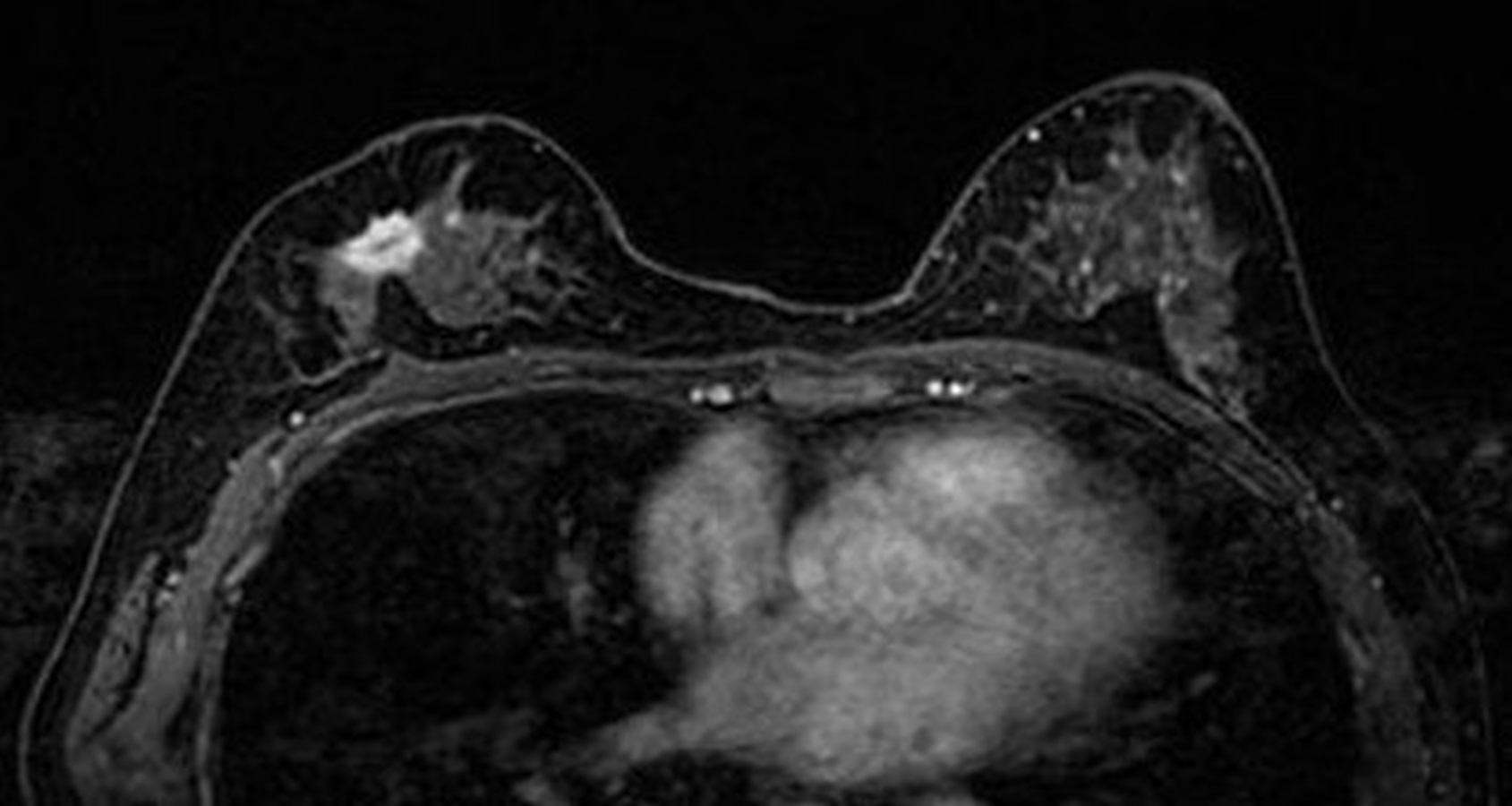
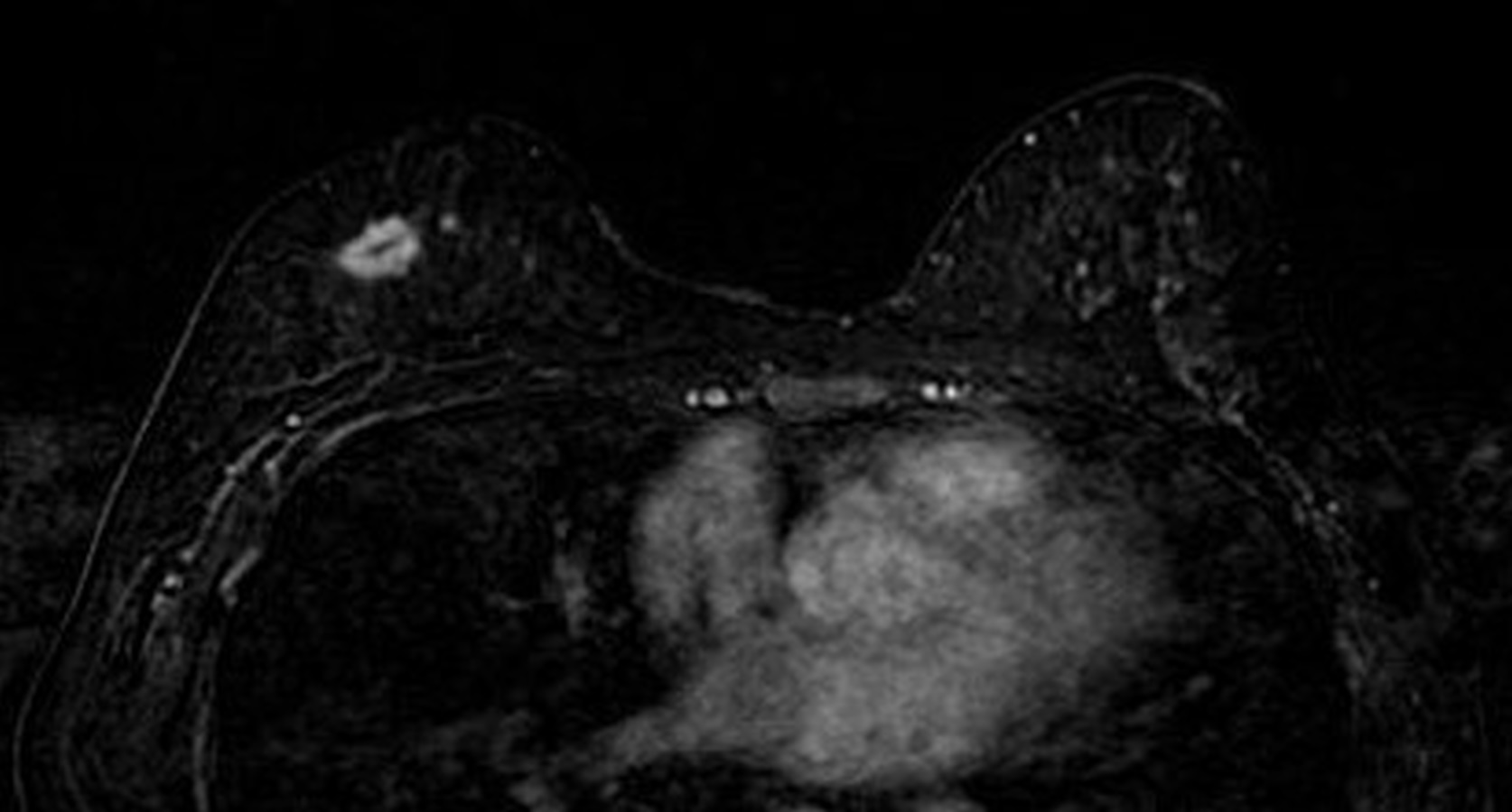
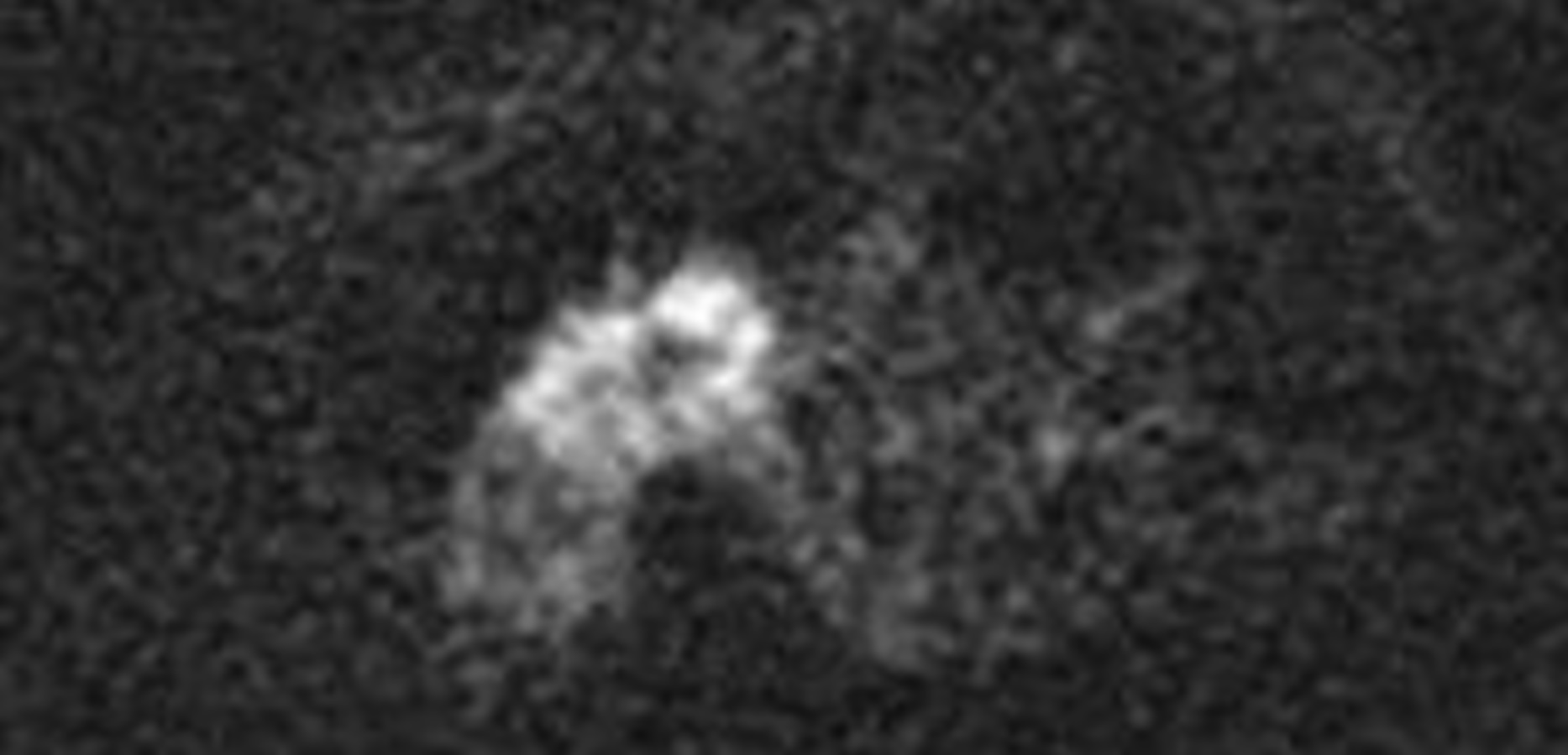
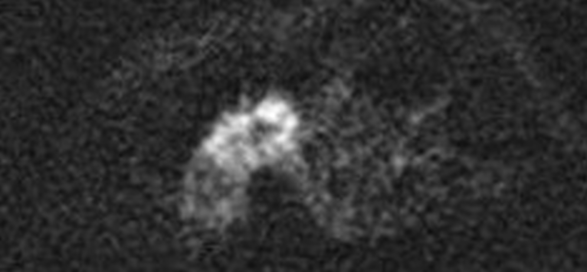
Totally, 130 biopsy-proven breast cancers from 130 women (age, 34-87 years; mean, 52.7 years) who underwent breast MRI from April 2017 to December 2017 were included. The image-sets of conventional rFOVA and rFOVS were reviewed and analyzed by comparing with DCE MRI. The rFOVS were reformatted by calculation of the apparent diffusion coefficient curve obtained from conventional rFOVA b=0 s/mm2 and b=500 s/mm2. Visual assessment of the image quality of rFOVA b=1000 s/mm2, rFOVS, and DCE MRI was done using a 4-point grading system. Morphologic analyses of the shape, margin, and size of the index cancer was performed on rFOVA, rFOVS and DCE MRI. For quantitative analysis, the signal-to-noise ratio (SNR), contrast-to-noise ratio (CNR), and contrast of tumor-to-parenchyma (TPC) were calculated.Results
Image quality scores with rFOVA, rFOVS, and DCE MRI were not significantly different (P=0.357). Lesion analysis of shape, margin, and size of the index cancer also did not show significant differences among the three sequences (P=0.858, P=0.242, and P=0.858, respectively). SNR, CNR, and TPC of DCE MRI were significantly higher than those of rFOVA and rFOVS (P<0.001, P=0.001, and P=0.016, respectively). Statistically significant differences were not found between the SNR, CNR, and TPC of rFOVA and those of rFOVS (P>0.999, P>0.999, and P>0.999, respectively).Discussion
In this study, we found that morphologic analysis and size measurement of the tumor could be performed well with rFOV DWI. In addition, with rFOVS, images equivalent to rFOVA could be obtained even with a relatively short image acquisition time. These results suggest that using rFOV DWI with computed DWI technique can provide efficient and accurate analysis of the breast tumor even with the short image acquisition time.Conclusion
In conclusion, both the rFOV DWI and rFOV DWI with computed DWI technique showed nearly equivalent levels of image quality required for analysis of the tumors and for lesion conspicuity compared with DCE MRI. The rFOV DWI with computed DWI technique especially, has the potential to have a useful clinical role in morphological evaluation of the breast tumor due to the relatively short image acquisition time and avoidance of contrast agent.Acknowledgements
No acknowledgement found.References
1. Partridge SC, Nissan N, Rahbar H, Kitsch AE, Sigmund EE. Diffusion-weighted breast MRI: Clinical applications and emerging techniques. J Magn Reson Imaging 2017; 45(2):337-355.
2. Yeom KW, Holdsworth SJ, Van AT, et al. Comparison of readout-segmented echo-planar imaging (EPI) and single-shot EPI in clinical application of diffusion-weighted imaging of the pediatric brain. Am J Roentgenol 2013; 200(5):W437-443.
3. Le Bihan D, Poupon C, Amadon A, Lethimonnier F. Artifacts and pitfalls in diffusion MRI. J Magn Reson Imaging 2006; 24(3):478-488.
4. Ma C, Li YJ, Pan CS, et al. High resolution diffusion weighted magnetic resonance imaging of the pancreas using reduced field of view single-shot echo-planar imaging at 3 T. Magn Reson Imaging 2014; 32(2):125-131.
5. Kim H, Lee JM, Yoon JH, et al. Reduced Field-of-View Diffusion-Weighted Magnetic Resonance Imaging of the Pancreas: Comparison with Conventional Single-Shot Echo-Planar Imaging. Korean J Radiol 2015; 16(6):1216-1225.
6. Korn N, Kurhanewicz J, Banerjee S, Starobinets O, Saritas E, Noworolski S. Reduced-FOV excitation decreases susceptibility artifact in diffusion-weighted MRI with endorectal coil for prostate cancer detection. Magn Reson Imaging 2015; 33(1):56-62.
7. Singer L, Wilmes LJ, Saritas EU, et al. High-resolution Diffusion-weighted magnetic resonance imaging in patients with locally advanced breast cancer. Acad radiol 2012; 19(5):526-534.
8. Dong H, Li Y, Li H, Wang B, Hu B. Study of the reduced field-of-view diffusion-weighted imaging of the breast. Clin Breast Cancer 2014; 14(4):265-271.
9. Blackledge MD, Leach MO, Collins DJ, Koh DM. Computed diffusion-weighted MR imaging may improve tumor detection. Radiology 2011; 261(2):573-581.
Figures