4101
Dynamic 3D lung MRI using the stack-of-stars sequence with SI navigation1Institute for Medical Imaging Technology, School of Biomedical Engineering, Shanghai Jiao Tong University, Shanghai, China, 2Shanghai United Imaging Healthcare Company, Ltd., Shanghai, China
Synopsis
Lung MRI has great potential in clinical early lung cancer screening and staging as it has a high detection rate for pulmonary nodules measuring. However, conventional lung MRI was limited by the physiological motion including respiration and heart beating. In this study, a free-breathing 3D golden-angle radial stack-of-stars pulse sequence with superior-inferior (SI) navigation was proposed for 3D dynamic lung MRI. Lung images in different respiratory stages were obtained. The proposed method provided higher SNR, CNR (between pulmonary vessels and the neighboring background in lung) and fewer motion artifacts than that without SI navigation.
Introduction
Lung MRI with ultrashort echo time (UTE) technique has a high detection rate of 83% for pulmonary nodules (4–17 mm) measuring[1], and has a 100% detection rate for nodules ≥ 5 mm[2]. Accurate detection of pulmonary nodules ≥ 5 mm is clinical relevant for early detection of lung cancer.[1, 2]
However, conventional lung MRI has been limited by substantial physiological motions including respiration and heart beating. Therefore, in free-breathing lung imaging, respiratory belt and ECG monitor are often needed as motion gating during the scan, which may cause patients uncomfortable and increase the total scan time.
The aim of this study was to propose a free-breathing 3D golden-angle radial stack-of-stars pulse sequence with superior-inferior (SI) navigation to trace the respiratory and cardiac motions. These motion signals will be subsequently used as retrospective self-gating signals for 3D dynamic lung imaging.
Materials and Methods
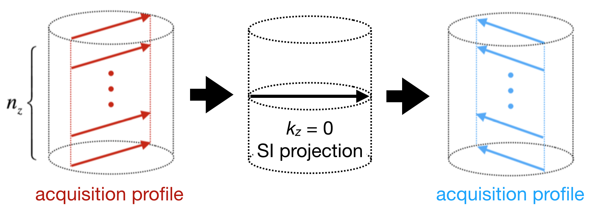
A free-breathing 3D golden-angle radial stack-of-stars pulse sequence with SI navigation was proposed. One SI projection was inserted between two consecutive acquisition profiles in coronal scans as shown in Figure 1. The SI projections cantrace the periodical change of MR signal of the entire excited volume including respiration and heart beating, and thus canbe used for extracting both respiratory and cardiac motion signals by filtering according to the different frequency ranges.In Figure 1, nz denotes the number of second phase-encoding in 3D radial stack-of-stars sequence.
Totally 5 healthy volunteers (4 males and 1 females) with ages ranged from 22 to 25 years (median age 24 years) were scanned in this study. The 3D free-breathing dynamic lung scans were performed on a 3.0 T uMR780 scanner (Shanghai United Imaging Healthcare Co., Ltd., Shanghai, China). A total of 24-channel body and spine phased-array coils were used for signal collection. The imaging parameters were: TR/TE = 3/1.3 ms, flip angle = 10◦, bandwidth = 600 Hz/pixel, slice thickness = 5 mm, number of slices = 32, spokes per slice = 2000, readouts = 256, FOV = 350 × 350 mm2, and total acquisition time ≈ 3 min 12 s.
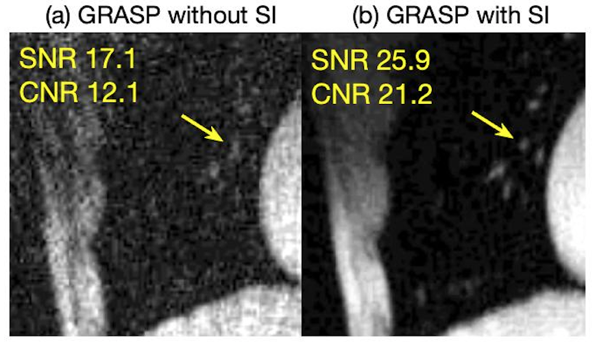
In the reconstructed images, the SNR was calculated by dividing the intensity of pulmonary vessels with the standard deviation of noise in air. The CNR was calculated by dividing the signal difference between a pulmonary vessel and the neighboring background in lung with the standard deviation of noise in air.
Results
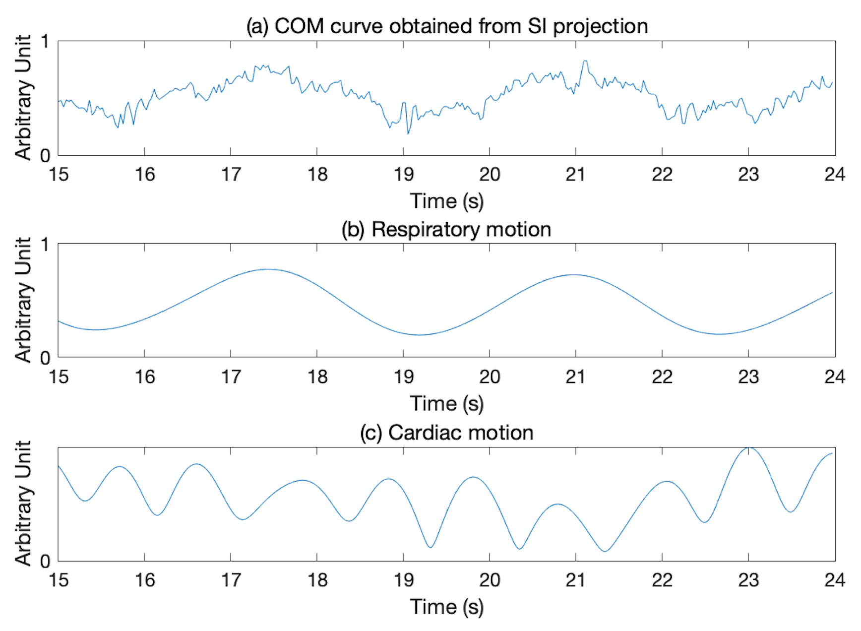
Figure 2 shows the self-gating signals obtained from the SI projections, (a), (b) and (c) show the center of mass (COM) curve, separated respiratory motion (0.26 Hz) and cardiac motion (0.98 Hz), respectively.
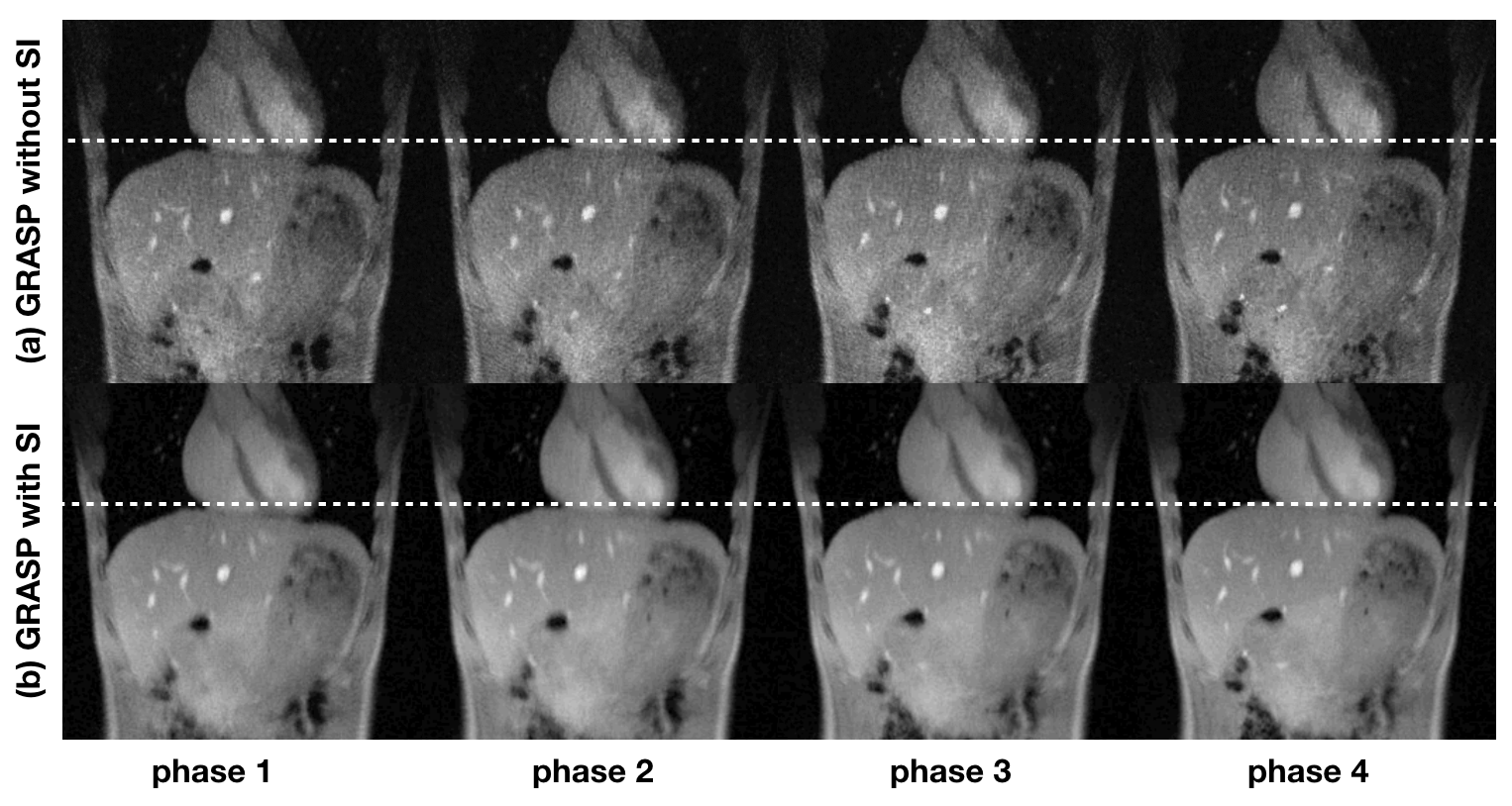
Figure 3 shows the reconstructions based on GRASP[3] without SI navigation (a) and with SI navigation (b). Four respiratory phases were selected, the white dashed line represents the same position of liver in four images for reference, phase 1 and phase 4 show the end-inspiration and end-expiration states respectively.
Figure 4 shows the comparison of reconstructed right lung images in end-expiration state between GRASP without SI navigation (a) and with SI navigation (b). In this figure, pulmonary vessels were indicated by yellow arrows.
Discussion
In the proposed 3D golden-angle radial stack-of-stars with SI navigation sequence, the reconstructed lung images have higher SNR and CNR than those without SI navigation based on GRASP as shown in Figure 4. The temporal resolution of motion gating signals was 96 ms, determined by nz·TR. The COM curve in Figure 2 (a) contains both respiratory and cardiac motions, which have different frequency ranges (0.1 ~ 0.5 Hz for respiratory motion and 0.6 ~ 3 Hz for cardiac motion). Respiratory and cardiac motion signals were separated after filtering. Therefore, lung images with different respiratory phases were reconstructed to minimize motion artifacts. Dynamic lung MRI has potential to be an alternative to conventional CT in oncology patients who are at risk of ionizing radiation exposure.Conclusion
The proposed 3D golden-angle radial stack-of-stars pulse sequence with SI navigation can provide both respiratory and cardiac motion signals. These signals can be used to minimize motion artifacts in 3D dynamic lung imaging for early detection of lung cancer. Future works will focus on self-gated pulmonary nodules imaging combined with the UTE technique.Acknowledgements
The authors are thankful for the assistance of United Imaging Healthcare, and also thank Feng Li for providing GRASP codes.References
[1] Burris N S, Johnson K M, Larson P E, Hope M D, Nagle S K, Behr S C, et al. Detection of Small Pulmonary Nodules with Ultrashort Echo Time Sequences in Oncology Patients by Using a PET/MR System. Radiology. 2016;278(1):239-46.
[2] Cha M J, Park H J, Paek M Y, Stemmer A, Lee E S, Park S B, et al. Free-breathing ultrashort echo time lung magnetic resonance imaging using stack-of-spirals acquisition: A feasibility study in oncology patients. Magn Reson Imaging. 2018;51:137-43.
[3] Feng L, Axel L, Chandarana H, Block K T, Sodickson D K, Otazo R. XD-GRASP: Golden-Angle Radial MRI with Reconstruction of Extra Motion-State Dimensions Using Compressed Sensing. Magnet Reson Med. 2016;75(2):775-88.
Figures