4082
Magnetization transfer and diffusion-weighted imaging in differential diagnosis of solid pulmonary masses: A comparison studyShan Dang1, Haifeng Duan1, Dong Han1, Nan Yu1, Qi Yang1, and Shaoyu Wang2
1Department of Radiology, Affiliated Hospital of Shaanxi University of Chinese Medicine, Xian Yang, China, 2MR Scientific Marketing, Siemens Healthineers, Shang hai, China
Synopsis
In this study, magnetization transfer (MT) and
diffusion-weighted imaging(DWI) techniques were used to differentiate the solid
pulmonary masses. It was found that magnetization transfer can be used to
distinguish benign and malignant pulmonary masses. Combining magnetization
transfer and diffusion-weighted imaging, the accuracy of differentiation can be
further improved.
Introduction
The accurate diagnosis of solid pulmonary masses can reduce the use of invasive examinations and help clinicians to choose the optimal treatment plan. Nowadays, it is still difficult to differentiate some lung masses using multi-slice spiral CT. Many previous publications have confirmed that diffusion-weighted imaging (DWI) is feasible in differentiating benign from malignant solid pulmonary masses. Both magnetization transfer (MT) and DWI can provide physiological and functional information of the organ. Meanwhile, MT imaging has been shown to be a valuable tool in detecting several diseases by reflecting the increase of macromolecules in the tissue. However, there have been relatively few studies of the MT on solid pulmonary masses. This study aims to investigate the value of MT and DWI in differentiating benign and malignant solid pulmonary masses.Methods
A total of 66 patients (18 females, 48 males; age range, 29-89 years), including 25 benign and 41 malignant cases, were enrolled in this study. HASTE (half fourier acquisition single shot turbo spin echo), radial VIBE (free-breathing radial 3D fat-suppressed T1-weighted gradient echo, radial volumetric interpolated breath-hold examination), T2 BLADE, MT and DWI sequences were performed on a 3T MR scanner (MAGNETOM Skyra, Siemens Healthcare, Erlangen, Germany). The DWI and MT images were independently evaluated by two radiologists. The apparent diffusion coefficient (ADC) and MT ratio (MTR) were calculated in the workstation. Intra-class correlation coefficient (ICC) was computed to investigate the consistency of the measurements between the two physicians. Mann-Whitney U test was used to compare the differences between the two groups; multivariate logistic regression analysis was used to evaluate the diagnostic value of the functional information. ROC curve was used to evaluate the diagnostic efficacy of MTR-only, ADC-only and the combination of MTR with ADC values for benign and malignant solid pulmonary masses.Results
The distribution of MTR and ADC values were not Gaussian (p=0.004, p<0.001), and the consistency of MTR and ADC values measured by the two radiologists were 0.927 and 0.944 (p<0.05), respectively. The difference of MTR and ADC values between the two groups was statistically significant (p=0.002, p<0.001) (Table 1). The MTR threshold is 18.73%, and the ADC threshold is 1069×10-6 mm2/s. Area under the curve (AUC) of combination of MTR and ADC was higher than that of ADC-only or MTR-only (Table 2). Malignant mass showed lower ADC and higher MTR values than benign mass. The analysis results of sensitivity, specificity and AUC of MTR-only, ADC-only and the combination of ADC and MTR were shown in Table 3.Disscusion
In this study, MT and DWI showed statistical significance in differentiating benign from malignant solid pulmonary masses. Although the MTR-only showed slightly lower performance than the ADC-only, while their combination yields the best AUC results, and indicate that the combination of two techniques could be a potential way to improve differentiation accuracy of solid pulmonary mass.Acknowledgements
No acknowledgement found.References
No reference found.Figures
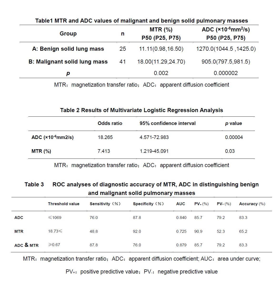
MTR:magnetization transfer ratio;ADC:apparent diffusion coefficient; AUC:area under curve; PV+:positive predictive value;PV-:negative predictive value
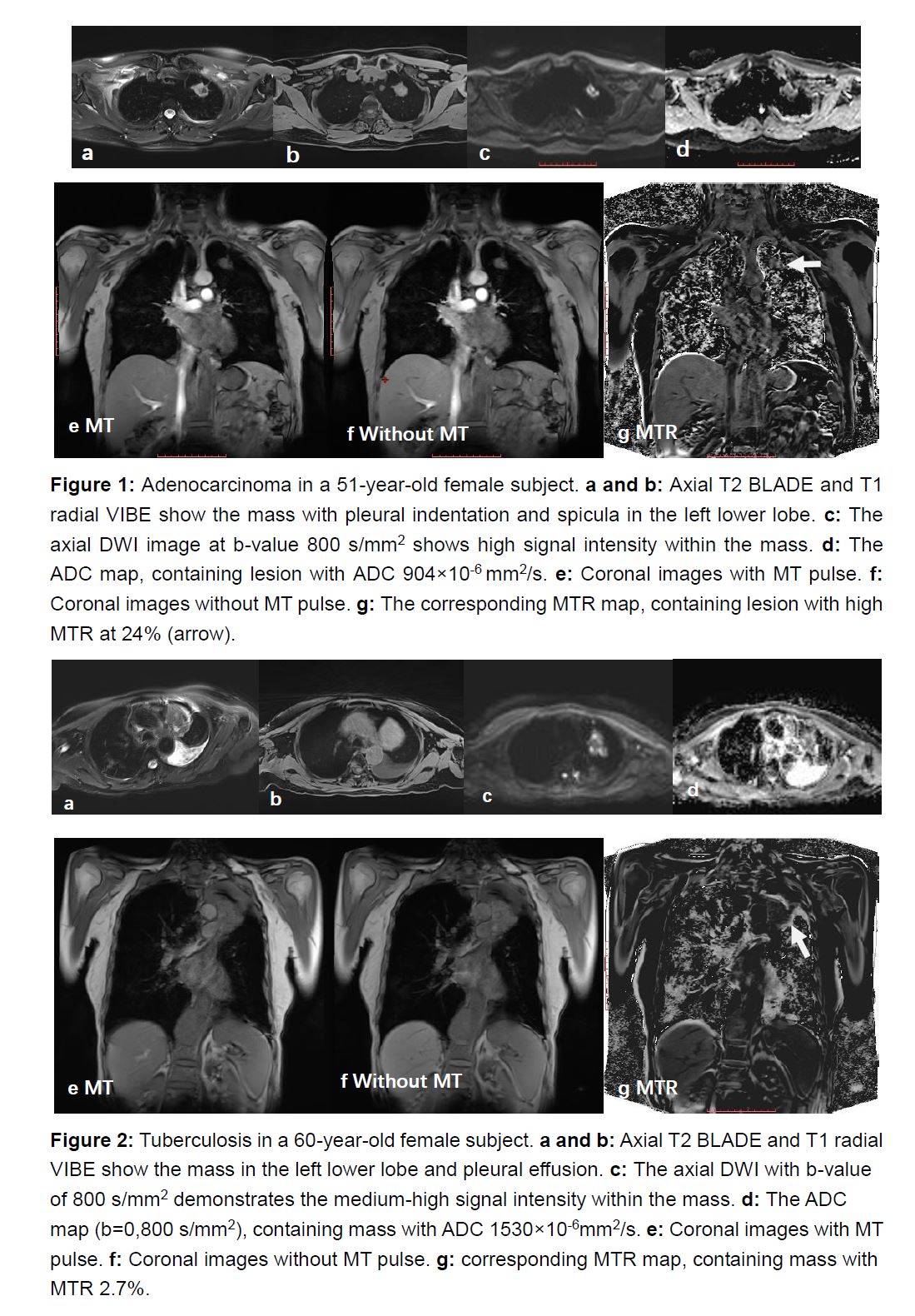
Figure 1: Adenocarcinoma
in a 51-year-old female subject. a and
b: Axial T2 BLADE and T1 radial VIBE show the mass with pleural indentation
and spicula in the left lower lobe. c: The
axial DWI image at b-value 800 s/mm2 shows high signal intensity
within the mass. d: The ADC map,
containing lesion with ADC 904×10-6 mm2/s. e: Coronal images with MT pulse. f: Coronal images without MT pulse. g: The corresponding MTR map,
containing lesion with high MTR at 24% (arrow).