4080
Free-Breathing Self-Gated 4D-Lung MRI with wave-CAIPI1Department of Diagnostic and Interventional Radiology, University Hospital Würzburg, Würzburg, Germany, 2Comprehensive Heart Failure Center Würzburg, Würzburg, Germany
Synopsis
The wave-CAIPI technique was applied to self-gated dynamic Lung MRI during free breathing. To prevent synchronization between respiratory motion and phase-encoding, the order of the phase-encoding steps was randomized using non-uniformly distributed, two-dimensional random numbers which oversample the k-space center. Breathing motion was gated using an additionally recorded DC signal. A healthy volunteer was investigated using the proposed method and a standard Cartesian scan for reference. For a scan time of 10:46 min, both methods exhibit comparable image quality. However, for accelerated scans, the wave-CAIPI technique clearly shows reduced residual image artifacts after applying parallel imaging.
Target audience
Clinicians and MR physicists interested in free-breathing lung MRI.Purpose
Long scan times are usually required to perform dynamic MR imaging of the lung with sufficient spatial and temporal resolution, e.g., for radiotherapy treatment planning. Standard Parallel Imaging (PI) can be used to accelerate the image acquisition, however, high acceleration factors typically lead to an unacceptable decrease of the Signal-to-Noise ratio (SNR). Recently, wave-CAIPI has proven to be an optimized method for accelerating volumetric acquisitions without significant reduction of image quality1 . We introduce a wave-CAIPI accelerated imaging technique for self-gated dynamic 4D lung MRI.Methods
The wave-CAIPI k-space trajectory was implemented in a 3D FLASH pulse sequence by playing out phase-shifted sinusoidal gradient oscillations on the two phase-encoding directions during data acquisition. The resulting k-space trajectory consists of helix-shaped readouts, compared to straight lines in the Cartesian case. Non-uniformly distributed, two-dimensional random numbers were used to randomize the order of the phase-encoding steps, in order to avoid synchronization between phase-encoding and breathing motion2,3 . The probability distribution of phase-encoding steps was larger in the k-space center than in the periphery. Gradient imperfections were corrected by means of the Gradient Impulse Response Function (GIRF)4 of the MR scanner. For respiratory gating, a DC signal was recorded directly after the excitation pulse. A single coil element near the lung/liver interface was manually selected, the signal was low-pass filtered and used to separate the breathing motion into 8 breathing states. The non-Cartesian data was resampled onto a Cartesian grid of size 512 x 512 x 512 by means of convolutional gridding5. To compensate missing k-space lines, Conjugate Gradient SENSE6 was applied to each breathing state. For this purpose, coil sensitivity maps were calculated from a time-averaged data set. All experiments were performed on a 3T clinical scanner (MAGNETOM Prisma, Siemens Healthcare GmbH, Erlangen, Germany). The following parameters were used in an exemplary investigation of a healthy volunteer: field of view 500 x 500 x 240 mm with readout in head-foot direction, matrix size 256 x 256 x 96, TE = 1.0 ms, TR = 2.6 ms, flip angle 5°, number of sinusoidal wave cycles during readout Nw = 5, maximum gradient wave amplitude Aw = 4 mT/m. In addition to that, a self-gated, randomized Cartesian measurement with identical parameters was performed for comparison. The total number of phase-encoding steps was set, such that the measurement time was 10:46 min in both cases. Retrospective acceleration of both acquisitions to 2:09, 2:42, 3:14, 4:19 and 5:23 min was performed. Including all memory-loading operations, the reconstruction time for all breathing states was about one hour in total.Results
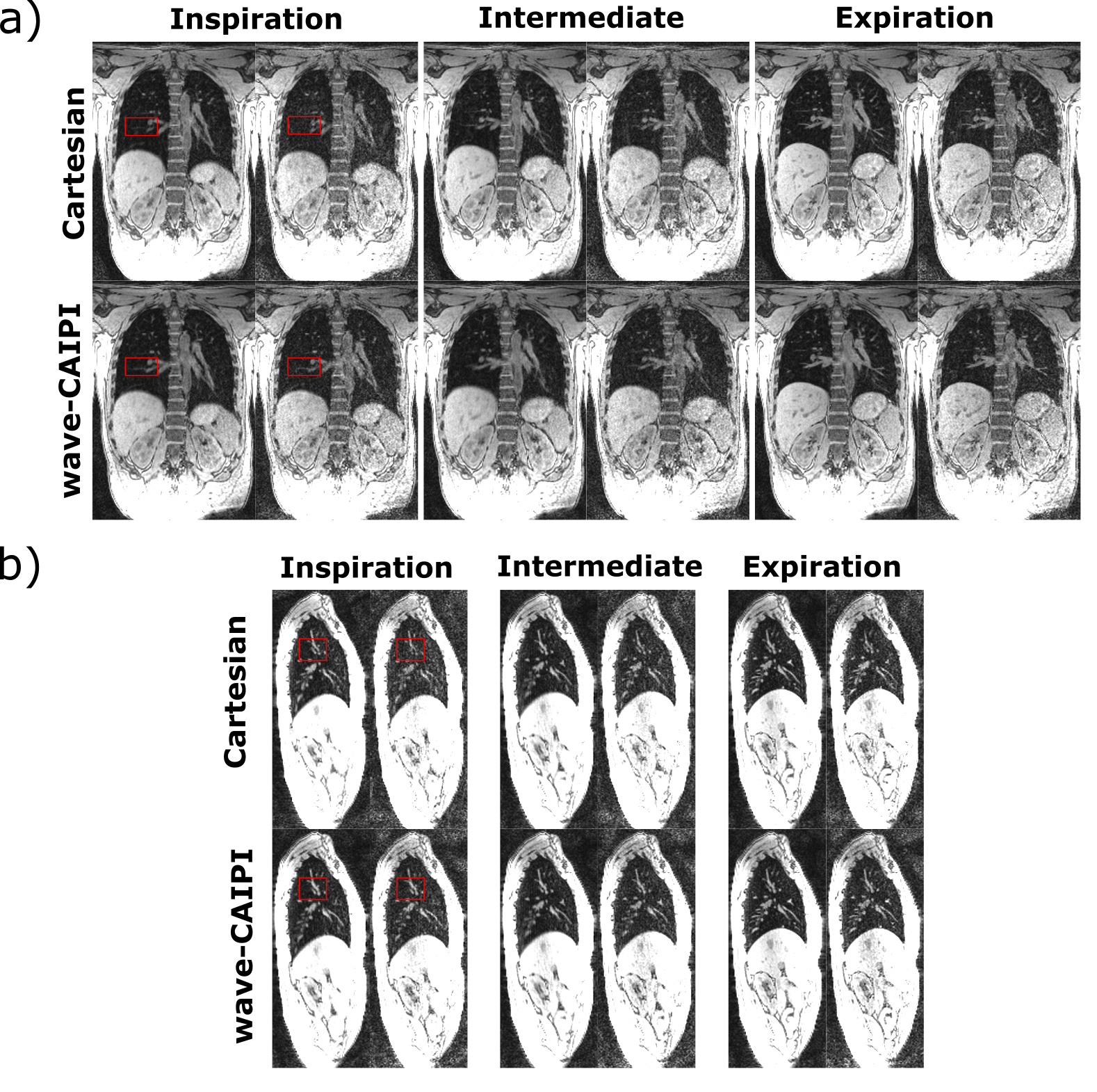
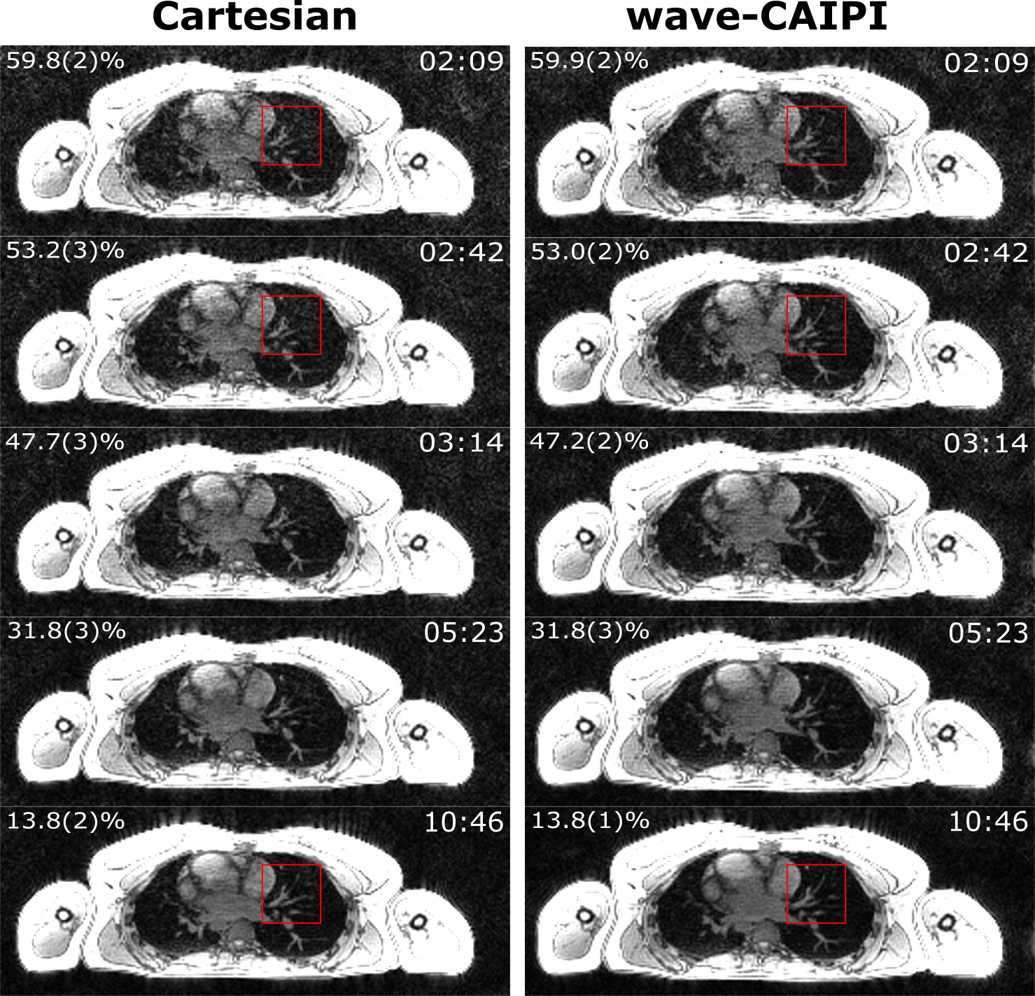
Fig. 1 shows three exemplary breathing states in coronal (Fig. 1a) and sagittal (Fig. 1b) orientation for both the Cartesian reference scan and wave-CAIPI using all data acquired (10:46 min). In the non-accelerated case (left images, respectively), the quality of both measurements is comparable. However, when reducing the acquisition time to 2:42 min (right images of Fig. 1), the apparent loss in SNR is higher for the Cartesian images. Fig. 2 shows an axial slice of a Cartesian and a wave-CAIPI image and illustrates the development of apparent SNR for different scan times (10:46, 5:23, 3:14, 2:42 and 2:09 min). Again, for shorter scan times, image quality is superior for the wave-CAIPI acquisitions, especially for lung tissue and vessels. In particular, small blood vessels are hardly visible in the accelerated Cartesian images, but are still accurately represented in the wave-CAIPI measurements (see regions in red rectangle). The percentage of missing k-space lines can be found in Fig. 2 (top left corner of each image).Discussion & Conclusion
The sinusoidal gradient oscillations during readout generate a wide spread of aliasing artifacts, thereby exploiting coil sensitivity variations in all three spatial dimensions, which ultimately leads to a more stable Parallel Imaging reconstruction. It is beneficial to reduce the acquisition time as much as possible, thereby decreasing the risk of patient movement in addition to respiratory motion. We demonstrate that by using the wave-CAIPI technique instead of the standard Cartesian sampling scheme, full coverage of the lung can be achieved in less than 3 minutes.Funding
Comprehensive Heart Failure Center Würzburg, Grant BMBF 01EO1504Acknowledgements
No acknowledgement found.References
1. Bilgic B, Gagoski BA, Cauley SF,et al. Wave-CAIPI for highly accelerated 3D imaging. Magn Reson Med. Jun 2015, Bd. 73, 6, S. 2152-62.
2. Weick S, Völker M, Hemberger K, Meyer C, Ehses P, Polat B, Breuer FA, Blaimer M, Fink C, Schad LR, Sauer OA, Flentje M, Jakob PM. Desynchronization of Cartesian k-space sampling and periodic motion for improved retrospectively self-gated 3D lung MRI using quasi-random numbers. Magn Reson Med. Feb 2017, Bd. 77, 2, S. 787-793.
3. Breuer K, Meyer CB, Breuer FA, Richter A, Exner F, Weng AM, Ströhle S, Polat B, Jakob PM, Sauer OA, Flentje M, Weick S. Stable and efficient retrospective 4D-MRI using non-uniformly distributed quasi-random numbers. Phys Med Biol. Mar 2018, Bd. 63, 7, S. 075002 (12pp).
4. Stich M, Wech T, Slawig A, Ringler R, Dewdney A, Greiser A, Ruyters G, Bley TA, Köstler H. Gradient waveform pre‐emphasis based on the gradient system transfer function. Magn Reson Med. Oct 2018, Bd. 80, 4, S. 1521-1532.
5. Jackson JI, Meyer CH, Nishimura DG, Macovski A. Selection of a convolution function for Fourier inversion using gridding [computerised tomography application]. IEEE Trans Med Imaging. 1991, Bd. 10, 3, S. 473-8.
6. Boesiger, Klaas P. Pruessmann Markus Weiger Peter Börnert Peter. Advances in sensitivity encoding with arbitrary k-space trajectories. Magn Reson Med. Oct 2001, Bd. 46, 4, S. 638-51.
Figures