4078
Comparison of Quantitative Pulmonary Ventilation Imaging using 3D-UTE under breath-holding and free-breathing in patients with structural lung disease at 3 T.1Department of Diagnostic and Interventional Radiology, University Hospital Würzburg, Würzburg, Germany, 2Application Development, Siemens Healthcare GmbH, Erlangen, Germany
Synopsis
UTE-MRI allows morphologic examination of lung parenchyma and acquisition of different breathing states enables determination of quantitative pulmonary ventilation (QV). In 4 patients with lung disease imaging with a prototypical UTE Spiral VIBE sequence during breath-holding was compared with a free-breathing 3D-UTE koosh-ball technique. Different breathing states were acquired, and QV values and the air volume fraction (AVF) were calculated. Both techniques yielded clinically sufficient image quality and comparable results for QV and AVF values. While the breath-hold approach acquires the data in a fraction of time but is dependent on patient compliance, the self-gated free-breathing technique overcomes that problem with the drawback of a longer acquisition time.
Introduction
So far magnetic resonance imaging (MRI) of the lung is impeded by the low proton density per volume, fast signal decay and respiratory motion. Ultrashort-echo-time (UTE) methods were developed in order to minimize the time between excitation and readout in order to overcome the short T2* and the fast signal decay in the lungs.1,2,3 Respiratory motion artifacts can be addressed by breath-hold techniques with shortened acquisition times or by gating techniques under free-breathing. Recently developed UTE-imaging of the lungs provides morphologic images with an image quality compared to CT without exposure to radiation, and simultaneous quantitative pulmonary ventilation (QV) imaging when different breathing states are acquired, adding valuable information for monitoring of lung diseases.3Methods
Four patients with different lung diseases and one healthy volunteer underwent functional 3D-UTE MR-imaging of the lung on a 3T MR-scanner (Magnetom Prisma, Siemens Healthcare, Erlangen, Germany). UTE-imaging was performed with a prototypical UTE Spiral VIBE sequence4 employing a 3D stack-of-spirals trajectory with a dual-density trajectory5, in-plane iPAT factor=2, and SPIRiT reconstruction6 with the following parameters: TE = 0.05ms; TR = 2.35ms; flip angle = 5°; in-plane resolution = 2.3mm x 2.3mm; slice thickness = 2.3 mm; number of spiral readouts per partition = 132; number of partitions: 118 ± 12 (depending on the size of the thorax). For functional information, data was acquired during breath-holding acquiring 5 different breathing states. For comparison, data was acquired with a free-breathing 3D UTE technique2 employing the following parameters: koosh-ball trajectory; TE = 0.03ms; TR = 1.49ms; flip angle = 2°; in-plane resolution = 2.3mm x 2.3mm, slice thickness = 2.3 mm and number of projections = 350,000; total acquisition time: 8.7 minutes. Eight different breathing states were reconstructed using data-driven gating. For both techniques, the different breathing states were registered to one intermediate breathing state7 for calculation of QV maps. Subsequently, lung volumes were segmented semi-automatically to obtain QV values as proposed earlier.2,8,10 Furthermore, air volume fraction (AVF) η of the lung was estimated from a single breathing state on a voxel-by-voxel basis according to the following equation:
$$ \eta \approx 1 - \frac{S_p}{S_{p, blood}}$$
where Sp denotes the signal in pulmonary parenchyma and Sp, blood the signal of pure blood under the assumption of a pure proton-density-weighted dataset and that the blood pool consists of 100 % water. Presented values are mean ± standard deviation for the entire lungs.
Results
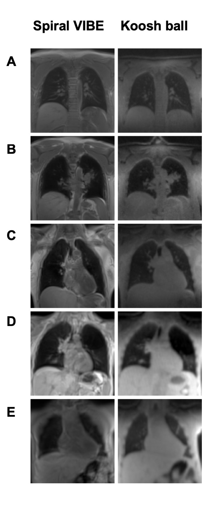
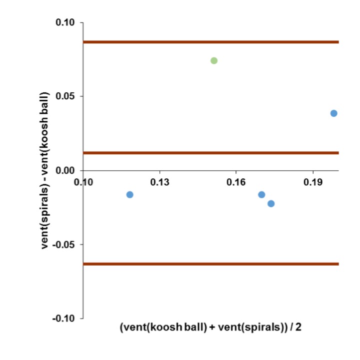
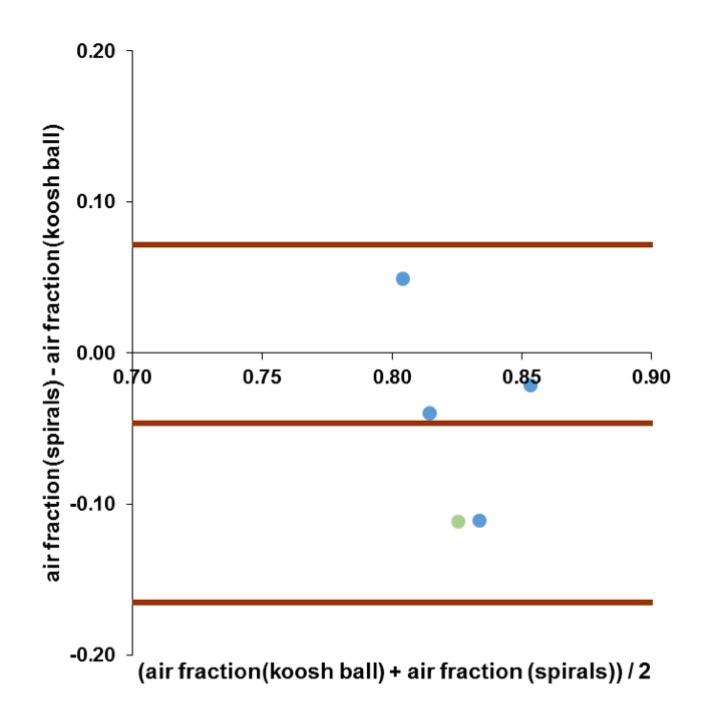
Figure 1 provides representative morphologic images of both imaging techniques for direct comparison of each dataset. Visual inspection of the images yielded good image quality with either technique and absence of prominent artifacts. Obtained QV and AVF values were in good agreement for both approaches. The mean difference and the interval of confidence were: QV: 0.01 mlgas/mlparenchym; [-0.06, 0.09]; AVF: -0.05 %; [-0.17, 0.07 ]. Figure 2 shows the Bland-Altman plot for the comparison of the QV values of the two techniques. Figure 3 presents the Bland-Altman plot for the obtained values for AVF.Discussion
Both presented techniques yielded clinically sufficient morphologic image quality for the detection of lung diseases as well as comparable QV and AVF values. In spite of shorter TE and TR, no significant increase of parenchymal signal was observed with the free-breathing 3D-UTE sequence. Regardless of the used approach QV maps were successfully obtained. However, the stack-of-spirals approach allowed acquisition of the thorax volume and 5 breathing states by a multi-breath-hold approach within 2 min. Yet, when the dataset of a single breathing state is afflicted with artifacts by interrupted breath-hold, incorrect QV data is obtained (green data), whereas the free-breathing 3D-UTE self-gating approach provides stable QV data regardless of the subject’s breathing condition. In contrast, AVF is determined from the dataset of a single breathing state; therefore, reliable mapping can be obtained as long as the respective dataset is free of artifacts.Conclusion
3D-UTE MR imaging is a powerful method for morphologic imaging of the lung which delivers higher resolution images in shorter acquisition times as compared to established imaging techniques in spite of low proton density in short acquisition times. Importantly, functional imaging as QV mapping and AVF add valuable information and may enhance the diagnosis and monitoring of lung diseases. Our data suggests that shorter TEs (0.03 vs. 0.05 ms) do not provide further enhancement of image quality. However, with regard to the patient’s condition the respective acquisition technique is of higher importance, since the breath-hold approach acquires the data in a fraction of time but is dependent on compliance, whereas the self-gated free-breathing technique overcomes that problem with the drawback of longer acquisition times.
Acknowledgements
No acknowledgement found.References
1. Qian Y, Boada FE. Acquisition-Weighted Stack of Spirals for Fast Echo Time MR Imaging. 2008;60:135–145.
2. Mendes Pereira L, Wech T, Weng AM, et al. UTE-SENCEFUL: first results for 3D high-resolution lung ventilation imaging. Magn. Reson. Med. 2018.
3. Mendes Pereira L, Wech T, Weng AM, et al. Self-gated ultra-short echo time lung MRI for quantitative ventilation assessment. Proc. Intl. Soc. Mag. Reson. Med. ; 2017. p. 3317.
4. Mugler JP, Meyer CH, Pfeuffer J, et al. Accelerated Stack-of-Spirals Breath-hold UTE Lung Imaging. ISMRM 2017.
5. Meyer CH, Zhao L, Lustig M, et al. Dual-Density and Parallel Spiral ASL for Motion Artifact Reduction. Proc. Intl. Soc. Mag. Reson. Med.
6. Lustig M, Pauly JM. SPIRiT: Iterative self-consistent parallel imaging reconstruction from arbitrary k-space. Magn. Reson. Med. 2010.
7. Kroon DJ, Slump CH. MRI modalitiy transformation in demon registration. Proceedings - 2009 IEEE International Symposium on Biomedical Imaging: From Nano to Macro, ISBI 2009.
8. Veldhoen S, Weng AM, Knapp J, et al. Self-gated Non-Contrast-enhanced Functional Lung MR Imaging for Quantitative Ventilation Assessment in Patients with Cystic Fibrosis. Radiology 2016;283:242–251.
9. Fischer A, Weick S, Ritter CO, et al. SElf-gated Non-Contrast-Enhanced FUnctional Lung imaging (SENCEFUL) using a quasi-random fast low-angle shot (FLASH) sequence and proton MRI. NMR Biomed. 2014;27:907–17.
Figures