4075
Fetal Brain Response to an Auditory Stimulus1Biomedical Engineering, Western University, London, ON, Canada, 2Medical Biophysics, Western University, London, ON, Canada, 3Division of Maternal, Fetal and Newborn Health, Children’s Research Institute, London, ON, Canada, 4Obstetrics and Gynaecology, Western University, London, ON, Canada, 5Brain and Mind Institute, Professor of Engineering, Western University, Lodon, ON, Canada, 6Clinical Neurological Sciences, Western University, London, ON, Canada
Synopsis
Functional magnetic resonance imaging (fMRI) was used in conjunction with an auditory task to assess fetal activation in the primary auditory cortex. Activation was found in the fetal right Heschl’s gyrus in response to an auditory task. This is a first report of fetal brain response to an “internal” auditory stimulus.
Introduction
Functional MRI (fMRI) is a non-invasive method to investigate the neural correlates of brain development. This project is part of a larger study looking at connectivity networks in high risk populations, to evaluate neurological development in utero, and to understand the differences between healthy and compromised fetuses. Fetal fMRI is challenging mainly because of random fetal movement, the small fetal brain, high water content in the fetal brain compared to adults, and the fact that the head of the fetus is deep within the mother, in a highly susceptible volume far from the receive coils. Fetal brain fMRI typically is limited to resting state fMRI, however, resting states can be influenced by multiple factors, including whether the fetus is awake or sleeping1, or hypercapnia2,3. Since neither can be controlled in a fetus, we sought to investigate a more reliable paradigm to study the development of fetal brain networks. Previous fetal fMRI task-based studies have demonstrated temporal lobe activation in response to an auditory stimulus4-7, however, since these studies have been published, there had been recommendation not to apply a direct stimulus to the mother’s abdomen8. An alternative to this direct auditory stimulus is to have the mother hum or sing, and we had postulated that this “internal” auditory task would result in activation in the fetal primary auditory cortex. Alternately, this study should reveal whether the superior temporal gyrus is implicated, if localization of the activation proves to be difficult. This would allow researchers to have a foundation of base line responses from a reliable paradigm to carry further studies and compare healthy verses at risk groups.Methods
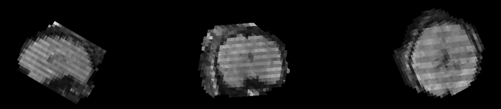
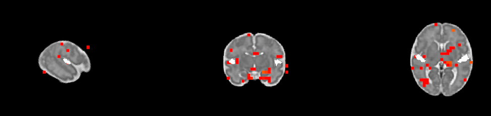
Four volunteers carrying singleton fetuses with a gestational age (GA) of 36-37 weeks were imaged on either a 3T (GE MR750), or a 1.5T (GE MR450w) MRI. T2-weighed anatomical images (SSFSE– TR >1200 ms, TE 80 ms, voxel size 0.98*1.96*5 mm3) were acquired before performing fMRI. Two task-based block design BOLD fMRI (TR 2 ms, TE 45-60ms(3T) /60ms (1.5T), flip angle 70º, voxel size 3.75*3.75*4 mm3) series were acquired where the mother was singing or humming a lullaby during the task phase. The data was converted into NIFTI format and each volume was assessed for unpredictable fetal motion. Initial manual reorientation was done for each volume to preserve the maximum amount of data. The volumes were then co-registered to the CRL fetal 36 GA atlas9 (Fig. 1). Brain extraction was done using FSL’s (v5.0.11)10 BET11 and segmentation was completed using the FAST12 tool (Fig. 2). The same process was done for the anatomical images, which were then co-registered with the functional images. The segmented functional data were analyzed using SPM 12 (v7219)13 as a task fMRI (p < 0.05). The respective GA regional atlas was deconstructed to assess which regions specific regions of the brain were active during a task. Each region was overlaid onto the activation map (which was co-registered to the atlas (Fig. 3)) to determine exactly which areas in the brain had activation during a task.Results
Our preliminary results suggest that there are 20 regions (postcentral and left precentral, right superior frontal gyrus, frontal middle gyrus, Rolandic operculum, right supplementary motor region, right medial frontal gyrus, right insula, right cingulum anterior gyrus, left internal capsule, white matter, fornix, corpus callosum) consistently activated by the four fetuses when they were exposed to the acoustic stimulus. Specifically, regions known to be part of the auditory network such as the right Heschl’s gyrus (Fig. 4), the right middle cingulate cortex (MCC), the left MCC (3/4 subjects) and the left putamen.Discussion
This preliminary study demonstrates that even without an external acoustic stimulus, and just having the mother hum or sing for blocks of time, one can activate the auditory network of the fetus. This can consequently be a tool to analyze the developing auditory cortex in the fetal brain. Much is known about activation in the Heschl’s gyrus for sound activation in adults14 and there is evidence for preterm neonatal activation in the right primary auditory cortex15. Fetal studies have reported that there is activation in the temporal lobe4-7, however, this is the first fetal study to report activation in the primary auditory cortex during an auditory task. Our results are consistent with studies on preterm infants that have shown activation in the left and right primary cortex, the left MCC, and the putamen15.Conclusion
We demonstrate that fetal primary auditory cortex and auditory network activation can be achieved by having the mother sing or hum.Acknowledgements
Grant support from Canada First Research Excellence Fund to BrainsCAN.References
1. G. Ferrazzi, M. Kuklisova Murgasova, T. Arichi, C. Malamateniou, M.J. Fox, A. Makropoulos, J. Allsop, M. Rutherford, S. Malik, P. Aljabar, J.V. Hajnal. Resting State fMRI in the moving fetus: A robust framework for motion, bias field and spin history correction. Neuroimage 101:555–568, 2014.
2. O. Marshall, J. Uh, D. Lurie, H. Lu, M.P. Milham, Y. Ge. The influence of mild carbon dioxide on brain functional homotopy using resting‐state fMRI. Hum. Brain Mapp., 36: 3912-3921, 2015.
3. Cousins, L. Fetal oxygenation, assessment of fetal well-being, and obstetric management of the pregnant patient with asthma. J Allergy Clin Immunol., 103(2)2: 343-349, 1999.
4. J. Hykin, R. Moore, K. Duncan, S. Clare, P. Baker, I. Johnson, R. Bowtell, P. Mansfield, P. Gowland. Fetal brain activity demonstrated by functional magnetic resonance imaging. Lancet 354, 645–646. 1999.
5. J. Fulford, S.H. Vadeyar, S.H. Dodampahala, S. Ong, R.J. Moore, P.N. Baker, D.K. James, P. Gowland. Fetal brain activity and hemodynamic response to a vibroacoustic stimulus. Hum. Brain Mapp. 22:116–121 106, 2004.
6. R. Jardri, D. Pins, V. Houfflin-Debarge, C. Chaffiontte, N. Rocourt, J.P. Pruvo, M. Steinling, P. Delion, P. Thomas. Fetal cortical activation to sound at 33 weeks of gestation: a functional MRI study. Neuroimage 42:10–18, 2008.
7. R.J. Moore, S. Vadeyar, J. Fulford, D.J. Tyler, C. Gribben, P.N. Baker, D. James, P.A. Gowland. Antenatal determination of fetal brain activity in response to an acoustic stimulus using functional magnetic resonance imaging. Hum. Brain Mapp. 12, 94–99, 2001.
8. C. Kruger, E. Horesh, B.A. Crossland. Safe sound exposure in the fetus and preterm infant. J. Obstet. Neonatal Nurs. 41(2)166–170, 2012.
9. A. Gholipour, C.K. Rollins, C. Velasco-Annis, A. Ouaalam, A. Akhondi-Asl, O. Afacan, C. Ortinau, S. Clancy, C. Limperopoulos, E. Yang, J.A. Estroff, and S.K. Warfield. A normative spatiotemporal MRI atlas of the fetal brain for automatic segmentation and analysis of early brain growth, Scientific Reports 7, Article number: 476, 2017.
10. M. Jenkinson, C.F. Beckmann, T.E. Behrens, M.W. Woolrich, S.M. Smith. FSL. NeuroImage, 62:782-90, 2012.
11. S.M. Smith. Fast robust automated brain extraction. Hum. Brain Mapp., 17(3):143-155, 2002.
12. Y. Zhang, M. Brady, and S. Smith. Segmentation of brain MR images through a hidden Markov random field model and the expectation-maximization algorithm. IEEE Trans Med Imag, 20(1):45-57, 2001.
13. R.S.J. Frackowiak, K.J. Friston, C.D. Frith, R.J. Dolan, and J.C. Mazziotta. Hum. Brain Function. Academic Press USA, 1997.
14. T.D. Griffiths, C. Buchel, R.S. Frackowiak, R.D. Patterson. Analysis of temporal structure in sound by the human brain. Nat Neurosci 1:422–427, 1998.
15. L. Lordier, S. Loukas, F. Grouiller, A. Vollenweider, L. Vansung, D.E. Meskaldij, F. Lejeune, M.P. Pittet, C. Borradori-Tolsa, F. Lazeyras, D. Grandjean, D. Van De Ville, P.S. Huppi. Music processing in preterm and full-term newborns: A psychophysiological interaction (PPI) approach in neonatal fMRI. Neuroimage. 1053–8119, 2017. In Press, Corrected Proof.
Figures