4063
Fetal dynamic phase-contrast angiography using Doppler ultrasound gating and comparison with Doppler ultrasound measurements1Diagnostic and Interventional Radiology, University Medical Center Hamburg-Eppendorf, Hamburg, Germany, 2NortHH Medical GmbH, Hamburg, Germany, 3Diagnsotic and Interventional Radiology, University Medical Center Hamburg-Eppendorf, Hamburg, Germany, 4UCSF Benioff Children's Hospital Oakland, Oakland, CA, United States, 5Gynaecology & Obstetrics, University Medical Center Hamburg-Eppendorf, Hamburg, Germany
Synopsis
In this prospective study 2D PC-MRI angiography was performed in nine human fetuses (gestational age 32.2 ± 2.4 weeks) using a novel MR compatible DUS device for gating of the fetal heart at a 1.5T MR scanner. The novel MR compatible DUS device for fetal cardiac gating allowed successful application of PC-MRI angiography and quantification of hemodynamics in the fetal AoD. This new technique for direct gating of the fetal heart demonstrates the diagnostic potential of PC-MRI for the quantification of fetal hemodynamics.
Objective
To perform fetal phase-contrast (PC)- MRI angiography of the fetal descending aorta (AoD) using a novel MR compatible Doppler Ultrasound (DUS) device for fetal cardiac gating and to compare velocimetry to Doppler ultrasound measurements.Methods
In this prospective study 2D PC-MRI angiography was performed in nine human fetuses (gestational age 32.2 ± 2.4 weeks) using a novel MR compatible DUS device for gating of the fetal heart at a 1.5T MR scanner. A T2 turbo spin echo (TSE) sequence was performed for acquisition of transversal, coronal and sagittal images of the fetal thorax (TE 90 ms, TR 1600 ms, FoV 430 x 360 mm, FA 90°, slice thickness 5 mm, matrix size 256 x 147, scan duration 32 sec.), Sense factor 2. Following these scout images a 2D phase-contrast angiography sequence was applied with the following parameters: TE 3 ms, TR 4,9 ms, FoV 250 mm, FA 15°, slice thickness 5 mm, acquisition matrix size 124 x 115 mm (reconstructed matrix size 192x192 mm), slice thickness 5mm, cardiac phases 10, velocity encoding factor 150 cm/s, NSA 2. Dependent on fetal heart rates temporal resolution was 36-46 ms and scan duration was 12 s. Measurements were performed in the fetal descending aorta (AoD) between the confluens of the arterious duct and aorta and the diaphragmatic entering of the aorta. Gating of the phase-contrast cine angiography sequence was realized using a newly developed MR compatible Doppler ultrasound (DUS) transducer that proved successful gating of the fetal hear. Peak flow velocities in the fetal AoD were compared to DUS measurements performed on the same day. Reproducibility of velocimetry was tested by repeated PC-MRI measurements in five fetuses.
Results
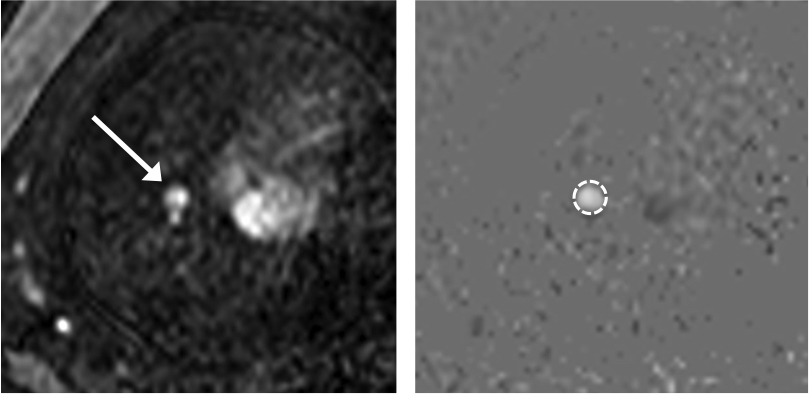
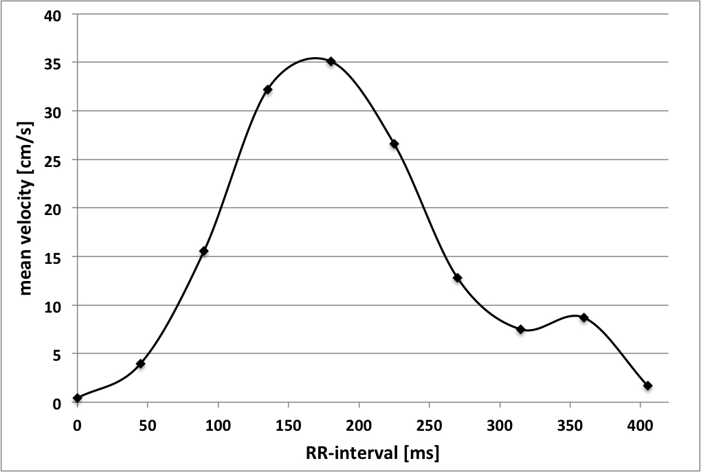
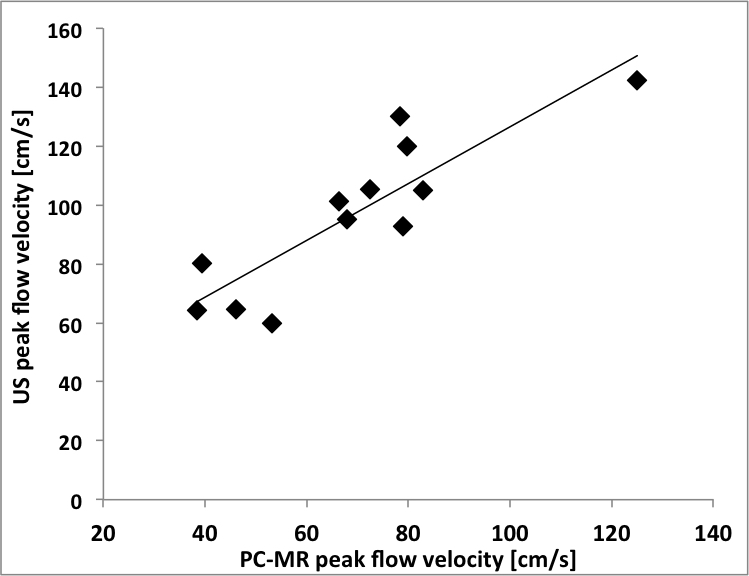
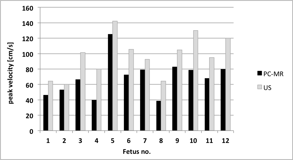
Dynamic PC-MRI in the fetal AoD was successfully performed in all fetuses using the novel DUS device for fetal cardiac gating, with fetal heart rates ranging from 131 to 163 bpm. In DUS gated PC-MRI cine images of the fetal AoD the cross-sectional aortic lumen could be identified in all cases and ROIs in the vessel lumen could be reliably defined (Figure 1). No phase aliasing was present with the selected velocity encoding factor of 150 cm/s. Mean vessel diameter of the AoD was 6.2 ± 0.6 mm. The assessed time-velocity curves revealed characteristic biphasic arterial flow waveform patterns with a strong early systolic peak and continuously positive low diastolic blood flow (Figure 2). The mean peak velocity was 67 ± 27.4 cm/s (range: 39 – 125 cm/s) and average mean flow velocity and average mean flux were 17.8 ± 5.5 (range 8.6 – 25.4) cm/s and 7.5 ± 3.5 (range 3.2 – 14.6) ml/s, respectively. Mean stroke volumes were 3.2 ± 1.3 ml, there was no regurgitation volume in all fetuses, i.e. no pathological waveforms were observed. The time-velocity curves revealed typical arterial bloodflow patterns, no pathological backward flow or regurgitation was observed. Peak velocities were higher for PC-MRI angiography than for Doppler ultrasound measurements (67 ± 27.4 vs. 90.6 ± 26.5 cm/s, p<0.001). PC-MRI derived peak flow velocities were generally lower than peak flows assessed by Doppler ultrasound in each fetus (67 ± 27.4 cm/s and 90.6 ± 26.5 cm/s; p<0.001) (Figure 3). Pearson correlation revealed strong correlation of Doppler ultrasound and PC-MRI peak flow velocities (r=0.92, p=0.002) (Figure 4). There was a positive correlation for average mean flux and mean flow velocities assessed by PC-MRI with gestational age, with a stronger correlation revealed for average mean flux and gestational age (r=0.65, p=0.06 and r=0.56, p=0.1). PC-MRI revealed good reproducibility with similar flow parameters of peak velocity (72.5 cm/s and 75.3 cm/s; p=0.12), mean flux (7.8 ml/s and 7.1 ml/s; p=0.3), mean velocity (p=19.9 cm/s and 19.9 cm/s; p=0.6) and SV (3.4 ml and 3 ml; p=0.2).PC-MRI and DUS peak velocities revealed strong correlation (r=0.92, p=0.002). The average PC-MRI mean flow velocity and mean flux were 17.8 ± 5.5 cm/s and 7.5 ± 3.5 ml/s, respectively. Reproducibility of velocimetry was high with no statistical differences.Discussion
For the first time, this study demonstrates successful application of fetal PC-MRI angiography for prenatal assessment of blood flow hemodynamics using a novel MR compatible DUS device for fetal cardiacgating. Comparison with Doppler ultrasound measurements revealed high correlation of peak flow velocities in the fetal AoD, with generally lower blood flow velocities for PC-MRI measurements. The technical development of direct andnon-invasive fetal cardiac gating allowed dynamic functional imaging of the fetal vascular system and offers a new possibility to assess blood flow measurements in the prenatal period. This may enhance the diagnostic potential in the evaluation of fetal hemodynamics or congenital vascular abnormalities and improve planning of therapeutic decision in prenatal care.Conclusion
The novel MR compatible DUS device for fetal cardiac gating allowed successful application of PC-MRI angiography and quantification of hemodynamics in the fetal AoD. This new technique for direct gating of the fetal heart demonstrates the diagnostic potential of PC-MRI for the quantification of fetal hemodynamics.Acknowledgements
No acknowledgement found.References
No reference found.Figures