4058
Diffusion weighted imaging evaluation of cerebral changes in twin pregnancies with one fetus demise1Radiology, Peking University Third Hospital, Beijing, China
Synopsis
Diffusion-weighted imaging (DWI) has been applied to fetal MRI and provides important information in evaluating the microstructures. Apparent diffusion coefficient (ADC) value is derived from DWI sequence. The aim of this study is to compare ADC values of brain in surviving fetus of one fetus demise with normal fetuses using diffusion weighted imaging and to find the differences and underlying cerebral microstructure changes. Decrease of ADC values were detected in surviving fetus of one twin demise. DWI and ADC values are very useful for detecting underlying changes, even changes of signal are not visible on conventional sequences.
Introduction:
Demise of one twin is a common complication of monoamniotic twins. Demise of one twin usually has a negligible effect on the surviving fetus which accounts for increasing in morbidity and mortality. The underlying mechanism involves the loss of circulatory equilibrium, with shunting of blood flow. The surviving fetus might present with hypotension and hypoperfusion. Diffusion-weighted imaging (DWI) has been applied to fetal MRI and provides important information in evaluating the microstructure and biophysical status of tissues and intracranial lesions[1]. Apparent diffusion coefficient (ADC) value is derived from DWI sequence. Recent studies have shown the feasibility of fetal diffusion imaging and fetal cerebral ADC determination [2]. The aim of this study is to compare ADC values of brain in surviving fetus of one fetus demise with twin controls and single fetus controls using diffusion weighted imaging and to find the differences among three groups and underlying cerebral microstructure changes.Method:
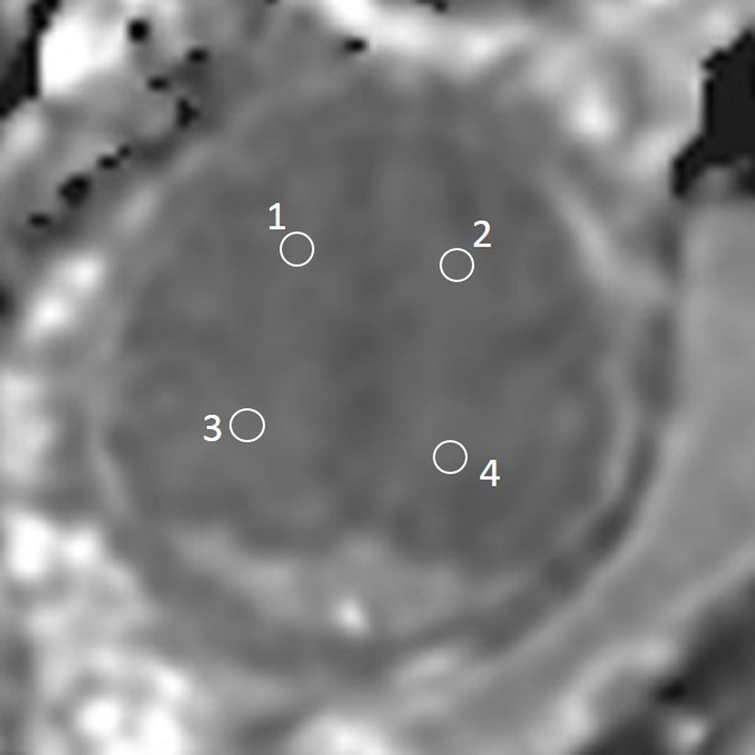
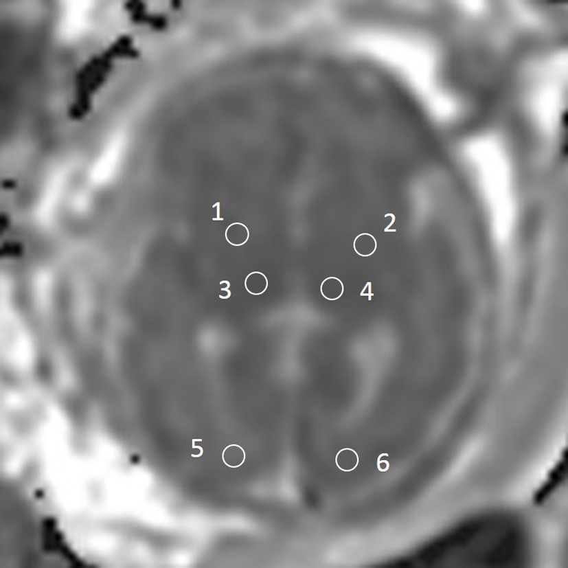
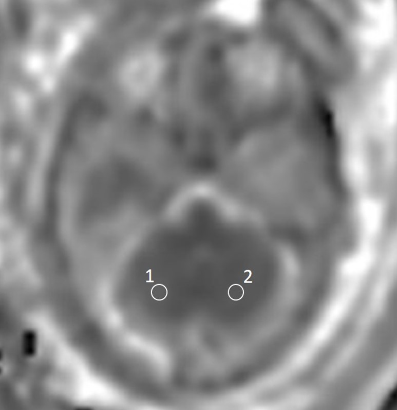
Twenty pregnant women with twins who were diagnosed as demise of one fetus (mean maternal age 28.8±3.8 years, mean gestational age 30.9±3.2 weeks) were recruited. Fifteen pregnant women with twins who were normal in brain (mean maternal age 30.2±3.6 years, mean gestational age 30.7±3.1 weeks)were chosen for twins control subjects. Fifteen pregnant women with single fetus (mean maternal age 30.2±3.5 years, mean gestational age 31.1±3.4weeks) were chosen for control subjects were recruited. MRI was acquired within 1~5days after ultrasound diagnosis. DWI was performed on a 1.5-T MR scanner (360 Optima, GE Healthcare). DWI parameters: TR 2831ms, TE77ms, slice thickness: 5 mm, slice interval: 1 mm, FOV: 320 mm, b values (0 and 600 s/mm2), scanning time: 16~20s. ADC maps were reconstructed automatically on post-processing software after DWI sequence scanning. The best images with no or least motion artifacts were used for ADC measurements. ADC values were calculated directionally from the ADC map, in circular regions of interest (ROIs) drawn on the regions. ROIs were placed bilaterally and over the desired anatomical areas. Average ADC values were calculated directionally on each region. White matter of frontal lobes, parietal lobes, temporal lobes and occipital lobes, basal ganglia, thalamus and cerebellum were selected for analysis. Statistical analyses were performed. ADC values among surviving fetuses, twin controls and single fetus controls in corresponding regions were assessed by performing variance analysis. ADC values in left regions of surviving fetuses were compared with ADC values in right regions. Correlations between ADC values and time of one fetus demise, gestational age and maternal age were calculated.Results:
1. There were no significant differences of gestational ages and maternal ages among three groups (P>0.05). Average time of one twin demise was (17.3±12.7) days.
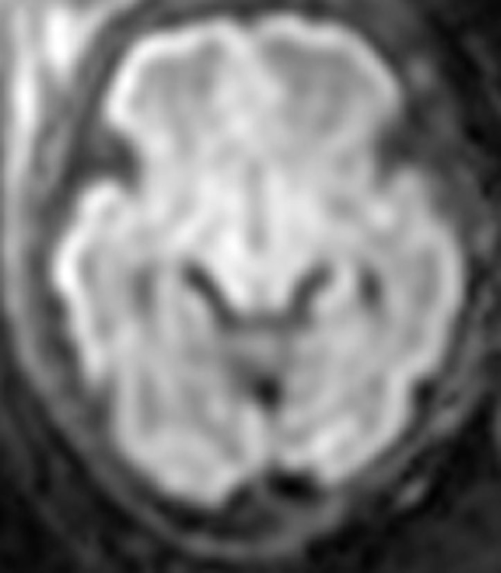
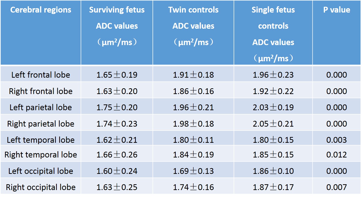
2. No significant signal abnormalities of all subjects were seen on DWI sequence. There were significant differences of ADC values among three groups in bilateral frontal lobes, parietal lobes, temporal lobes and occipital lobes. ADC values decreased in these regions in surviving fetuses.
3. No significant differences between left and right hemisphere were detected (P>0.05).
4. There was no correlation between time of one twin demise and ADC values. Maternal age were not correlated with ADC values in cerebral regions. ADC values of bilateral occipital lobes were negatively correlated with gestational age in surviving fetuses (L: r=-0.48, P=0.033; R: r=-0.57, P=0.009). ADC values of left occipital lobe( r=-0.55, P=0.041), left basal ganglia( r=-0.56, P=0.037) and right cerebellum ( r=-0.55, P=0.042) were negatively correlated with gestational age in twin controls.
5. An ROC curve was generated for ADC values in left frontal lobe of surviving fetuses and twin controls to determine the optimal cutoff value at a sensitivity of 95.0%. The area under the ROC curve was 0.864 (95% confidence interval). A sensitivity of 64.3% resulted in a specificity of 95.0% and an optimal cutoff value for ADC in left frontal lobe of 1.87μm2/ms. At this cutoff value, the positive predictive value was 0.79 and the negative predictive value was 0.90.
Conclusions:
Decrease of ADC values were detected in surviving fetus of one twin demise. DWI is a very useful sequence for detecting underlying changes. ADC value might be more sensitive to detect the subtle anomalies, even changes of signal are not visible on conventional sequences. Well-controlled and long-term studies are needed to reveal the relationship between reduction of the ADC values and postnatal outcome.Acknowledgements
No.References
[1] Rutherford MA. Magnetic resonance imaging of the fetal brain. Curr Opin Obstet Gynecol, 2009; 21(2):180-186.
[2] Hoffmann C, Weisz B, Yinon Y, Hogen L, Gindes L, Shrim A, et al. Diffusion MRI findings in monochorionic twin pregnancies after intrauterine fetal death. AJNR Am J Neuroradiol.2013; 34(1):212-216.
Figures