4048
MR features including ADC values of Uterine CarcinosarcomaMeng-yao Wang1, Mei-yu Sun2, Xu Han2, and Rui Fan2
1radiology, the first Affiliated Hospital of Dalian Medical University, dalian, China, 2the First Affiliated Hospital of Dalian Medical University, dalian, China
Synopsis
Uterine carcinosarcoma(UCS) is a rare and aggressive tumor with universally poor prognosis. To reinforce the knowledge of uterine carcinosarcoma(UCS) and improve the preoperative diagnostic accuracy on magnetic resonance imaging(MRI). A retrospective imaging review was performed of the MR images of 12 patients with uterine carcinosarcoma. In this study, we summarized its MRI features.
Introduction
Uterine carcinosarcoma(UCS) is a very rare aggressive neoplasm with rapidly progressing and poor prognosis. Patient usually presents with postmenopausal vaginal bleeding. Surgery combined of radiotherapy and chemotherapy is the main therapeutic approach but it is prone to poor response to treatment and relapse. Hence, the reliable diagnosis of UCS is a major unmet clinical need. MRI is a commonly used technique for preoperative evaluation of uterine malignancies. Therefore, the purpose of this present study is to reinforce the knowledge of UCS and improve the preoperative diagnostic accuracy on MR imaging.Methods
From July 2010 to August 2017, 12 women aged 46-82 years (mean age, 66years) with UCS underwent preoperative MR imaging, including T1- and T2-weighted imaging, DCE MR imaging and diffusion-weighted imaging (DWI) sequence. MR features of UCS were interpreted by two senior radiologists in consensus, including lesions localization, tumor dimensions, shape, border, depth of myometrial invasion (DMI), interior signal, enhancement characteristics, ADC values and other uterine diseases combined.Results
The MRI characteristics are detailed in Table 1. The mean ADC values (ADCmean) of our twelve lesions was (1.15±0.15) ×10-3mm2/s. The inter-observer consistency was good (ICC=0.82). These lesions were suspected as malignant tumor in 4 cases, endometrial carcinoma in 6 cases, cervical carcinoma in 1 case and degenerated leiomyoma with hemorrhage in 1 case. Ultimately UCSs was confirmed by postoperative pathology in all tumors. Four cases of them contained chondrosarcomatous elements, meaning heterologous differentiation.Discussion
Clinically, UCS is rare but highly malignant and invasive disease[1]. Microscopically UCS consists of epithelial(or carcinomatous) and mesenchymal(or sarcomatous) elements. On the basis of the clonal origin of both tumor components, carcinosarcomas are currently thought to be metaplastic carcinomas rather than uterine sarcomas. UCS usually demonstrates a well-demarcated intrauterine mass locating in uterine corpus or cervix on MRI, invading <50% of DMI even if with large volume; iso- or hypo- intense on T1-weighted imaging and heterogeneously hyper- or slight hyper- intense on T2-weighted imaging relative to myometrial signal, hyperintesne on DW images; enhances equal to or greater than myometrial enhancement or reveals delayed enhancement on MRI.Conclusion
UCS is a rare and aggressive tumor with universally poor prognosis. Radiologists should consider UCS in daily work. And MR functional imaging techniques, such as DWI, may have noticeable potential for identifying it.Acknowledgements
No acknowledgement found.References
[1] HU Pinɡ-pinɡ1, XU Yue2, CHEN Miɑo3, FAN Qin-he4. Carcinosarcomas of uterus: a clinicopathologic analysis of 9 cases. J Diag Pathol, 2016, 23(4): 27-282.Figures
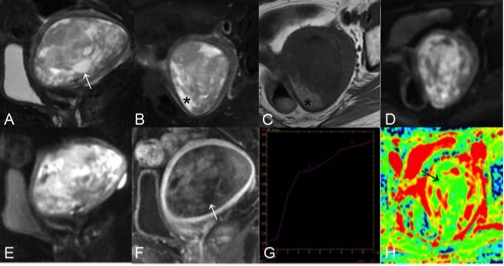
Figure 1. A: sag T2WI, B: axi T2WI, C: axi T1WI,
D: axi DWI, E: sag DWI, F: sag DCE image, G: signal intensity-time(SI-T) curve
map, H: ADC map. Seventy-nine-year-old-woman with vaginal discharge. (A) demonstrated a tumor growing exophytically within the endometrial
cavity. It was heterogeneously hyperintense on the T2WI(A,B), hypointense on
the T1WI(C), including necrotic portions(white arrows) and hemorrhage(*). The
neoplastic parenchyma showed heterogeneous enhancement slight weaker on the DCE images(F) and delayed enhancement on SI-T curve
map(G). The black arrow on ADC map(H, value 1.27×10⁻³mm2/s
) showed the diseased area.
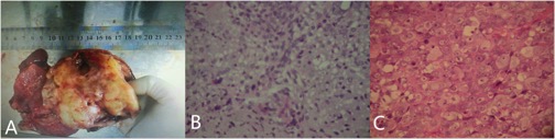
Figure
2. At laparotomy, the uterus was enlarged as 60 gestational days. Macroscopic view(A) of the resected specimen demonstrated
a large exophytic mass from the posterior uterine wall, occupying the whole
uterine cavity and combining with hemorrhage and necrosis. Tumor size was 9.5
cm × 8 cm × 3.5 cm. Histological specimen ( hematoxylin / eosin stain,
high-power field ) revealed myxoid degeneration in cytoplasm and cartilage
lacunar-like structure in sarcoma region (B: chondrosarcoma ). The carcinoma
region demonstrated tumor cells presented widespread with clear
nucleoli and abundant cytoplasm, the nuclei were oval to round in shape and the
nuclear-to-cytoplamic ratio was consistently high (C: endometrial
undifferentiated carcinoma).
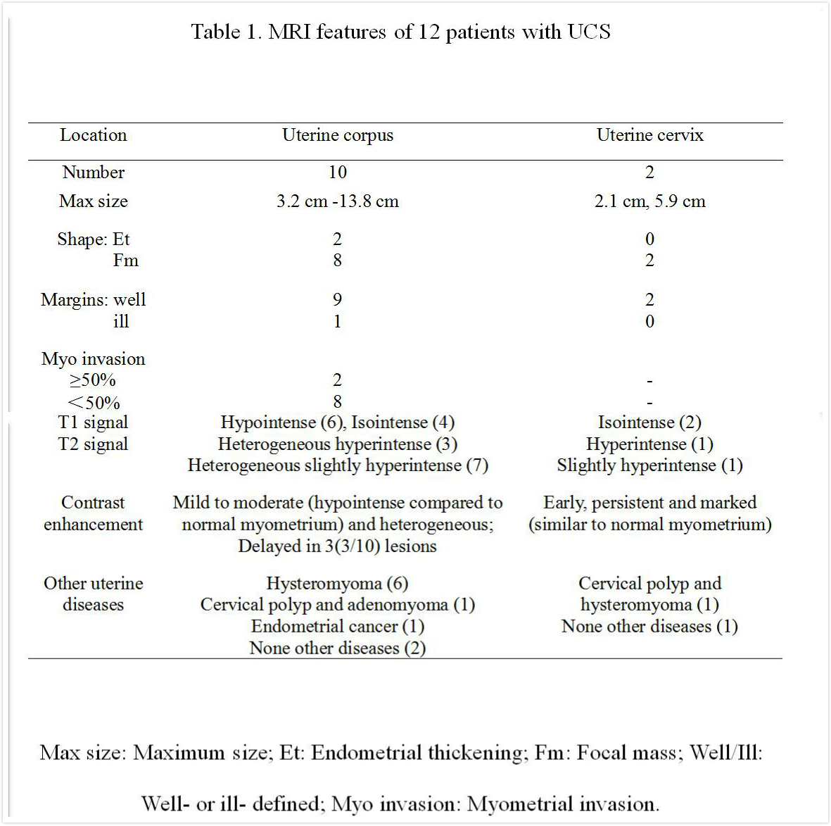
Table 1