4043
Evaluation of DWI with lymphovascular space invasion and without lymphovascular space invasion in the stage I endometrial Carcinoma1the first Affiliated Hospital of Dalian Medical University, Dalian, China, 2the First Affiliated Hospital of Dalian Medical University, Dalian, China, 3GE Healthcare, Beijing, China
Synopsis
Endometrial cancer is one of the most commonly diagnosed gynecologic malignancies in developed countries. The fundamental extent of the operation involves hysterectomy and bilateral salpingo-oophorectomy for the endometrial carcinoma. The presence of LVSI is associated with lymph node metastases. However, Lymphadenectomy can’t improve the survival rate and has a reported higher rate of postoperative complications in endometrial cancer patients. So lymphadenectomy is a controversial issue in recent years. LVSI is a crucial factor in determining the lymphadenectomy and cannot be detected on traditional MR imaging.
Purpose
The current study was designed to investigate and compare the value of apparent diffusion coefficient (ADC) among lymphovascular space invasion (LVSI ) and without LVSI in the Federation of Gynecology and Obstetrics (FIGO) stage I endometrial carcinoma.Materials and Methods
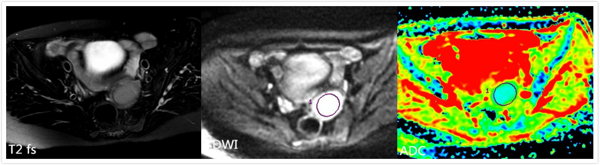
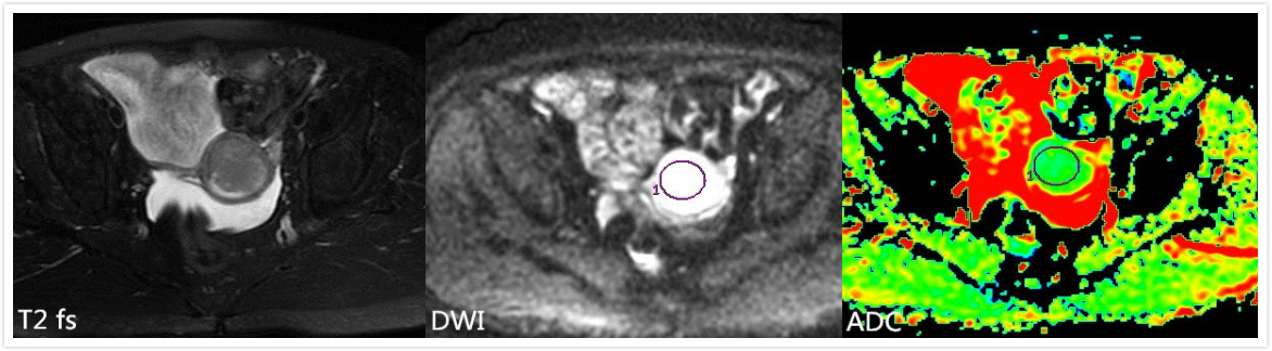
Thirty-six females were collected in the current study, including 16 patients with LVSI and 20 patients without LVSI in the stage I endometrial carcinoma confirmed by pathology between January 2013 and July 2018. Regions of interest were drawn to obtain apparent diffusion coefficient (ADC). ROI placement and maps were demonstrated in the Figure.1 and Figure.2. The ADC values were measured among the patients with LVSI and without LVSI. The ADC values of 16 patients with LVSI(LVSI group) and 20 patients without LVSI(without LVSI group) in the stage I endometrial carcinoma were retrospectively analyzed. Independent sample T-test was applied to analyzed significant difference of the results.Results
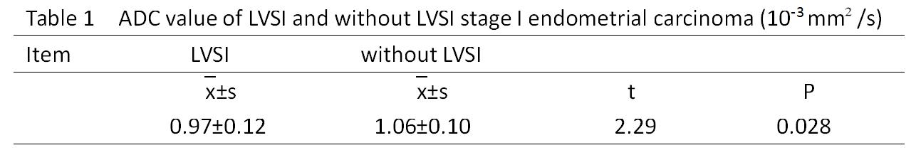
The mean ADC value of the LVSI group was (0.97±0.12)×10-3 mm²/s, which was smaller than that of without LVSI group was (1.06±0.10)×10-3 mm²/s, as shown in Figure 3. ADC value were significantly different between the LVSI group and without LVSI group (P<0.05).Conclusion
DWI has shown promising application for prediction of LVSI in recent years. DWI can be used as a significant technique to differentiate the LVSI and without LVSI in the stage I endometrial carcinoma. DWI is an effective method for the evaluation of LVSI.Acknowledgements
No acknowledgement found.References
[1] Nougaret S, Reinhold C, Alsharif SS, et al. Endometrial cancer: combined MR volumetry and diffusion-weighted imaging for assessment of myometrial and lymphovascular invasion and tumor grade. Radiology, 2015,276(3):797-808.
[2] Ueno Y, Forghani B, Forghani R, et al. Endometrial carcinoma:MR imaging-based texture model for preoperative risk stratification-a preliminary analysis. Radiology,2017,284(3):748-757.
Figures